What is Horner syndrome?
Horner syndrome (oculosympathetic paresis, Bernard–Horner syndrome) is the clinical picture produced when the sympathetic supply to one eye and one side of the face is interrupted anywhere along its long route from the hypothalamus, down the brainstem and spinal cord, over the apex of the lung and back up the neck along the carotid artery. Because the parasympathetic supply is intact, the eye shows the effects of unopposed parasympathetic tone.
The classic triad is ptosis, miosis and anhidrosis on the same side as the lesion. Horner syndrome itself is not dangerous, but its cause can be — a lung apex cancer, a carotid artery dissection, a brainstem stroke or, in a child, neuroblastoma. That is why exams test not just recognition but localisation.
What is the three-neuron oculosympathetic pathway?
The sympathetic supply to the eye is a three-neuron chain. Lesions are named by the neuron they hit: first-order (central), second-order (preganglionic) or third-order (postganglionic).

| Neuron | Starts in | Course | Ends at |
|---|---|---|---|
| First-order (central) | Posterolateral hypothalamus | Descends uncrossed through the lateral brainstem (midbrain, pons, lateral medulla) and cervical cord | Ciliospinal centre of Budge in the intermediolateral grey column, C8–T1 (some sources C8–T2) |
| Second-order (preganglionic) | Ciliospinal centre (exits mainly with T1) | Through the white ramus into the sympathetic chain, over the pulmonary apex, through the stellate ganglion, up the cervical sympathetic chain close to the carotid sheath | Superior cervical ganglion (at about C2–C3, near the carotid bifurcation / angle of the jaw) |
| Third-order (postganglionic) | Superior cervical ganglion | Along the internal carotid artery into the cavernous sinus (briefly with the abducens nerve), then with the ophthalmic division of the trigeminal nerve (nasociliary nerve) and long ciliary nerves into the orbit | Iris dilator muscle, Müller (superior tarsal) muscle; sweat fibres to most of the face leave early along the external carotid artery |
What are the clinical features of Horner syndrome?

| Sign | Mechanism | Exam detail |
|---|---|---|
| Ptosis (mild, ~1–2 mm) | Paralysis of Müller's (superior tarsal) muscle, a sympathetically supplied smooth muscle | Levator palpebrae (CN III) is intact, so ptosis is only partial and the lid still elevates on upgaze |
| Upside-down (reverse) ptosis | Denervation of the smooth-muscle lower-lid retractors | Lower lid sits slightly higher, narrowing the palpebral fissure |
| Miosis | Weak iris dilator; unopposed sphincter (parasympathetic) | Anisocoria is greater in dim light; both pupils still react briskly to light |
| Dilation lag | Loss of active dilator pull | When lights go off, the Horner pupil dilates several seconds later than the normal pupil |
| Anhidrosis | Loss of sudomotor fibres | Extent depends on lesion level (see table above) |
| Apparent enophthalmos | Narrowed palpebral fissure (upper ptosis + raised lower lid) | Not true enophthalmos — measurements show no real backward displacement |
| Iris heterochromia | Sympathetic input is needed for iris pigmentation during infancy | Lighter iris on the affected side in congenital Horner; not seen in recently acquired Horner |
Because the problem is a small pupil, the anisocoria is exaggerated in darkness (the normal pupil dilates, the Horner pupil cannot keep up) and reduced in bright light. This is the reverse of a third-nerve lesion or a pharmacologically dilated pupil, where bright light exaggerates the difference.
How do you localise the lesion in Horner syndrome?
Localisation comes mostly from the company the Horner syndrome keeps — the associated symptoms and signs. In a large series (Kanagalingam & Miller), of the cases with an identified cause, about 13% were central, 44% preganglionic and 43% postganglionic.
| Level | Key causes | Associated clues |
|---|---|---|
| First-order (central) | Lateral medullary (Wallenberg) syndrome from PICA or vertebral artery occlusion; other brainstem strokes; multiple sclerosis; syringomyelia; cervical cord trauma/tumour; Arnold–Chiari malformation | Crossed sensory loss (ipsilateral face, contralateral body pain/temperature), vertigo, nystagmus, dysphagia/hoarseness (nucleus ambiguus), ipsilateral ataxia; long-tract signs in cord lesions |
| Second-order (preganglionic) | Pancoast (superior sulcus) tumour of the lung apex; neck/thyroid surgery and large goitre; brachial plexus injury including Klumpke (lower trunk) birth palsy; subclavian aneurysm; central line or chest-drain trauma; neuroblastoma in children | Shoulder pain, pain/paraesthesia along the medial arm and ring/little fingers, wasting of small hand muscles (C8–T1 roots); hoarseness (recurrent laryngeal nerve) |
| Third-order (postganglionic) | Internal carotid artery dissection; cavernous sinus lesions; internal carotid aneurysm; cluster headache; Raeder paratrigeminal syndrome; herpes zoster | Ipsilateral head, face or neck pain (dissection), other cranial nerve palsies (III, IV, V1, VI in cavernous sinus), minimal anhidrosis |
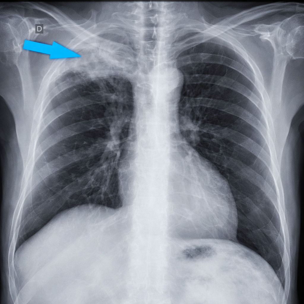
Pancoast tumour anatomy is a favourite: the tumour at the lung apex invades the lower brachial plexus (C8, T1) and the sympathetic chain/stellate ganglion. On the neck of the first rib the sympathetic trunk lies most medially, with the superior intercostal artery lateral to it and the first thoracic nerve most laterally; the cervicothoracic (stellate) ganglion sits directly in front of the rib neck. That is why an apical tumour so often catches the sympathetic chain. A smoker with shoulder pain radiating down the inner arm to the little finger plus ptosis and miosis is a Pancoast tumour with a preganglionic Horner syndrome.

In children, a Horner syndrome without a clear history of birth trauma or surgery must be investigated for neuroblastoma (urinary catecholamine metabolites and MRI of brain, neck and chest). A congenital Horner syndrome is suspected when there is heterochromia (the affected iris is lighter).
Which eye-drop tests confirm and localise Horner syndrome?
Eye-drop tests answer two separate questions: (1) Is this Horner syndrome or just physiological anisocoria? (cocaine, apraclonidine) and (2) Is the lesion postganglionic or not? (hydroxyamphetamine).
| Drug | Mechanism | Normal pupil | Horner pupil | Use |
|---|---|---|---|---|
| Cocaine (2–10%) | Blocks re-uptake of noradrenaline at the nerve ending | Dilates | Dilates poorly or not at all (little noradrenaline released) | Confirms Horner (post-drop anisocoria ≥ 0.8 mm); cannot localise; hard to obtain and shows in urine drug tests |
| Apraclonidine (0.5–1%) | Strong α2, weak α1 agonist | No change or slight constriction | Dilates (denervation supersensitivity of α1 receptors) — reversal of anisocoria; ptosis often improves | Now the preferred confirmatory test; may be negative very early, before supersensitivity develops |
| Hydroxyamphetamine (0.5–1%) | Releases stored noradrenaline from the third-order nerve terminal | Dilates | Dilates if third-order neuron intact (first/second-order lesion); fails to dilate in a third-order (postganglionic) lesion | Localises: separates postganglionic from central/preganglionic |
Apraclonidine is avoided in infants because of reported central nervous system and respiratory depression. Imaging then follows the clinical localisation: MRI brain and cervical cord for central lesions, CT chest/neck for preganglionic lesions, and CT or MR angiography of the neck vessels for a painful or postganglionic Horner syndrome.
How do you approach a patient with a new Horner syndrome?
- Confirm it is Horner syndrome. Compare pupils in bright and dim light: if the anisocoria increases in the dark and the smaller pupil shows dilation lag, suspect Horner syndrome. Look for the mild upper-lid ptosis and the slightly raised lower lid. If unsure, use apraclonidine (reversal of anisocoria) or cocaine (failure of the small pupil to dilate).
- Decide how urgent it is. An acute, painful Horner syndrome (head, face or neck pain) — especially after neck trauma or with stroke symptoms — needs same-day vascular imaging for carotid dissection.
- Localise clinically. Brainstem signs (vertigo, crossed sensory loss, dysphagia, ataxia) → first-order. Arm pain, hand wasting, smoking history, previous neck or chest surgery → second-order. Headache with other cranial nerve palsies (III, IV, V1, VI) → cavernous sinus, third-order.
- Localise pharmacologically if needed. Hydroxyamphetamine separates a third-order lesion (no dilation) from a first- or second-order lesion (dilation).
- Image the right region. MRI brain (with contrast) for central and many postganglionic causes; CT chest/neck for preganglionic causes such as an apical lung tumour; CT or MR angiography of the neck vessels for dissection. In a child without a clear history of birth trauma or surgery: examination, urinary catecholamine metabolites and MRI of brain, neck and chest to exclude neuroblastoma.
Treatment is that of the cause — for example, oncological treatment of an apical tumour or antithrombotic treatment of a carotid dissection. The Horner syndrome itself usually needs no treatment; the mild ptosis can be corrected surgically if it is troublesome (apraclonidine drops also have a measurable lid-elevating effect, which is used diagnostically).
How is Horner syndrome different from other causes of ptosis and a small pupil?
| Condition | Pupil | Ptosis | Key differentiator |
|---|---|---|---|
| Horner syndrome | Small, reacts to light; anisocoria worse in dark | Mild (1–2 mm) | Anhidrosis, dilation lag, reverse ptosis; apraclonidine reverses anisocoria |
| Third-nerve palsy | Large (if pupil involved), poor light reaction | Marked or complete | Eye 'down and out'; anisocoria worse in bright light |
| Physiological anisocoria | Difference usually < 1 mm (seen in up to 20% of people), both react | None | Same difference in light and dark; varies day to day; drop tests negative |
| Argyll Robertson pupil | Small, irregular, usually bilateral | No | Light–near dissociation; neurosyphilis (tabes dorsalis) |
| Adie tonic pupil | Large, poor light response, slow tonic constriction to near | No | Light–near dissociation; slow redilation; ciliary ganglion damage |
| Aponeurotic (senile) or myasthenic ptosis | Normal | Variable | No anisocoria; myasthenia is fatigable |
What are the high-yield exam traps on Horner syndrome?
- Ptosis muscle: the paralysed muscle is Müller's (superior tarsal), not levator palpebrae superioris (CN III) and not orbicularis oculi (CN VII).
- Enophthalmos is apparent, not real — the narrowed fissure only makes the eye look sunken.
- Heterochromia = congenital (or very long-standing) Horner syndrome; it does not appear after an acute lesion.
- Lateral medullary syndrome (PICA or vertebral artery) gives an ipsilateral Horner syndrome with crossed pain–temperature loss — Horner syndrome is present in the large majority of these patients.
- Pancoast tumour causes a second-order (preganglionic) Horner syndrome; carotid dissection causes a third-order (postganglionic) one.
- Hydroxyamphetamine failure to dilate means a postganglionic lesion; cocaine and apraclonidine confirm but do not localise.
- Large goitre can compress the cervical sympathetic chain (Horner), trachea (dyspnoea), oesophagus (dysphagia) and recurrent laryngeal nerve (hoarseness) — a common match-the-following.
- Stellate ganglion block produces a temporary Horner syndrome — a sign that the block has worked.