NEET-PG Anatomy Previous Year Questions (2017–2026)

92 NEET-PG Anatomy PYQs from 2017 to 2026, tagged by topic, with an explanation for every question. Anatomy had 11 of the 180 questions in NEET-PG 2026.

Last verified: 24 September 2026 against Kinase question bank and NEET-PG 2026 recall paper. natboard.edu.in · nmc.org.in

Quick Answer

NEET-PG Anatomy had 11 of 180 questions (6.1%) in the NEET-PG 2026 paper — up to 44 marks. Kinase has 92 NEET-PG Anatomy PYQs from 2017 to 2026; the most-asked topic is Head and Neck (18 questions).

Anatomy in NEET-PG at a glance

Questions in NEET-PG 202611 of 180 (6.1%)
Maximum marks at stake44 of 720
NEET-PG Anatomy PYQs on Kinase92 (5.5% of all NEET-PG PYQs)
Years covered2017–2026
INICET Anatomy PYQs on Kinase213

Most-asked Anatomy topics in NEET-PG PYQs

TopicNEET-PG PYQsShare of subject
Head and Neck1820%
Embryology1516%
Neuroanatomy1516%
Abdomen and Pelvis1213%
Thorax910%

Across NEET PG and INI-CET papers: Most repeated Anatomy topics →

Sample NEET-PG Anatomy PYQs with answers

Question 1 · NEET PG Aug 2026

During a routine bimanual examination of a healthy young nulliparous woman, the cervix is felt pointing backward while the fundus tips forward to rest over the bladder - the normal anteverted, anteflexed lie of the uterus. Her tutor points out that a single fibromuscular cord runs from each uterine cornu, through the deep inguinal ring and inguinal canal, to end in the labium majus, and asks: which structure is chiefly responsible for holding the fundus forward in anteversion?

  1. A.Cardinal ligament
  2. B.Pubocervical ligament
  3. C.Round ligament
  4. D.Uterosacral ligament
Show answer and explanation

Answer: C. Round ligament

Correct answer: C. Round ligament. The round ligament of the uterus is the principal structure maintaining uterine anteversion.

Each round ligament passes from the uterine cornu through the deep inguinal ring and inguinal canal to blend into the labium majus. Its forward and downward pull tilts the fundus anteriorly over the bladder, producing anteversion. By contrast the uterosacral ligament pulls the cervix backward and chiefly resists retroversion, so for anteversion specifically the round ligament is the best answer.
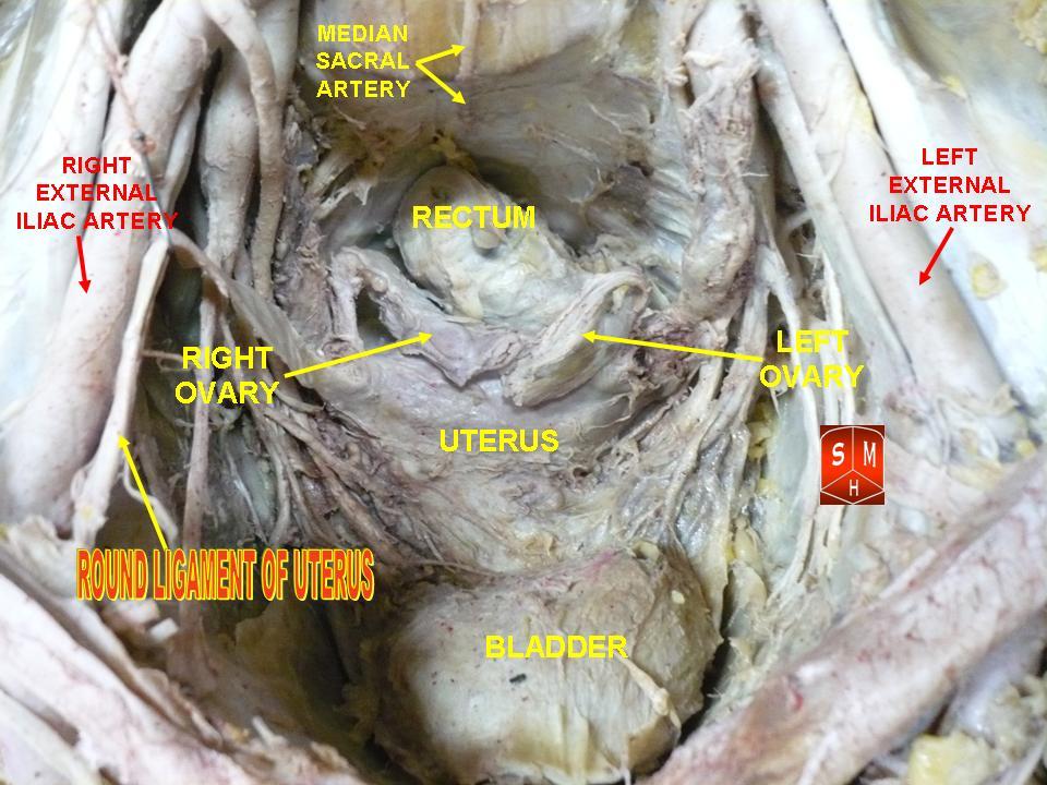
Cadaveric female pelvis with the round ligament of the uterus labelled

Cadaveric dissection: the round ligament runs forward from the uterine cornu toward the deep inguinal ring, tilting the fundus into anteversion.

Incorrect Options:

  • A. Cardinal ligament: the transverse cervical (Mackenrodt) ligament gives lateral support at the cervix and vaginal vault and prevents uterine descent; it does not create anteversion. Pelvic support diagram labelling the cardinal ligament (CL)
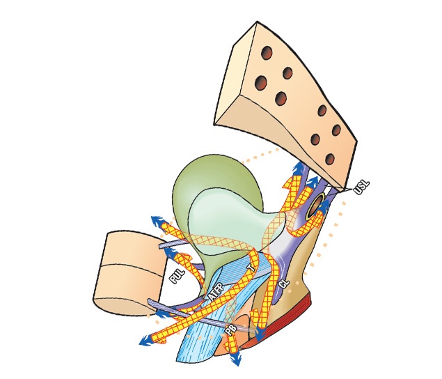
    Pelvic support ligaments; the cardinal ligament (CL) anchors the cervix laterally.

  • B. Pubocervical ligament: part of the anterior endopelvic (levator) fascia supporting the bladder neck and cervix; it has no role in tilting the fundus forward. Sagittal diagram of pelvic floor and endopelvic fascia
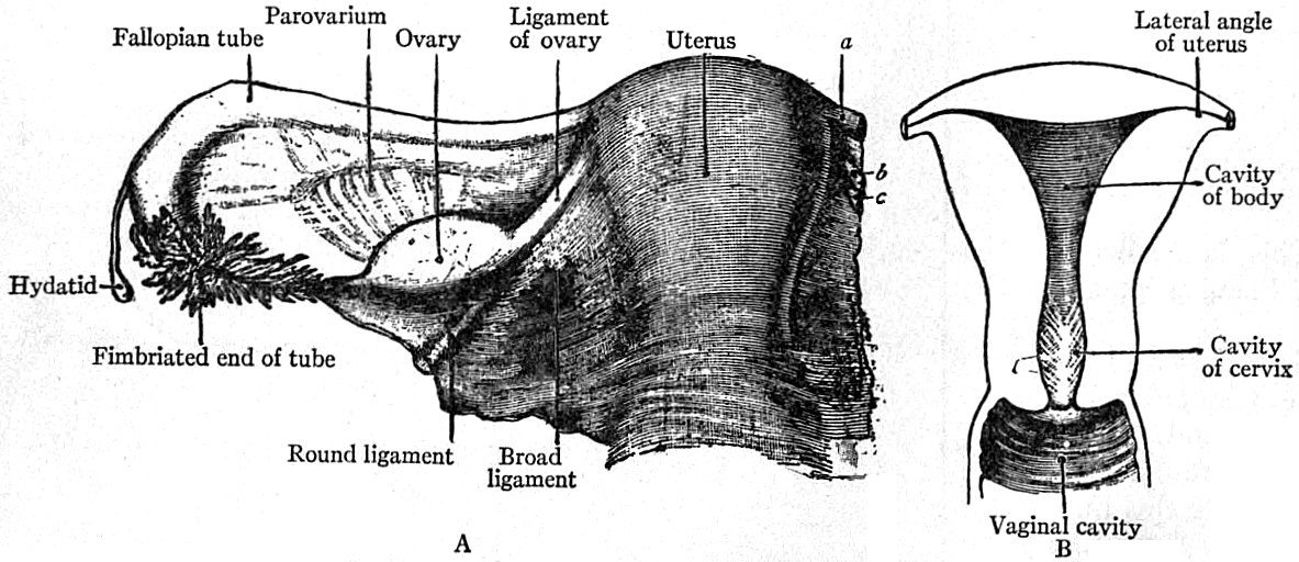
    Levator ani and endopelvic (pubocervical) fascia providing anterior support.

  • D. Uterosacral ligament: runs from the cervix to the sacrum and pulls the cervix backward, so it chiefly prevents retroversion rather than producing anteversion. Pelvic support diagram labelling the uterosacral ligament (USL)
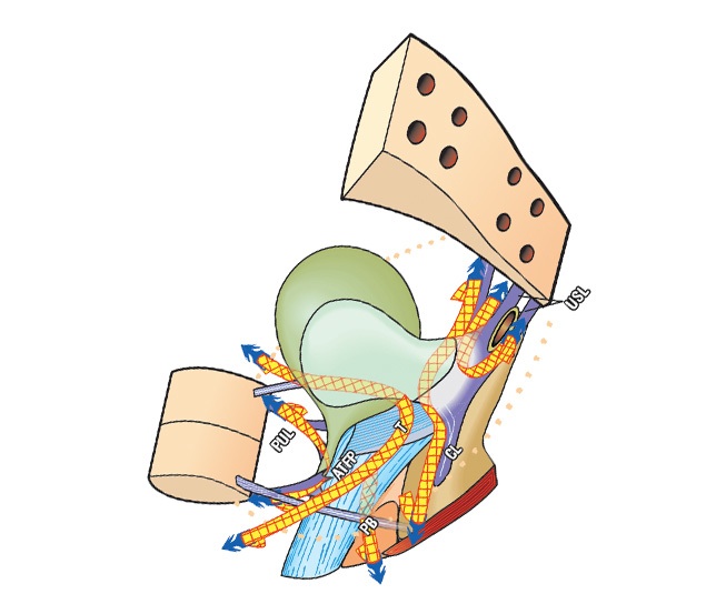
    Pelvic support ligaments; the uterosacral ligament (USL) tethers the cervix posteriorly.

High-Yield Facts:

  • Maintains anteversion: round ligament is greater than uterosacral ligament.
  • Prevents retroversion: uterosacral ligament is greater than round ligament.
  • The round ligament is the female remnant of the gubernaculum and travels with the canal of Nuck.
  • The cardinal (Mackenrodt) ligament transmits the uterine artery and is the main lateral cervical support.
Question 2 · NEET PG Aug 2026

A 60-year-old man with long-standing hypertension suddenly develops a drooping right upper eyelid; the right eye rests down-and-out with a dilated pupil, while his LEFT arm and leg show coarse flinging involuntary movements and a tremor, together with loss of pain and temperature sensation on the left. MRI reveals a paramedian infarct of the right midbrain tegmentum (Benedikt syndrome). Damage to which structure best accounts for the contralateral tremor and involuntary movements?

  1. A.Substantia nigra
  2. B.Red nucleus
  3. C.Medial lemniscus
  4. D.Spinal lemniscus
Show answer and explanation

Answer: B. Red nucleus

Correct answer: B. Red nucleus. In Benedikt syndrome the contralateral tremor and involuntary movements come from injury to the red nucleus.

The red nucleus lies in the midbrain tegmentum and is a relay in the dentato-rubro-thalamic (cerebellar) circuit. Damage to it, together with adjacent superior cerebellar peduncle fibres, produces contralateral tremor, ataxia and choreoathetosis. The nearby oculomotor (cranial nerve three) fascicles are also caught, giving the ipsilateral down-and-out eye with ptosis and a dilated pupil, while medial lemniscus/spinothalamic involvement explains the contralateral sensory loss.

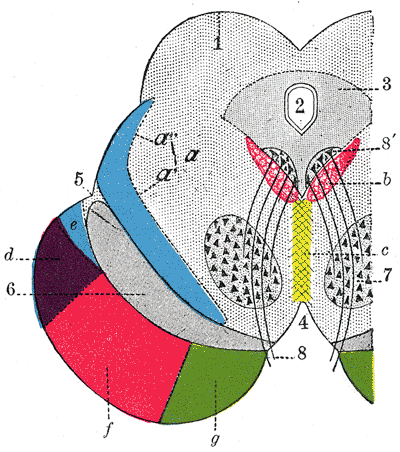
Coloured transverse section of the midbrain showing the paired red nuclei in the tegmentum

Transverse midbrain section; the paired red nuclei sit in the tegmentum, ventral to the cerebral aqueduct.

Incorrect Options:

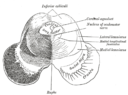
  • A. Substantia nigra: a dopaminergic nucleus; its loss causes parkinsonism (rest tremor, rigidity, bradykinesia), not the rubral tremor of Benedikt syndrome. Midbrain section labelling the substantia nigra

    Midbrain at the inferior colliculi; the substantia nigra lies between the crus cerebri and tegmentum.

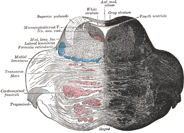
  • C. Medial lemniscus: carries fine touch, vibration and proprioception; its damage gives contralateral loss of these modalities, not a tremor. Brainstem section labelling the medial lemniscus

    Brainstem section; the medial lemniscus conveys dorsal-column sensation upward.

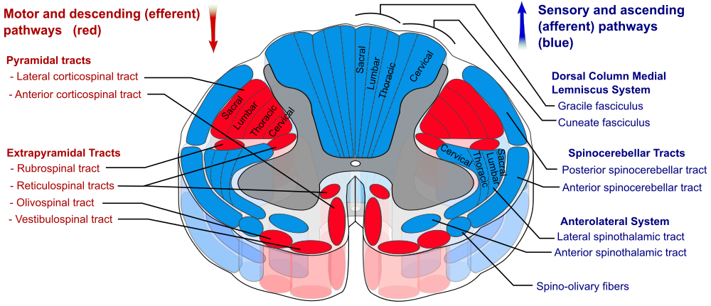
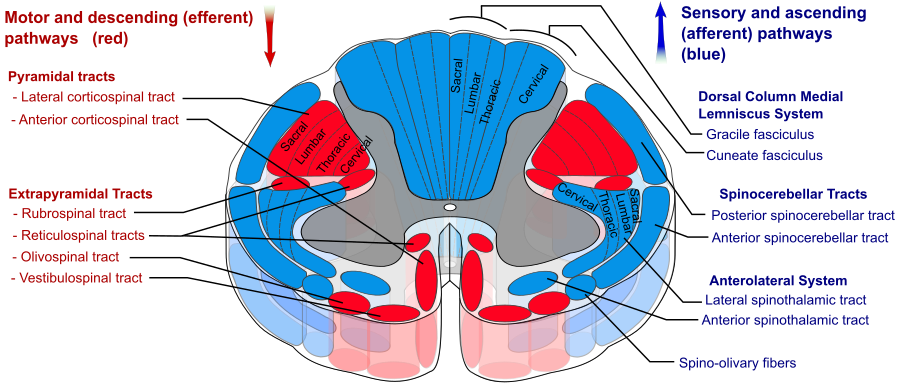
  • D. Spinal lemniscus: the brainstem continuation of the spinothalamic tract carrying pain and temperature; its involvement explains the contralateral pain/temperature loss, not the movements. Spinal cord tracts diagram highlighting the anterolateral spinothalamic system

    The anterolateral (spinothalamic) system continues as the spinal lemniscus in the brainstem.

High-Yield Facts:

  • Benedikt syndrome = red nucleus plus cranial nerve three fascicle (paramedian midbrain tegmentum).
  • Weber syndrome = crus cerebri: cranial nerve three palsy with contralateral hemiplegia.
  • Claude syndrome = red nucleus plus superior cerebellar peduncle: cranial nerve three palsy with contralateral ataxia.
  • The red nucleus is a key relay of the dentato-rubro-thalamic tremor circuit.
Question 3 · NEET PG Aug 2026

A young man is brought to the emergency department after a knife wound to the right side of his back. Below the level of injury he has, on the RIGHT (same) side, weakness together with loss of vibration and joint-position sense, while on the LEFT (opposite) side he has lost pain and temperature sensation starting a couple of segments lower. Which cord syndrome does this dissociated, one-sided pattern represent?

  1. A.Central cord syndrome
  2. B.Anterior cord syndrome
  3. C.Posterior cord syndrome
  4. D.Brown-Sequard syndrome
Show answer and explanation

Answer: D. Brown-Sequard syndrome

Correct answer: D. Brown-Sequard syndrome. A clean hemisection of the cord produces this crossed pattern of deficits.

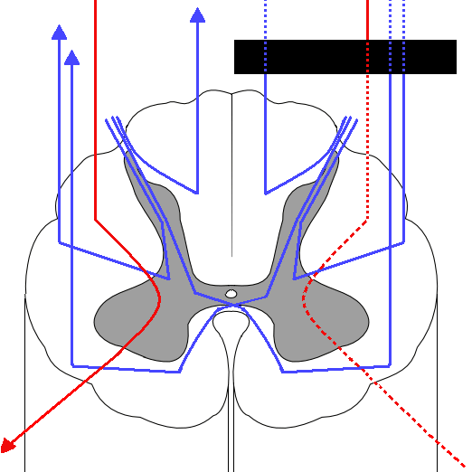
Hemisection damages the ipsilateral dorsal column (uncrossed) giving ipsilateral loss of vibration and proprioception, and the ipsilateral lateral corticospinal tract (already crossed in the medulla) giving ipsilateral weakness. The spinothalamic tract carries pain and temperature that have decussated one to two segments above entry, so its interruption causes contralateral pain and temperature loss beginning a little below the lesion.

Spinal cord cross-section showing ascending dorsal column and crossed spinothalamic pathways

Cord tracts: dorsal columns (blue, uncrossed) and spinothalamic fibres (red, crossed) explain the dissociated deficits of hemisection.

Incorrect Options:

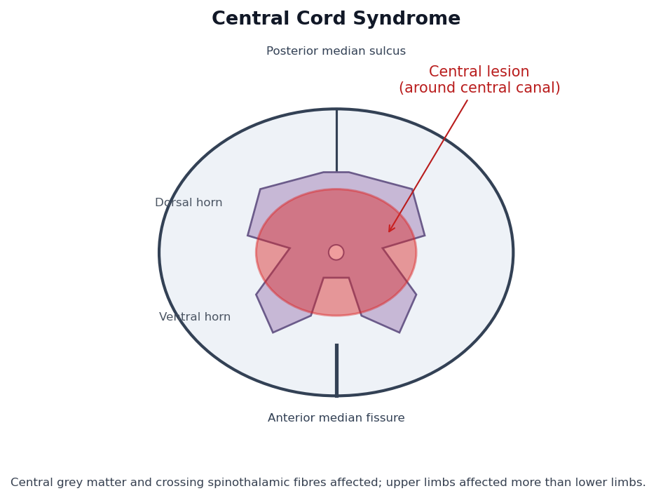
  • A. Central cord syndrome: damage around the central canal (often hyperextension injury) affecting crossing spinothalamic fibres, with upper limbs weaker than lower limbs - not a hemi-pattern. Cord cross-section schematic with the central region shaded

    Central cord syndrome: central grey and crossing fibres affected; arms more than legs.

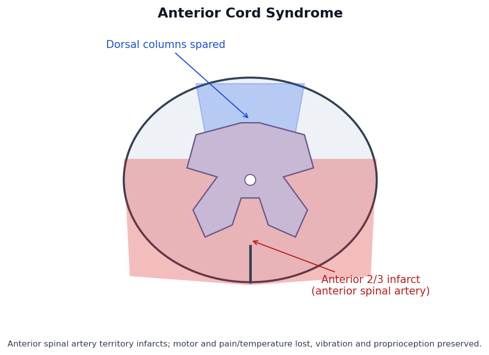
  • B. Anterior cord syndrome: anterior spinal artery infarct loses motor power and pain/temperature bilaterally while the dorsal columns are spared. Cord cross-section schematic with the anterior two-thirds shaded

    Anterior cord syndrome: anterior two-thirds infarcted, dorsal columns preserved.

  • C. Posterior cord syndrome: isolated dorsal column loss gives loss of vibration, proprioception and fine touch with sensory ataxia - rare and not one-sided motor loss. Spinal cord tracts diagram highlighting the dorsal column medial lemniscus system

    The dorsal column medial lemniscus system (gracile and cuneate fasciculi) is lost in posterior cord syndrome.

High-Yield Facts:

  • Brown-Sequard: ipsilateral motor and dorsal-column loss with contralateral pain and temperature loss.
  • The spinothalamic tract decussates within one to two segments of entry, so the crossed loss starts just below the lesion.
  • Dorsal columns ascend ipsilaterally; the lateral corticospinal tract has already crossed in the medulla.
  • Most common causes are penetrating trauma (stab or gunshot) and lateral cord compression.

Anatomy concept guides

Browse the full concept library and clinical image atlas.

Practise the rest: open the Kinase question bank, choose Anatomy, set the exam to NEET-PG and switch on PYQs only. Questions you get wrong go into Fix My Weakness for targeted revision.

Frequently Asked Questions

How many Anatomy questions were in NEET-PG 2026?▼
11 of the 180 questions in the NEET-PG 2026 paper (30 August 2026) were Anatomy — 6.1% of the paper, worth up to 44 marks at +4 per correct answer. This is from the recall paper in the Kinase question bank.
How many NEET-PG Anatomy PYQs does Kinase have?▼
Kinase has 92 NEET-PG Anatomy previous year questions from 2017 to 2026, each with an explanation, plus 213 INICET Anatomy PYQs for extra practice.
What are the most-asked Anatomy topics in NEET-PG?▼
In Kinase's NEET-PG Anatomy PYQs, the most-asked topics are Head and Neck, Embryology, Neuroanatomy. Head and Neck alone accounts for 18 of the 92 questions.
Are NEET-PG Anatomy PYQs the same as INICET PYQs?▼
Both exams test the MBBS syllabus, so most Anatomy concepts overlap. The marking differs: NEET-PG is +4 / −1 and INICET is +1 / −1/3. Kinase lets you practise either exam's PYQs, or both together.
How should I practise Anatomy PYQs for NEET-PG?▼
Start with the most-asked topics above, do a daily set of Anatomy PYQs, and read every explanation — including the ones you got right. Kinase's Fix My Weakness then builds practice sets from the topics you miss most.
92 Anatomy PYQs
2017–2026 papers
Topic + year tagged

Ready to Start Practicing?

Anatomy was 11 questions (up to 44 marks) in NEET-PG 2026.

Start Free Practice

No credit card required