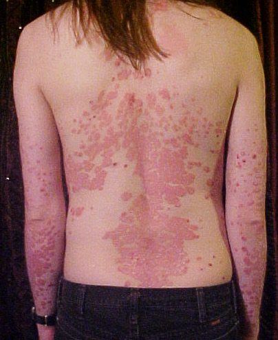
Dermatology


Ophthalmology
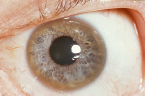
Kayser-Fleischer Ring
Clinical eye photograph
Herbert L. Fred, MD, Hendrik A. van Dijk · CC BY 3.0

Cherry-Red Spot (Central Retinal Artery Occlusion)
Fundus photograph
18mlk7 · CC BY-SA 4.0
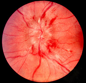
Papilloedema
Fundus photograph
Jonathan Trobe, M.D. – University of Michigan Kellogg Eye Center · CC BY 3.0
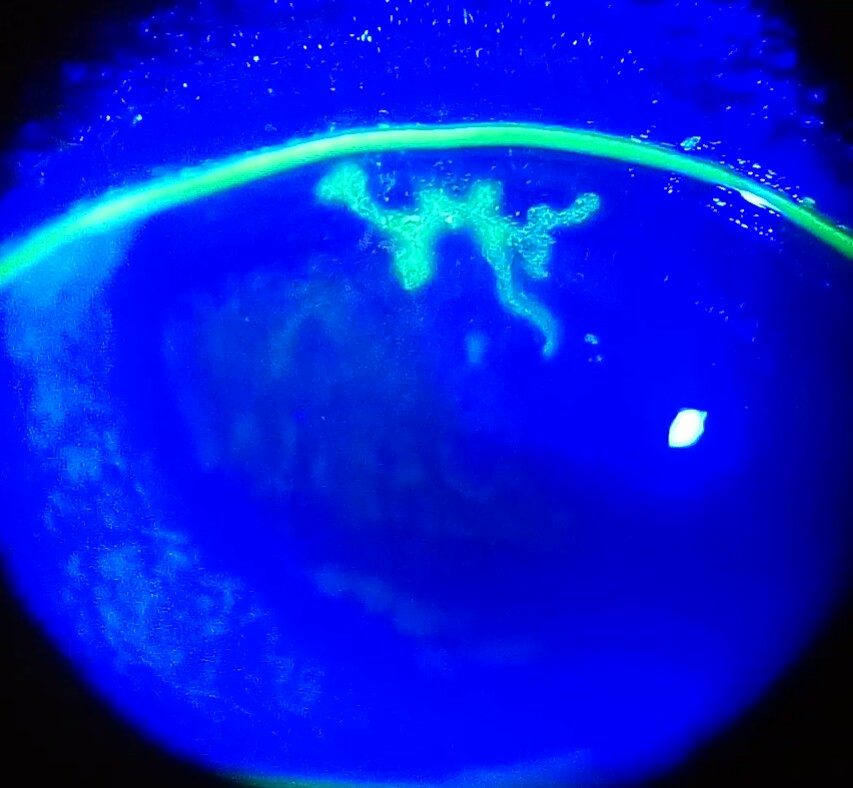
Dendritic Ulcer (Herpes Simplex Keratitis)
Slit-lamp photograph with fluorescein (cobalt blue light)
Imrankabirhossain · CC BY-SA 4.0

Horner Syndrome
Clinical photograph
Davplast · CC BY-SA 4.0
Radiology

Pneumoperitoneum (Air Under the Diaphragm)
Erect chest X-ray + CT abdomen
Cerevisae · CC BY-SA 4.0

Sigmoid Volvulus (Coffee Bean Sign)
Abdominal X-ray
Mont4nha · CC0

Boot-Shaped Heart (Tetralogy of Fallot)
Chest X-ray (PA)
Medicalpal · CC BY-SA 4.0

Extradural (Epidural) Haematoma on CT
Non-contrast CT head
James Heilman, MD · CC BY-SA 4.0
Haematology

Sickle Cells on Peripheral Smear
Peripheral blood smear
SpicyMilkBoy · CC BY-SA 4.0

Auer Rods (Acute Myeloid Leukaemia)
Peripheral smear / marrow (Wright stain)
Ed Uthman from Houston, TX, USA · CC BY 2.0

Hypersegmented Neutrophils
Peripheral blood smear
Prof. Osaro Erhabor · CC BY-SA 4.0
Microbiology

Plasmodium falciparum: Ring Forms and Gametocyte
Thin blood smear (Giemsa)
Jenkayaks · CC BY-SA 3.0

Acid-Fast Bacilli (Ziehl-Neelsen Stain)
Ziehl-Neelsen stained smear, ×1000
CDC / Dr. George P. Kubica · Public domain

Cryptococcus neoformans (India Ink Preparation)
India ink wet mount
CDC / Dr. Leanor Haley · Public domain
Clinical signs

For exam preparation and medical education only — not a substitute for clinical judgement, diagnosis or treatment of any patient.