NEET-PG Pathology Previous Year Questions (2017–2026)

120 NEET-PG Pathology PYQs from 2017 to 2026, tagged by topic, with an explanation for every question. Pathology had 13 of the 180 questions in NEET-PG 2026.

Last verified: 24 September 2026 against Kinase question bank and NEET-PG 2026 recall paper. natboard.edu.in · nmc.org.in

Quick Answer

NEET-PG Pathology had 13 of 180 questions (7.2%) in the NEET-PG 2026 paper — up to 52 marks. Kinase has 120 NEET-PG Pathology PYQs from 2017 to 2026; the most-asked topic is Hematopathology (31 questions).

Pathology in NEET-PG at a glance

Questions in NEET-PG 202613 of 180 (7.2%)
Maximum marks at stake52 of 720
NEET-PG Pathology PYQs on Kinase120 (7.2% of all NEET-PG PYQs)
Years covered2017–2026
INICET Pathology PYQs on Kinase264

Most-asked Pathology topics in NEET-PG PYQs

TopicNEET-PG PYQsShare of subject
Hematopathology3126%
General Pathology2218%
Gastrointestinal System1210%
Genitourinary System1210%
Cardiovascular System108%

Across NEET PG and INI-CET papers: Most repeated Pathology topics →

Sample NEET-PG Pathology PYQs with answers

Question 1 · NEET PG Aug 2026

A 12-year-old girl is investigated for heavy menstrual bleeding since menarche and easy bruising; her mother also has menorrhagia and a maternal uncle bleeds excessively after dental work. On testing, the platelet count is normal but the bleeding time is prolonged, the activated partial thromboplastin time is prolonged, the prothrombin time is normal, ristocetin-induced platelet aggregation is impaired, and factor VIII activity is reduced to about half of normal. What is the diagnosis?

  1. A.Von Willebrand disease
  2. B.Haemophilia A
  3. C.Bernard-Soulier syndrome
  4. D.Glanzmann thrombasthenia
Show answer and explanation

Answer: A. Von Willebrand disease

Correct answer: A. Von Willebrand disease.

The combination of mucocutaneous bleeding (menorrhagia, easy bruising), a prolonged bleeding time, prolonged aPTT with normal PT, impaired ristocetin-induced aggregation and a secondary fall in factor VIII is classic von Willebrand disease. Von Willebrand factor (vWF) does two jobs: it bridges platelets to subendothelium (so its deficiency prolongs the bleeding time and blunts ristocetin aggregation) and it chaperones factor VIII in plasma (so low vWF lowers factor VIII, prolonging the aPTT). The autosomal, both-sexes inheritance fits too.
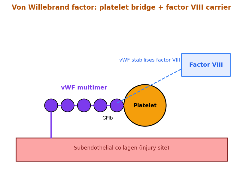
Von Willebrand factor multimers binding platelets
vWF bridges platelets and carries factor VIII; its lack causes this mixed picture.

Incorrect Options:

  • B. Haemophilia A: X-linked, affects males, gives deep joint and muscle bleeds with a normal bleeding time and normal ristocetin aggregation.
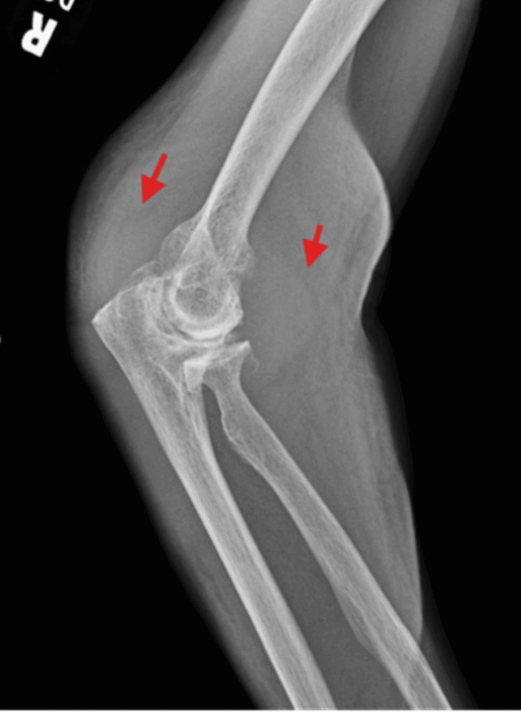
    Haemophilia haemarthrosis swollen knee joint
    Haemophilia A: deep haemarthroses, normal bleeding time; ristocetin normal.
  • C. Bernard-Soulier syndrome: GPIb defect gives giant platelets and thrombocytopenia; ristocetin fails but factor VIII is normal.
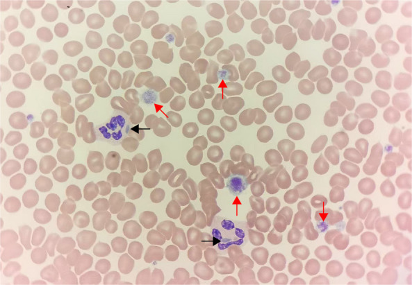
    Bernard-Soulier giant platelets blood smear
    Bernard-Soulier: giant platelets, low count; factor VIII is not reduced.
  • D. Glanzmann thrombasthenia: GPIIb/IIIa defect; aggregation fails to all agonists but ristocetin agglutination is preserved and factor VIII is normal.
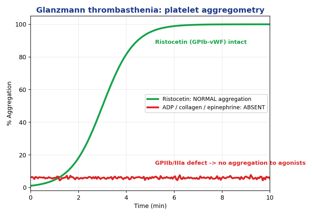
    Platelet aggregometry Glanzmann thrombasthenia
    Glanzmann: absent aggregation to ADP but intact ristocetin; factor VIII normal.

High-Yield Facts:

  • Von Willebrand disease is the commonest inherited bleeding disorder, autosomal dominant.
  • vWF stabilises factor VIII, so severe deficiency lowers factor VIII secondarily.
  • Ristocetin cofactor activity assay is the key functional test.
  • Desmopressin (DDAVP) raises vWF and factor VIII in type 1 disease.
Question 2 · NEET PG Aug 2026

A pathologist reviews a liver biopsy from a man with suspected iron overload. To confirm that the golden-brown intracellular pigment is truly haemosiderin rather than lipofuscin or bile, a special stain is ordered in which potassium ferrocyanide reacts with ferric iron to give an insoluble blue deposit. Which stain is this?

  1. A.Congo red
  2. B.Masson trichrome
  3. C.Perls Prussian blue
  4. D.Periodic acid-Schiff
Show answer and explanation

Answer: C. Perls Prussian blue

Correct answer: C. Perls Prussian blue.

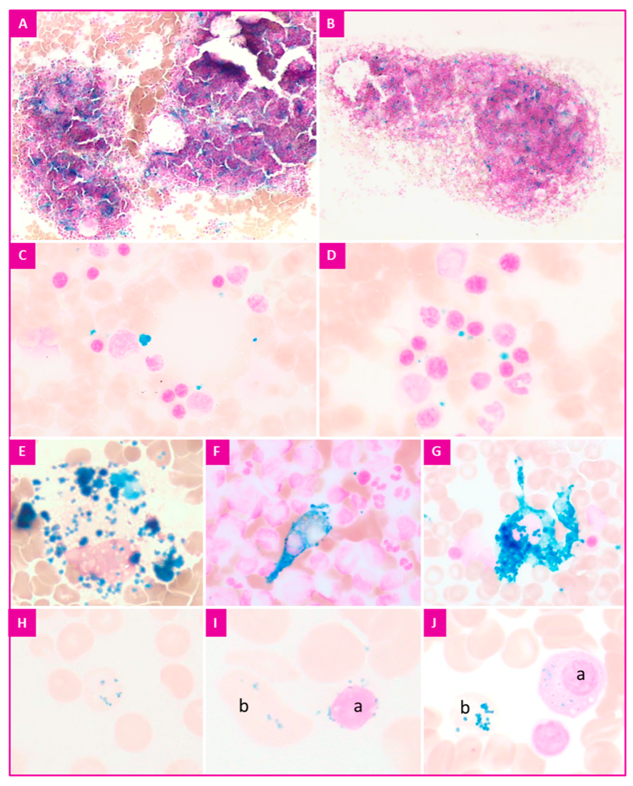
The Perls Prussian blue reaction is the specific stain for haemosiderin. In acid conditions, potassium ferrocyanide reacts with the ferric (Fe3+) iron released from haemosiderin to form an insoluble bright-blue ferric ferrocyanide pigment. This distinguishes true iron-containing haemosiderin from the look-alike golden pigments lipofuscin and bile, which stay negative.

Perls Prussian blue stain haemosiderin iron in tissue
Perls Prussian blue: ferric iron in haemosiderin stains vivid blue.

Incorrect Options:

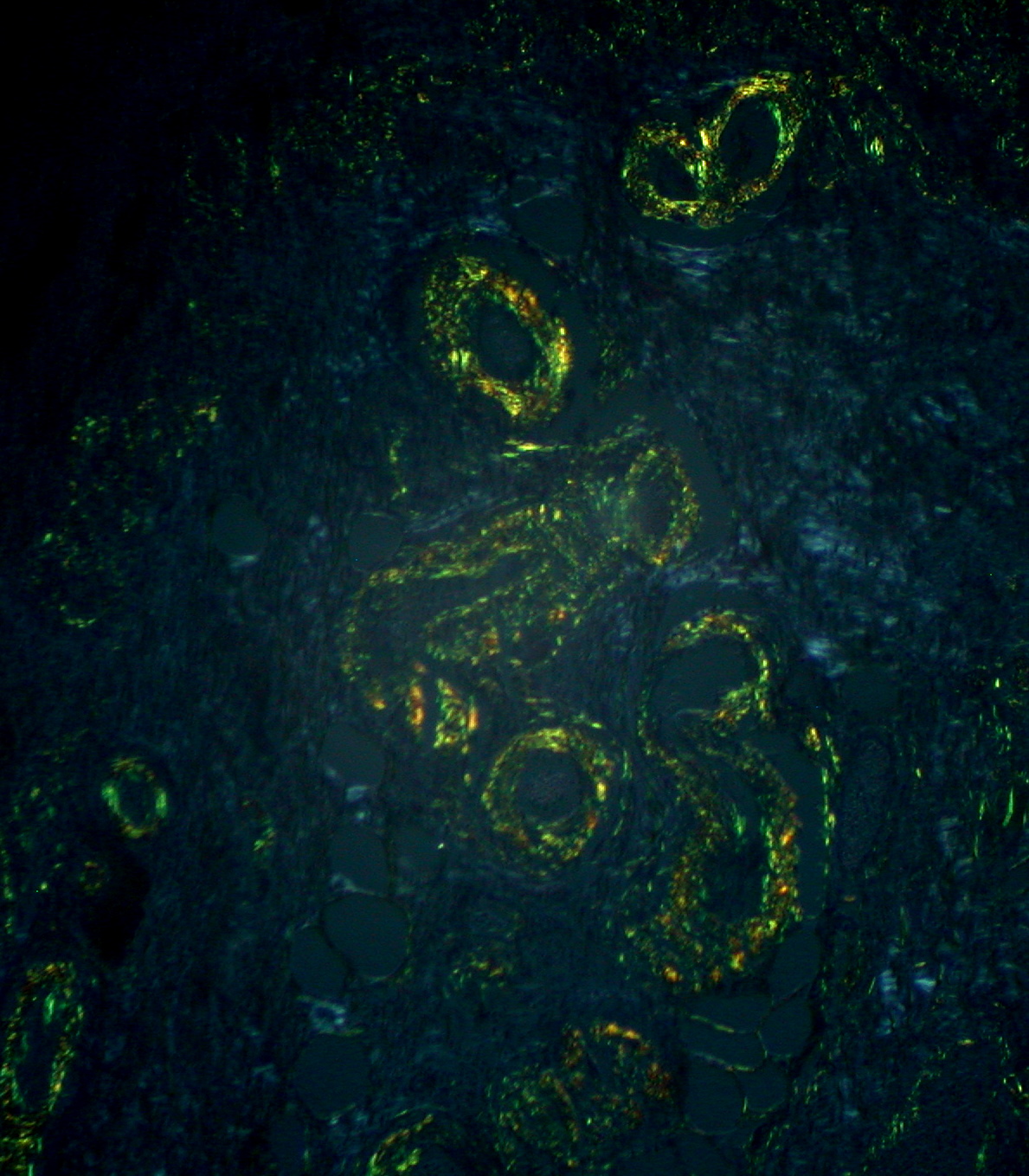
  • A. Congo red: Stains amyloid, giving apple-green birefringence under polarised light, not iron.
    Congo red amyloid apple green birefringence
    Congo red highlights amyloid, not haemosiderin.
  • B. Masson trichrome: Demonstrates collagen and fibrosis (blue or green), used to grade cirrhosis, not to detect iron.
    Masson trichrome stain collagen fibrosis
    Masson trichrome shows collagen/fibrosis, not iron pigment.
  • D. Periodic acid-Schiff: Stains glycogen, basement membranes and fungal walls magenta, not haemosiderin.
    Periodic acid Schiff PAS stain glycogen magenta
    PAS marks glycogen and basement membranes, not iron.

High-Yield Facts:

  • Perls Prussian blue is the classic special stain for haemosiderin (ferric iron).
  • Chemistry: potassium ferrocyanide plus ferric iron gives insoluble blue pigment.
  • Congo red = amyloid; Masson trichrome = collagen; PAS = glycogen.
  • Prussian blue positivity in urine (haemosiderinuria) indicates chronic intravascular haemolysis.
Question 3 · NEET PG Aug 2026

A 58-year-old woman with long-standing mitral stenosis and chronic left heart failure has intermittent blood-tinged sputum. A transbronchial lung biopsy shows alveoli filled with plump macrophages stuffed with golden-brown pigment that turns blue with Perls stain. What are these cells called?

  1. A.Anthracotic macrophages
  2. B.Heart failure cells
  3. C.Lipid-laden macrophages
  4. D.Foamy histiocytes
Show answer and explanation

Answer: B. Heart failure cells

Correct answer: B. Heart failure cells.

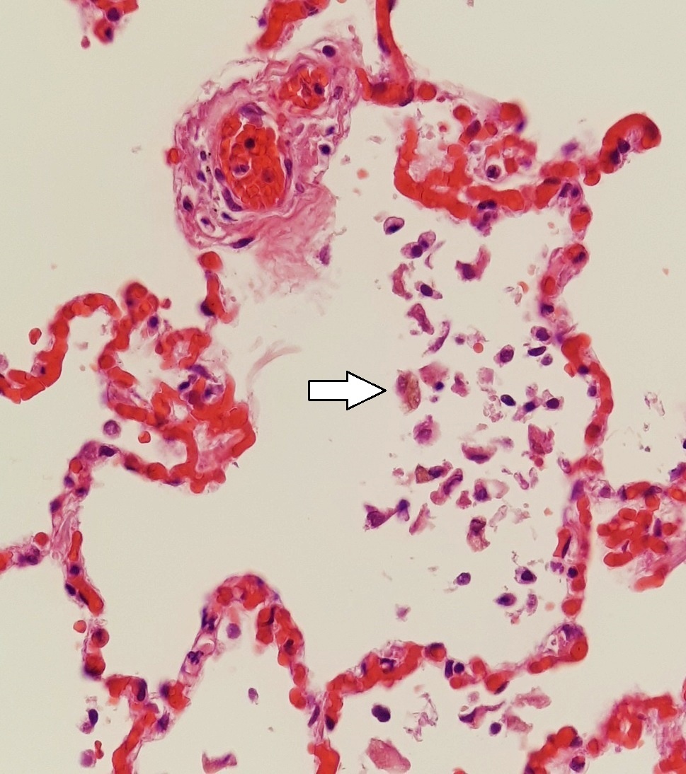
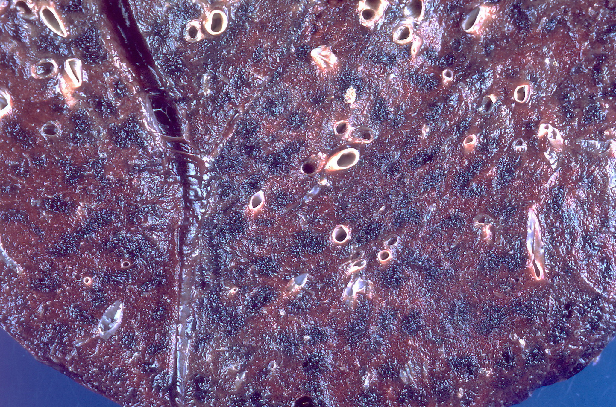
Chronic left heart failure and mitral stenosis raise pulmonary venous pressure, causing chronic pulmonary congestion and small alveolar micro-haemorrhages. Alveolar macrophages phagocytose the extravasated red cells and store the iron as haemosiderin, becoming heart failure cells. The golden-brown pigment stains blue with Perls Prussian blue, confirming iron. This is the microscopic hallmark of brown induration of the lung.

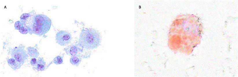
Heart failure cells haemosiderin-laden macrophages in alveoli
Heart failure cells: alveolar macrophages laden with haemosiderin.

Incorrect Options:

  • A. Anthracotic macrophages: Carry black carbon (soot) pigment from inhaled air pollution, not iron -- Perls is negative.
    Anthracosis black carbon pigment lung macrophages
    Anthracosis: black carbon pigment, Perls-negative, unrelated to heart failure.
  • C. Lipid-laden macrophages: Contain fat (as in lipoid pneumonia or fat embolism), staining with oil red O, not Perls.
    Lipid laden macrophages foamy vacuolated cytoplasm
    Lipid-laden macrophages hold fat, not iron pigment.
  • D. Foamy histiocytes: Lipid-filled macrophages seen in xanthomas and infections -- again not iron-laden congestion cells.
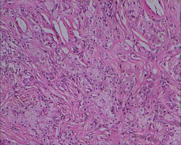
    Foamy histiocytes vacuolated macrophages
    Foamy histiocytes are lipid-rich, distinct from heart failure cells.

High-Yield Facts:

  • Heart failure cells are haemosiderin-laden alveolar macrophages of chronic pulmonary congestion.
  • They cause brown induration of the lung in chronic left heart failure and mitral stenosis.
  • Perls Prussian blue confirms the iron content.
  • Anthracosis (carbon) is Perls-negative -- the pigment source is inhaled soot.

Pathology concept guides

Pathology images to recognise

Browse the full concept library and clinical image atlas.

Practise the rest: open the Kinase question bank, choose Pathology, set the exam to NEET-PG and switch on PYQs only. Questions you get wrong go into Fix My Weakness for targeted revision.

Frequently Asked Questions

How many Pathology questions were in NEET-PG 2026?▼
13 of the 180 questions in the NEET-PG 2026 paper (30 August 2026) were Pathology — 7.2% of the paper, worth up to 52 marks at +4 per correct answer. This is from the recall paper in the Kinase question bank.
How many NEET-PG Pathology PYQs does Kinase have?▼
Kinase has 120 NEET-PG Pathology previous year questions from 2017 to 2026, each with an explanation, plus 264 INICET Pathology PYQs for extra practice.
What are the most-asked Pathology topics in NEET-PG?▼
In Kinase's NEET-PG Pathology PYQs, the most-asked topics are Hematopathology, General Pathology, Gastrointestinal System. Hematopathology alone accounts for 31 of the 120 questions.
Are NEET-PG Pathology PYQs the same as INICET PYQs?▼
Both exams test the MBBS syllabus, so most Pathology concepts overlap. The marking differs: NEET-PG is +4 / −1 and INICET is +1 / −1/3. Kinase lets you practise either exam's PYQs, or both together.
How should I practise Pathology PYQs for NEET-PG?▼
Start with the most-asked topics above, do a daily set of Pathology PYQs, and read every explanation — including the ones you got right. Kinase's Fix My Weakness then builds practice sets from the topics you miss most.
120 Pathology PYQs
2017–2026 papers
Topic + year tagged

Ready to Start Practicing?

Pathology was 13 questions (up to 52 marks) in NEET-PG 2026.

Start Free Practice

No credit card required