What is the pouch of Douglas?
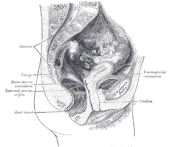
The pouch of Douglas — also called the rectouterine pouch, posterior cul-de-sac or rectovaginal pouch — is a deep, blind-ended extension of the peritoneal cavity that dips down between the uterus in front and the rectum behind. It is named after the Scottish anatomist James Douglas (1675–1742).
It forms because the pelvic peritoneum drapes over the pelvic organs like a sheet over furniture. In the midline, peritoneum runs down the back of the uterus and cervix, onto the upper posterior wall of the vagina next to the posterior fornix, and then turns back up onto the front and sides of the rectum. The fold it creates between uterus and rectum is the pouch of Douglas. In front of the uterus, a second, much shallower vesicouterine pouch lies between the uterus and the bladder.
What are the boundaries and relations of the pouch of Douglas?
| Boundary | Structure | Exam relevance |
|---|---|---|
| Anterior | Posterior surface of the uterus (body and cervix) and the posterior vaginal fornix / upper posterior vaginal wall | Only vaginal wall and peritoneum separate the fornix from the pouch — basis of culdocentesis and posterior colpotomy |
| Posterior | Anterior surface of the rectum (upper rectum) | Palpable through the anterior rectal wall on per-rectal examination (Blumer's shelf, pelvic abscess) |
| Lateral | Rectouterine folds of peritoneum, overlying the uterosacral ligaments | Uterosacral ligament nodularity is a classic sign of endometriosis |
| Floor (inferior) | Peritoneal reflection onto the rectum and vagina (rectovaginal fold); below it lies the extraperitoneal rectovaginal septum | The lowest point that intraperitoneal fluid can reach |
| Superior | Open above to the rest of the peritoneal cavity; loops of small bowel can slip down into it | Why an enterocele contains small bowel |
The uterosacral ligaments are condensations of pelvic fascia running from the cervix to the posterolateral pelvic wall; the sharp, sickle-shaped rectouterine folds are the peritoneum lying over them. Together they form the side-walls of the pouch — which is why pain from the pouch, and tenderness on moving the cervix, are so characteristic of pelvic peritonitis and endometriosis.
| Pouch | Sex | Between | Depth / importance |
|---|---|---|---|
| Rectouterine (Douglas) | Female | Uterus + posterior fornix (front) and rectum (back) | Deep; most dependent part of the female peritoneal cavity |
| Vesicouterine | Female | Bladder (front) and uterus (back) | Shallow; lies higher than the pouch of Douglas |
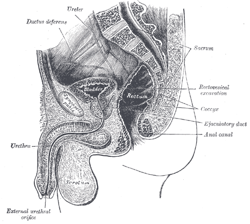
| Rectovesical | Male | Bladder in front and rectum behind | Most dependent part of the male peritoneal cavity; the male counterpart of Douglas |
Why is the pouch of Douglas the most dependent part of the peritoneal cavity?
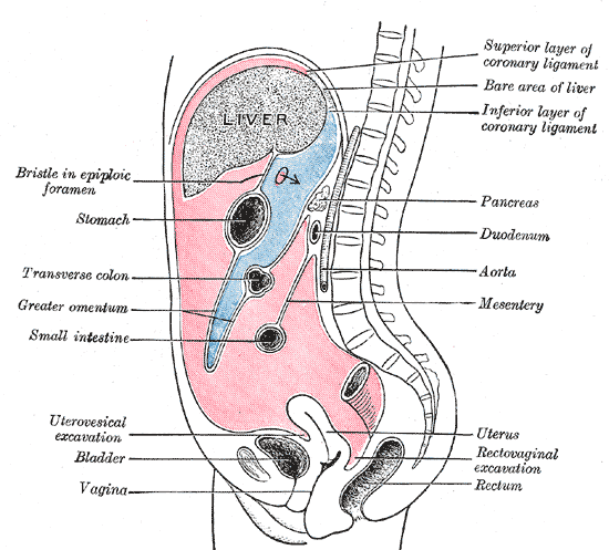
Because it is the lowest recess of the peritoneal cavity in the pelvis, gravity drains free intraperitoneal fluid into it. In a woman who is sitting or standing, the pouch of Douglas is unquestionably the lowest point of the whole abdominopelvic cavity. It is also the most dependent part of the pelvis in the supine position, which is why the pelvic window of a trauma ultrasound looks there.
| Position / region | Lowest recess | Clinical use |
|---|---|---|
| Upright, female | Pouch of Douglas | Blood, pus, ascites pool here; culdocentesis, TVS |
| Upright, male | Rectovesical pouch | Pelvic collections, drop metastases |
| Supine — pelvis | Pouch of Douglas (female) / rectovesical pouch (male) | FAST pelvic (suprapubic) view |
| Supine — upper abdomen | Hepatorenal recess (Morison's pouch) | FAST right-upper-quadrant view; subhepatic abscess |
A small amount of free fluid in the pouch of Douglas is physiological in women of reproductive age — it accumulates around ovulation and during menstruation. Larger, echogenic (blood-containing) or complex collections are pathological.
How is the pouch of Douglas examined — culdocentesis, TVS and FAST?
The pouch of Douglas cannot be palpated through the abdomen, but it lies just behind the posterior vaginal fornix and in front of the rectum, so it can be felt on per-vaginal (bimanual) and per-rectal examination, sampled with a needle through the fornix, and imaged well with transvaginal ultrasound.
- Culdocentesis — the cervix is steadied with a tenaculum and an 18-gauge spinal needle is passed through the posterior vaginal fornix, about 1 cm below the cervix, into the pouch of Douglas while aspirating.
- Positive tap: non-clotting blood — blood that has pooled in the peritoneal cavity has been defibrinated, so it no longer clots. This indicates haemoperitoneum (classically a ruptured ectopic pregnancy).
- Non-diagnostic tap: clotted blood (the needle probably entered a vessel) or no fluid at all. Serous fluid is a negative result.
- Contraindication: a mass or a retroverted uterus occupying the pouch of Douglas.
In trauma, the FAST examination has four windows — right upper quadrant (hepatorenal recess), left upper quadrant (splenorenal recess), subxiphoid (pericardium) and suprapubic/pelvic. The pelvic window looks for fluid in the rectovesical pouch in men and the rectouterine and vesicouterine pouches in women. A full bladder acts as an acoustic window, and a few hundred mL of free intraperitoneal fluid is usually needed before ultrasound reliably picks it up.
What diseases involve the pouch of Douglas?
| Condition | What happens in the pouch | How it shows up |
|---|---|---|
| Ruptured ectopic pregnancy / ruptured haemorrhagic cyst | Haemoperitoneum pools here | Echogenic free fluid on TVS; non-clotting blood on culdocentesis |
| Pelvic inflammatory disease, pelvic abscess | Pus collects in the most dependent recess | Tender fullness on PV/PR; fluid on TVS; can be drained through the vagina or rectum |
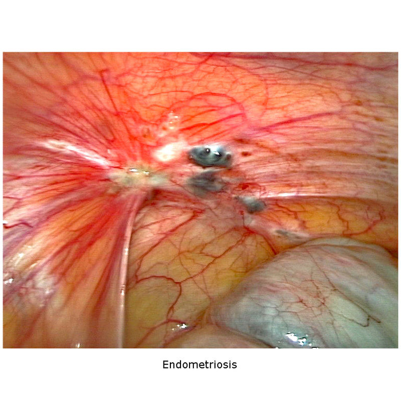
| Endometriosis | Implants on the posterior cul-de-sac and uterosacral ligaments; adhesions pull the uterus back | Dysmenorrhoea, deep dyspareunia, infertility; fixed retroverted uterus, uterosacral nodularity |
| Peritoneal (drop) metastases | Transcoelomic spread of tumour cells settles in the lowest recess | Blumer's (rectal) shelf — hard shelf-like mass felt on rectal or vaginal examination; gastric, colorectal, ovarian cancers |
| Ascites | Free fluid collects first in the pelvis | Fluid in the pouch on ultrasound |
| Enterocele | Hernia of the pouch of Douglas peritoneum containing small bowel, pushing into the rectovaginal space | Posterior-apical vaginal bulge; the only true hernia among pelvic-floor disorders |
Endometriosis deserves a closer look because it is asked every year. The ovaries are the most common site, followed by the posterior broad ligament, anterior and posterior cul-de-sac and the uterosacral ligaments. The classic triad is chronic pelvic pain/dysmenorrhoea, dyspareunia and infertility, and the classic examination is a fixed, retroverted uterus with tender nodularity of the uterosacral ligaments or cul-de-sac. Transvaginal ultrasound is the first-line imaging test; the definitive diagnosis rests on histology of lesions obtained at laparoscopy.
Surgery and procedures. A pelvic abscess in the pouch of Douglas can be drained through the vagina (posterior colpotomy) or the rectum without opening the abdomen, and image-guided transvaginal or transrectal drainage is usually successful. At vaginal hysterectomy, surgeons may approximate the uterosacral ligaments to close off (obliterate) the cul-de-sac to treat or prevent an enterocele. In peritoneal dialysis, the catheter tip is placed in this dependent space.
What does the pouch of Douglas feel like on PV and PR examination?
- Normal: the pouch is empty and impalpable; bowel slides freely behind the uterus.
- Tenderness / fullness in the posterior fornix — blood or pus in the pouch (ectopic pregnancy, PID, pelvic abscess). Cervical motion tenderness reflects irritated pelvic peritoneum.
- Nodularity of the uterosacral ligaments with a fixed retroverted uterus — endometriosis.
- Hard, shelf-like mass above the prostate or cervix felt through the anterior rectal wall — Blumer's shelf (peritoneal metastases).
- A mass occupying the pouch (e.g. retroverted gravid or fibroid uterus, ovarian tumour) — also the reason culdocentesis is contraindicated in that situation.
How does the pouch of Douglas appear in NEET PG and INI-CET questions?
Examiners rarely ask 'define the pouch of Douglas'. Instead the pouch is the clue inside a clinical stem. Recognise the pattern and the answer follows.
| Clue in the stem | Interpretation | Typical question and answer |
|---|---|---|
| Amenorrhoea 6 weeks, pain, shock, empty uterus, echogenic free fluid in the pouch of Douglas | Haemoperitoneum | Diagnosis → ruptured ectopic pregnancy; next step → resuscitate and operate |
| Stable, adnexal gestational sac, no free fluid in the pelvis, low hCG | Unruptured ectopic | Management → medical (methotrexate) if criteria are met; the absence of pouch fluid supports non-rupture |
| Dysmenorrhoea, deep dyspareunia, infertility, fixed retroverted uterus, cul-de-sac nodularity | Endometriosis | First investigation → TVS; confirmation → laparoscopy with histology |
| Fever, lower abdominal pain, PID, 'where will pus collect when she sits up?' | Most dependent recess | Answer → rectouterine pouch |
| Gastric cancer + hard mass felt anteriorly on per-rectal examination | Drop metastasis | Answer → Blumer's shelf |
| Bulge high on the posterior vaginal wall after hysterectomy containing bowel | Hernia of the pouch | Answer → enterocele; culdoplasty closes the cul-de-sac |
| Trauma patient, FAST pelvic window shows anechoic fluid behind the bladder | Free intraperitoneal fluid | Positive FAST; man → rectovesical pouch, woman → rectouterine pouch |
What are the exam traps on the pouch of Douglas?
| Trap | Correct fact |
|---|---|
| Pouch of Douglas lies between bladder and uterus | No — that is the vesicouterine pouch; Douglas is between uterus and rectum |
| Most dependent part of female pelvis = posterior fornix or ovarian fossa | The most dependent part of the peritoneal cavity is the rectouterine pouch; the fornix is part of the vagina, the gateway to the pouch |
| Most dependent recess in a supine patient is always Morison's pouch | Morison's pouch is the lowest recess of the upper abdomen; the pelvis (pouch of Douglas) is the lowest overall — read the region asked |
| Male equivalent is the rectoprostatic space | Male equivalent is the rectovesical pouch (a peritoneal pouch); below it, the extraperitoneal rectovesical septum separates prostate and bladder base from the rectum |
| Culdocentesis positive if blood clots | Non-clotting blood is positive; clotted blood suggests a vessel was hit |
| Blumer's shelf is an ovarian metastasis | Ovarian metastasis = Krukenberg; Blumer's shelf = pouch deposits felt on rectal exam |
| Most common site of endometriosis is the pouch of Douglas | Ovary is the most common; cul-de-sac and uterosacral ligaments follow |