What is Virchow's triad?
Virchow's triad is the classic framework for why blood clots inside a living vessel. It groups every thrombotic risk factor into three buckets: (1) abnormal blood flow — stasis or turbulence, (2) endothelial injury (vessel-wall damage or dysfunction), and (3) hypercoagulability — an increased tendency of the blood itself to clot. The concept is credited to the Berlin pathologist Rudolf Virchow, whose work on venous thrombosis in 1856 identified these factors.
Thrombosis is pathological haemostasis: the same platelets, coagulation factors and fibrin that seal a cut form a clot where there is no bleeding to stop. A single factor may be enough (a ruptured coronary plaque), but most real patients have two or three at once — the post-operative orthopaedic patient is immobile (stasis), has had tissue and vessel trauma (injury), and is in a post-surgical hypercoagulable state.
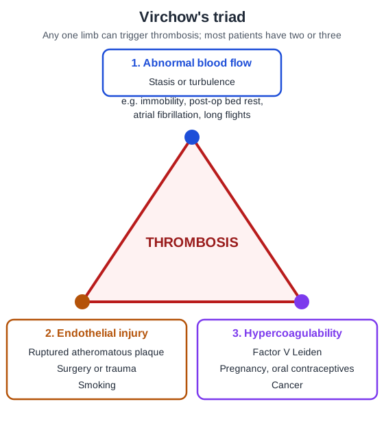
| Limb | What goes wrong | Classic examples asked in exams |
|---|---|---|
| Abnormal blood flow (stasis / turbulence) | Sluggish flow lets platelets touch the endothelium and slows washout of activated factors; turbulence damages endothelium and creates eddies of stasis | Immobility, long flights, post-op bed rest, atrial fibrillation with a dilated left atrium (mitral stenosis), ventricular aneurysm after MI, aortic aneurysm, hyperviscosity (polycythaemia vera), sickle cell disease |
| Endothelial injury | Exposed subendothelial collagen, von Willebrand factor and tissue factor; 'activated' endothelium shifts to a prothrombotic state | Ruptured atherosclerotic plaque, MI (endocardial injury), vasculitis, trauma, surgery, catheters, smoking, chronic hypertension, hyperlipidaemia, homocystinaemia |
| Hypercoagulability | Altered coagulation factors or natural anticoagulants | Factor V Leiden, prothrombin G20210A, protein C/S or antithrombin deficiency; cancer, pregnancy, oral contraceptives, antiphospholipid syndrome, heparin-induced thrombocytopenia, nephrotic syndrome |
Which component of Virchow's triad is most important?
It depends on the circulation. Robbins states that endothelial injury leading to platelet activation almost inevitably underlies thrombus formation in the heart and the arterial circulation, because high flow rates normally wash away clotting factors. That is why aspirin and other antiplatelet drugs are the backbone of coronary disease treatment.
In veins the picture flips: stasis is a major factor in venous thrombi, and hypercoagulability contributes infrequently to arterial or intracardiac thrombosis but is an important risk factor for venous thrombosis (Robbins). A young patient with recurrent DVT and no obvious trigger should make you think of an inherited thrombophilia; a smoker with an acute STEMI should make you think of plaque rupture.
| Site | Dominant limb | Typical thrombus | Main drug class |
|---|---|---|---|
| Coronary / cerebral arteries | Endothelial injury (plaque rupture) | Platelet-rich 'white' thrombus | Antiplatelets (plus anticoagulants/fibrinolytics acutely) |
| Heart chambers (mural thrombus) | Stasis + endocardial injury (AF, MI, aneurysm) | Mural thrombus, embolises to brain, kidney, spleen | Anticoagulants |
| Deep leg veins | Stasis + hypercoagulability | Fibrin- and red-cell-rich 'red' thrombus | Anticoagulants |
What are the inherited and acquired causes of hypercoagulability?
Robbins divides hypercoagulable states into primary (genetic) and secondary (acquired) disorders. Inherited causes should be considered in young patients (under about 50 years) with venous thrombosis, even when an acquired trigger is also present, because a carrier's risk multiplies with pregnancy, bed rest or long flights.
| Inherited (primary) | Key point | Acquired (secondary) | Key point |
|---|---|---|---|
| Factor V Leiden (F5 G1691A → Arg506Gln) | Most common inherited thrombophilia in people of European descent; factor Va resists cleavage by activated protein C (APC resistance) | Prolonged immobilisation / surgery / fracture / burns | Stasis plus tissue injury — the commonest real-world trigger |
| Prothrombin G20210A (3′-untranslated region) | Second most common; raises prothrombin levels | Cancer (esp. mucin-secreting adenocarcinoma) | Procoagulant tumour products; migratory thrombophlebitis |
| Antithrombin deficiency | Rare but high risk | Pregnancy, postpartum, oral contraceptives | Higher hepatic clotting-factor synthesis, lower antithrombin |
| Protein C deficiency | Warfarin-induced skin necrosis is the classic association | Antiphospholipid antibody syndrome | Both arterial and venous thrombosis; recurrent miscarriage |
| Protein S deficiency | Cofactor for activated protein C | Heparin-induced thrombocytopenia | Paradoxical thrombosis despite low platelets |
| Raised factor VIII, IX, XI or fibrinogen | Less common | Paroxysmal nocturnal haemoglobinuria, nephrotic syndrome, smoking, obesity, advancing age, AF, MI, prosthetic valves, DIC | PNH is an acquired clonal disorder — a favourite INI-CET distractor |
Hyperhomocysteinaemia sits in both camps: it can be inherited (e.g. homocystinuria from cystathionine β-synthase deficiency) or acquired (folate/B12 deficiency). If a question forces a choice between PNH and homocystinuria as 'acquired', choose PNH.
How do arterial and venous thrombi differ?
Both contain platelets, fibrin and trapped red cells — the proportions differ because of shear. Harrison explains that arterial thrombi are rich in platelets because of high shear in injured arteries and look white, whereas venous thrombi form under low shear, contain relatively few platelets and are mostly fibrin and trapped red cells, so they look red. StatPearls describes arterial thrombi as a platelet-rich 'white head' followed by a fibrin- and red-cell-rich 'red tail'.
| Feature | Arterial thrombus | Venous thrombus (phlebothrombosis) |
|---|---|---|
| Main trigger | Endothelial injury (plaque rupture) | Stasis ± hypercoagulability |
| Composition | Platelet-rich ('white') | Fibrin + red cells ('red', 'stasis' thrombus) |
| Growth | Retrograde from the point of attachment | Propagates toward the heart as a long cast |
| Occlusion | Frequently occlusive | Almost invariably occlusive |
| Commonest site | Coronary, cerebral, femoral arteries | Deep veins of the lower limb (~90% of venous thromboses) |
| Main consequence | Infarction downstream | Oedema, pain; pulmonary embolism |
| Mainstay drug | Antiplatelets | Anticoagulants |
Mural thrombi form in heart chambers or the aortic lumen. Abnormal myocardial contraction (arrhythmia, dilated cardiomyopathy, MI) or endocardial injury promotes cardiac mural thrombi; ulcerated plaques and aneurysms promote aortic ones. Robbins notes that about 80% of systemic emboli arise from intracardiac mural thrombi, two-thirds of them linked to left-ventricular infarcts.
How do you tell an antemortem thrombus from a postmortem clot?
This is a favourite pathology and forensic question. Lines of Zahn are grossly and microscopically visible laminations — pale bands of platelets and fibrin alternating with darker red-cell-rich layers. They form only in flowing blood, so they usually mark a thrombus formed before death.
| Feature | Antemortem thrombus | Postmortem clot |
|---|---|---|
| Lines of Zahn | Present (laminated) | Absent (bland, non-laminated) |
| Attachment | Attached (at least focally) to the vessel wall | Not attached; lifts out easily |
| Consistency | Firm, dry, friable, granular surface | Soft, gelatinous, rubbery, smooth and glistening |
| Colour pattern | Mixed white-tan to red | Dark red dependent part ('currant jelly') with a yellow upper part ('chicken fat') from red-cell settling |
| Shape | Does not fit the vessel perfectly; may be coiled | Forms a cast of the vessel lumen |

How does each clinical risk factor map onto the triad?
Most vignettes describe a patient, not a limb of the triad. Translating the scenario into stasis, injury or hypercoagulability — and noting the mechanism — is what separates a guess from a sure answer.
| Scenario | Main limb(s) | Mechanism |
|---|---|---|
| Post-operative / fracture / burns patient | All three | Immobility (stasis) + tissue and vessel trauma (injury) + post-injury hypercoagulability |
| Mitral stenosis with atrial fibrillation | Stasis | Dilated, non-contracting left atrium — a prime site for thrombi and systemic emboli |
| Acute MI / ventricular aneurysm | Stasis + endocardial injury | Non-contractile myocardium → cardiac mural thrombus |
| Ulcerated atherosclerotic plaque | Injury + turbulence | Exposed subendothelial matrix and turbulent flow |
| Pregnancy, oral contraceptives | Hypercoagulability | More hepatic synthesis of clotting factors, less antithrombin |
| Disseminated adenocarcinoma | Hypercoagulability | Procoagulant tumour products such as mucin |
| Advancing age | Hypercoagulability | More platelet aggregation, less endothelial prostacyclin (PGI2) |
| Polycythaemia vera | Stasis | Hyperviscosity slows small-vessel flow |
| Sickle cell anaemia | Stasis | Deformed red cells occlude vessels |
| Smoking, obesity | Hypercoagulability (smoking also causes endothelial injury) | Mechanism incompletely understood |
Inherited and acquired factors add up. Robbins points out that factor V Leiden carriers are at particular risk when an acquired trigger — pregnancy, prolonged bed rest, a long flight — is layered on top, and that among patients with recurrent DVT the frequency of the Leiden mutation approaches 60%. This is why a first DVT in a young patient, or an unusual-site thrombosis, justifies a thrombophilia work-up (protein C, protein S, antithrombin, APC resistance/factor V Leiden, prothrombin variant and antiphospholipid antibodies).
How does Virchow's triad apply to DVT and the Wells score?
Deep vein thrombosis of the leg is the textbook triad disease: the veins of the lower extremity account for about 90% of venous thromboses. Every major DVT risk factor maps to a limb — immobility and long travel (stasis), surgery and trauma (injury), cancer, pregnancy, oestrogens and thrombophilias (hypercoagulability). DVT is usually unilateral; clinical signs are unreliable, so objective testing (compression ultrasound) is needed, and a DVT may first declare itself as a pulmonary embolism.

The Wells criteria for DVT turn the history and examination into a pre-test probability. Each item scores 1 point, and one item subtracts 2:
| Clinical feature | Points |
|---|---|
| Active cancer (treatment within 6 months or palliative) | +1 |
| Paralysis, paresis or recent plaster cast of a lower limb | +1 |
| Bedridden ≥ 3 days, or major surgery within 12 weeks needing general/regional anaesthesia | +1 |
| Localised tenderness along the deep venous system | +1 |
| Entire leg swollen | +1 |
| Calf swelling ≥ 3 cm more than the other side (10 cm below tibial tuberosity) | +1 |
| Pitting oedema confined to the symptomatic leg | +1 |
| Collateral superficial (non-varicose) veins | +1 |
| Previously documented DVT | +1 |
| Alternative diagnosis at least as likely as DVT | −2 |
In the three-tier version, ≤ 0 = low, 1–2 = moderate and ≥ 3 = high probability.
Which other Virchow eponyms do students confuse with the triad?
Rudolf Virchow's name appears across pathology, surgery and neuroanatomy. Match-the-following questions love to mix them up:
| Eponym | What it is | Exam hook |
|---|---|---|
| Virchow's triad | Stasis + endothelial injury + hypercoagulability | Pathogenesis of thrombosis |
| Virchow's node | The most proximal left supraclavicular lymph node, near where the thoracic duct drains | Metastasis from abdominal cancer, classically gastric carcinoma |
| Troisier's sign | A palpable, enlarged Virchow's node | Advanced (often stage IV) GI malignancy; also lung, breast, pelvic, testicular cancers and lymphoma |
| Virchow–Robin spaces | Perivascular spaces around vessels entering the brain | Cryptococcal 'soap-bubble' lesions spread along them |
| Virchow's law | In craniosynostosis, skull growth stops perpendicular to the fused suture and increases parallel to it | Sagittal synostosis → long, narrow skull (scaphocephaly) |
How is Virchow's triad asked in NEET PG and INI-CET?
- Identify the limb — 'Atrial fibrillation contributes to thrombosis mainly by…' → stasis. 'Smoking…' → hypercoagulability and endothelial injury.
- Primary initiating factor of an arterial thrombus → endothelial injury (NEET PG 2020 pattern).
- Most common inherited thrombophilia → factor V Leiden (asked in INI-CET 2023 and 2024 and NEET PG 2018).
- Acquired vs inherited → PNH, antiphospholipid syndrome, cancer, OCPs are acquired (INI-CET 2024).
- Lines of Zahn → antemortem thrombus in flowing blood; postmortem clot is 'chicken fat and currant jelly', unattached.
- DVT statements → DVT is mostly unilateral; clinical assessment is unreliable; it can present first as PE (NEET PG 2019).
- NOT a risk factor questions → check each option against stasis, injury or hypercoagulability.