Last verified: 5 October 2026 against Wikimedia Commons (licence checked per file).
Quick Answer
For image questions, learn each instrument as feature → use → look-alike: Babcock's (smooth, atraumatic) vs Allis' (toothed); Cusco's (self-retaining) vs Sims' (assistant-held, anterior wall); Macintosh (curved, vallecula) vs Miller (straight, infants); LMA (supraglottic) vs cuffed endotracheal tube (protects against aspiration).
Look-alikes at a glance
| Pair | The difference examiners test |
|---|---|
| Babcock's vs Allis' | Fenestrated, toothless jaws for bowel vs interlocking teeth for fascia |
| Cusco's vs Sims' | Self-retaining, dorsal position vs assistant-held, anterior-wall view |
| Macintosh vs Miller blade | Curved, tip in the vallecula vs straight, lifts the epiglottis (infants) |
| LMA vs endotracheal tube | Sits above the larynx vs cuff inside the trachea, protects against aspiration |
| No. 3 vs No. 4 BP handle | Small blades (10–15) vs large blades (20–24) |
Surgery
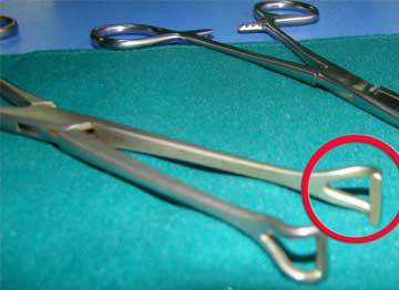
Babcock's forceps
Atraumatic holding of delicate tubular structures such as bowel, appendix and fallopian tube.
Exam angle: Atraumatic — contrast with Allis' forceps.
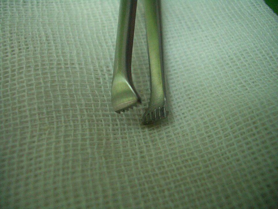
Allis' forceps
Holding tough tissue such as fascia and skin edges.
Exam angle: Traumatic grip, so not used on delicate bowel.
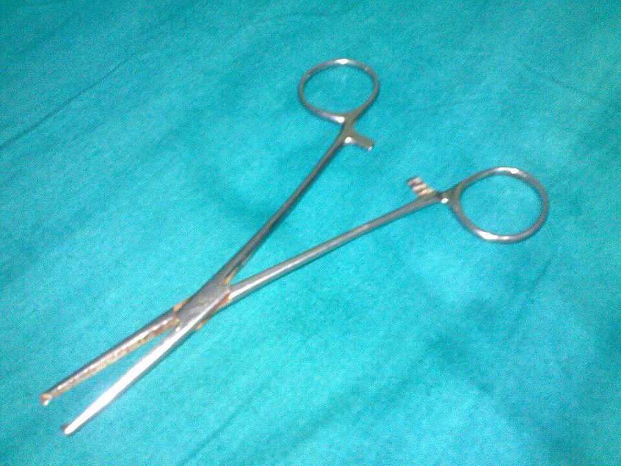
Kocher's forceps
Holding tough fibrous tissue such as the rectus sheath, and clamping pedicles.
Exam angle: Toothed tip distinguishes it from a plain artery forceps.
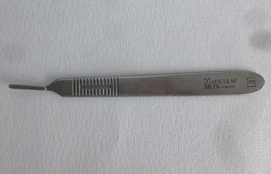
Bard-Parker (BP) handle
Scalpel handle that takes detachable blades.
Exam angle: No. 3 handle takes the small blades (10–15); No. 4 takes the large blades (20–25).
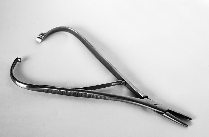
Needle holder (Mathieu pattern)
Holding a curved needle while suturing.
Exam angle: Grip the needle about two-thirds of the way from the tip.
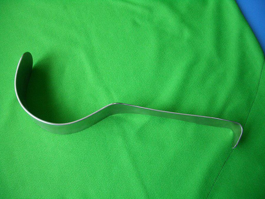
Deaver's retractor
Retracting deep abdominal structures, such as the liver or abdominal wall.
Exam angle: Hand-held deep retractor used in abdominal surgery.
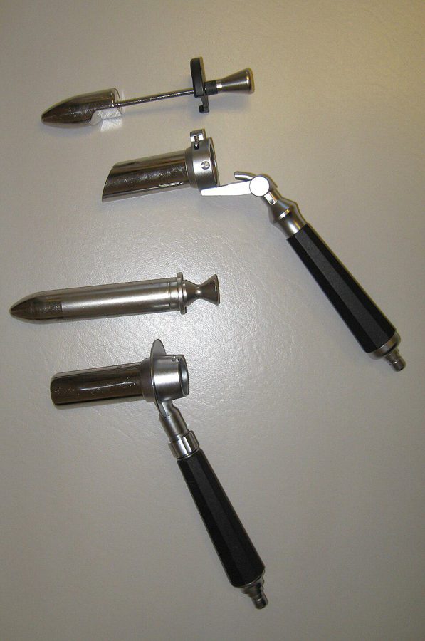
Proctoscope / anoscope
Examining the anal canal and lower rectum, and treating haemorrhoids (banding, injection).
Exam angle: Haemorrhoids are seen and treated through it.
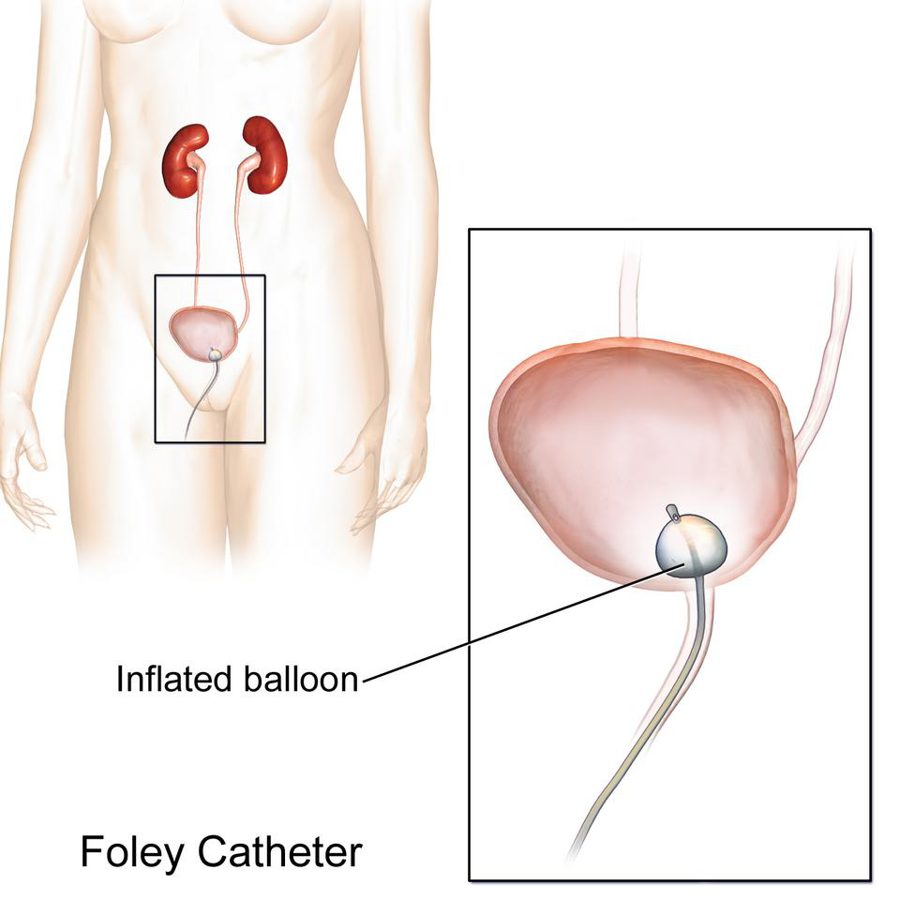
Foley catheter
Self-retaining urinary catheter for bladder drainage, held in place by an inflatable balloon.
Exam angle: Size is in French (1 Fr = 1/3 mm, so diameter ≈ Fr ÷ 3); the balloon is inflated with sterile water; a three-way catheter allows continuous bladder irrigation.
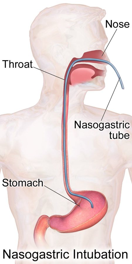
Ryle's tube (nasogastric tube)
Passed through the nose into the stomach for gastric aspiration or decompression and for feeding.
Exam angle: Insertion length is estimated from nose to earlobe to xiphisternum; position is checked by the pH of the aspirate, with a chest X-ray when that is not conclusive.
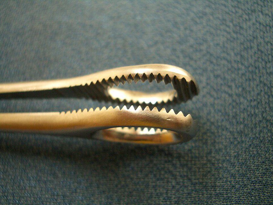
Sponge-holding forceps (Rampley's)
Holding gauze for skin preparation and swabbing deep areas.
Exam angle: Used for painting (skin preparation); ring jaws with serrations.
OBG
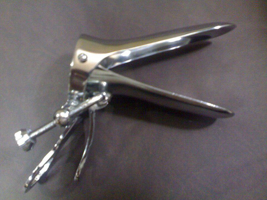
Cusco's speculum
Self-retaining bivalve vaginal speculum for per-speculum examination, Pap smear and IUD insertion, with the patient in the dorsal position.
Exam angle: Self-retaining, used in the dorsal position; contrast with Sims' speculum.
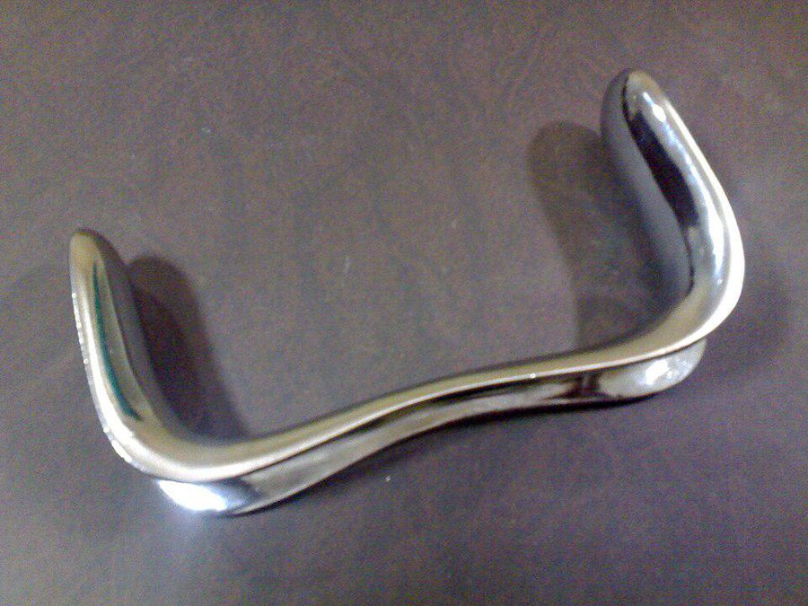
Sims' speculum
Double-ended vaginal speculum used in the Sims' (left lateral) or dorsal position; gives a good view of the anterior vaginal wall, e.g. in prolapse and fistula assessment.
Exam angle: Not self-retaining — an assistant must hold it.
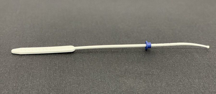
Uterine sound
Measures the length and direction of the uterine cavity before IUD insertion or D&C.
Exam angle: Sounding before IUD insertion; risk of uterine perforation.
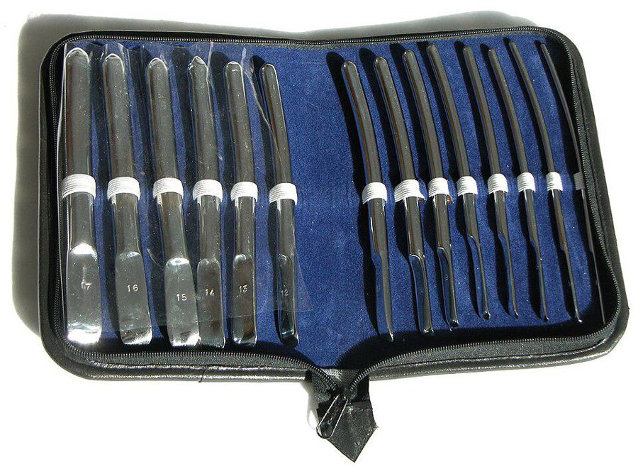
Hegar's dilators
Graduated cervical dilators for D&C and surgical abortion.
Exam angle: Dilators are sized by diameter in mm; dilate gradually, one size at a time.

Ayre's spatula
Scrapes cells from the cervix, around the squamocolumnar junction, for a Pap smear.
Exam angle: Cervical cancer screening sample from the transformation zone.
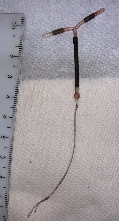
Copper T (IUCD)
Long-acting reversible contraception placed in the uterine cavity.
Exam angle: Copper acts mainly by preventing fertilisation; check threads after insertion.
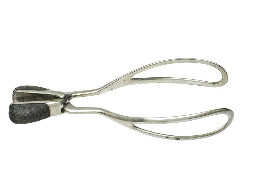
Obstetric forceps (Simpson's)
Assisted vaginal delivery when specific prerequisites such as full cervical dilatation and an engaged head are met.
Exam angle: Prerequisites for forceps delivery are a favourite exam question.
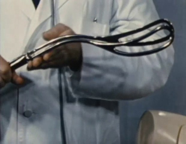
Piper's forceps
Delivering the after-coming head in a vaginal breech birth.
Exam angle: Breech only — applied once the after-coming head has entered the pelvis, with the baby's body held up in a towel; an alternative to the Mauriceau manoeuvre.
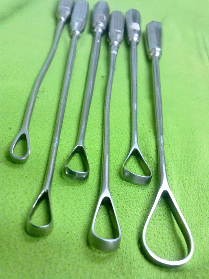
Uterine curette
Curettage in D&C — endometrial sampling when an office biopsy fails, and evacuating retained products of conception or an early pregnancy failure.
Exam angle: A gritty feel means the cavity is empty or adequately sampled; uterine perforation is the most common immediate complication of D&C.
Anaesthesia
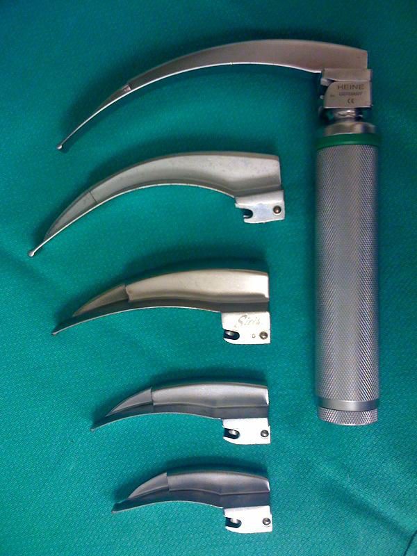
Laryngoscope (Macintosh blades)
Exposing the glottis for endotracheal intubation.
Exam angle: Curved Macintosh blade tip goes into the vallecula; straight (Miller) blade lifts the epiglottis directly and suits infants.
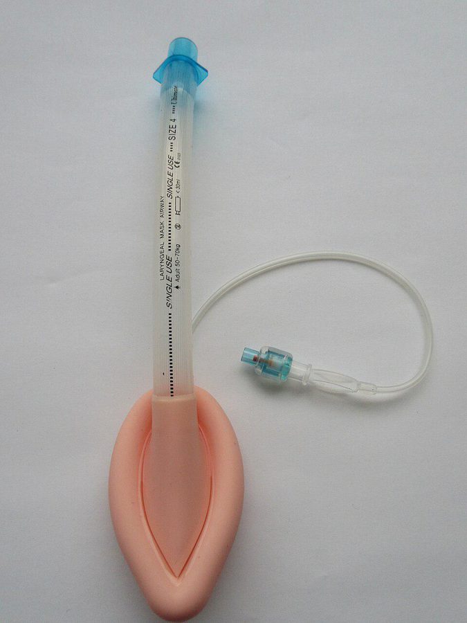
Laryngeal mask airway (LMA)
Supraglottic airway inserted without a laryngoscope; sits over the laryngeal inlet.
Exam angle: Does not reliably protect against aspiration, unlike a cuffed endotracheal tube.
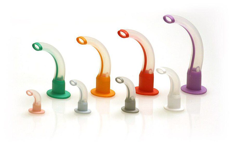
Oropharyngeal (Guedel) airway
Keeps the tongue off the posterior pharyngeal wall in an unconscious patient.
Exam angle: Size from the incisors to the angle of the jaw; not tolerated by a patient with an intact gag reflex.
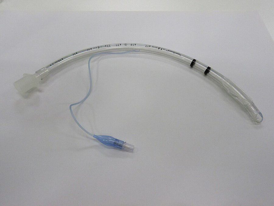
Cuffed endotracheal tube
Definitive airway for ventilation, protecting the lungs from aspiration.
Exam angle: Size is the internal diameter in mm; the cuff seals the trachea.
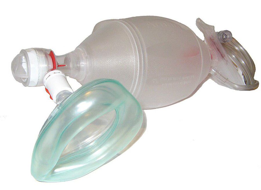
Bag-valve-mask (Ambu bag)
Manual ventilation of a patient who is not breathing adequately, through a face mask or an airway device.
Exam angle: Self-inflating, so it works without a gas supply; adding oxygen with a reservoir bag raises the delivered oxygen concentration.
ENT
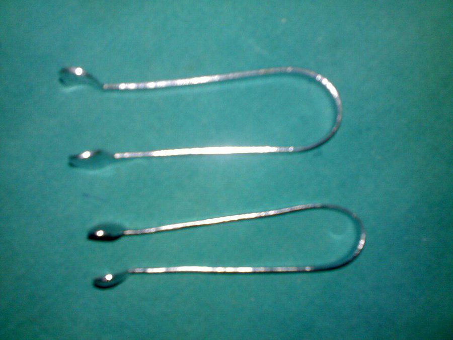
Thudichum's nasal speculum
Anterior rhinoscopy — opening the nostril to view the nasal cavity.
Exam angle: Anterior rhinoscopy instrument, used with a head mirror or light.
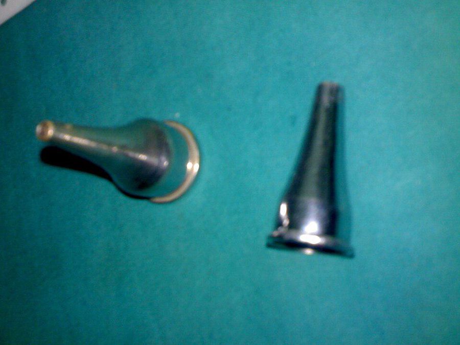
Aural (ear) speculum
Holding the external ear canal open to view the canal and tympanic membrane.
Exam angle: Pull the pinna up and back in adults before inserting.
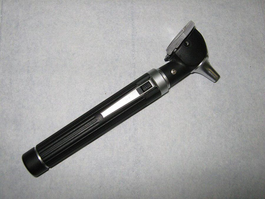
Otoscope
Illuminated, magnified examination of the ear canal and tympanic membrane.
Exam angle: Pneumatic otoscopy tests tympanic membrane mobility.
Photographs are credited under each image; CC BY-SA images are shared under the same licence. Practise more images in the clinical image atlas.
Frequently Asked Questions
How are instruments tested in NEET-PG and INI-CET?▼
Which laryngoscope blade is used in infants?▼
Why is a Sims' speculum used instead of a Cusco's?▼
Are these instrument photos free to use?▼
Ready to Start Practicing?
Start Free PracticeNo credit card required