Last verified: 5 October 2026 against Wikimedia Commons (licence checked per file).
Quick Answer
Name the view from the question: maxillary sinus → Waters'; frontal sinus → Caldwell; occipital bone → Towne's; zygomatic arch → submentovertical; scaphoid → scaphoid series; patellofemoral joint → skyline; acetabulum → Judet; free gas in a patient who cannot sit up → left lateral decubitus.
Skull & face
Waters' view (Occipitomental view)
Question it answers: Maxillary sinusitis, orbital floor (blow-out) and zygomatic fractures. Shows: Maxillary sinuses, orbital floor, zygoma.
Caldwell view (Occipitofrontal (PA with ~15° caudal tilt))
Question it answers: Frontal and ethmoid sinus disease. Shows: Frontal and ethmoid sinuses.
Towne's view (AP axial (fronto-occipital))
Question it answers: Occipital bone fractures. Shows: Occipital bone, foramen magnum.
Submentovertical view (Basal / 'jug-handle' view)
Question it answers: Zygomatic arch fractures. Shows: Skull base, zygomatic arches.
Schüller's view (Lateral oblique of the mastoid)
Question it answers: Mastoiditis and the extent of mastoid pneumatisation. Shows: Mastoid air cells.
Rhese view (Oblique view of the orbit)
Question it answers: Plain-film assessment of the optic canal. Shows: Optic canal (optic foramen), one side at a time.
Neck
Lateral soft-tissue neck
Question it answers: Retropharyngeal abscess; epiglottitis ('thumb sign'). Shows: Prevertebral soft tissue, epiglottis.
Chest
Lateral decubitus (chest)
Question it answers: Small pleural effusion; whether an effusion is free-flowing. Shows: Free pleural fluid layering along the dependent side.
Apical lordotic view
Question it answers: Apical lesions hidden by the clavicles, e.g. tuberculosis. Shows: Lung apices, clear of the clavicles.
AP chest view (vs PA) (Anteroposterior; the standard view is PA erect)
Question it answers: Sick or intubated patients who cannot stand for a PA film (bedside, mobile X-ray); heart size cannot be judged on it — the cardiothoracic ratio needs a PA erect film. Shows: Heart and mediastinum look magnified (heart further from the detector); scapulae often overlie the lungs.
Spine
Swimmer's view
Question it answers: Lower cervical spine when the shoulders obscure it on a lateral view. Shows: Cervicothoracic junction (C7–T1).
Upper limb
Scaphoid views (PA with ulnar deviation, plus obliques)
Question it answers: Suspected scaphoid fracture. Shows: The scaphoid along its length.
Stryker notch view (Arm resting on the head, beam 10–15° cephalad)
Question it answers: Hill-Sachs defect after anterior shoulder dislocation. Shows: Posterior humeral head.
Lower limb
Skyline view (Sunrise / Merchant view)
Question it answers: Patellar fractures and subluxation; also shows a knee effusion (the lateral knee view is the standard one for effusion). Shows: Patellofemoral joint, patella in axial profile.
Mortise view (AP ankle with ~15° internal rotation)
Question it answers: Ankle fractures and syndesmotic injury. Shows: The ankle mortise joint space all round.
Pelvis
Judet views (Iliac and obturator oblique views)
Question it answers: Acetabular fractures. Shows: Anterior and posterior columns of the acetabulum.
Frog-leg lateral view (Knees flexed and hips abducted ~45°, usually both sides)
Question it answers: Children: slipped capital femoral epiphysis (SUFE/SCFE) and Perthes disease. Shows: Hip joints and femoral necks (both hips on one film).
Abdomen
Left lateral decubitus (abdomen)
Question it answers: Pneumoperitoneum when the patient cannot sit up for an erect chest X-ray. Shows: Free gas collecting over the liver.
Radiographs and diagrams
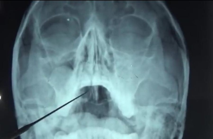
Waters' view. Waters' (occipitomental) view. The source file's description reports a right maxillary antrochoanal polyp in this patient.
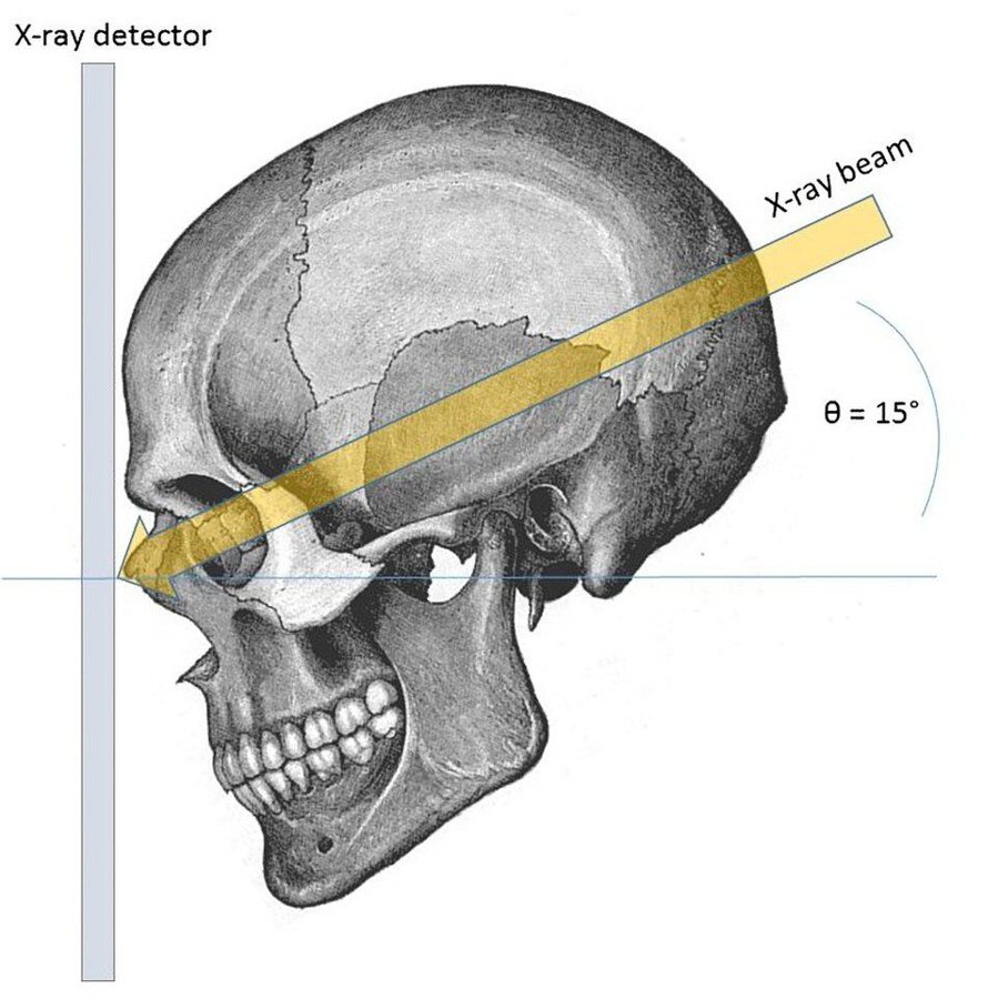
Caldwell view. Diagram of the Caldwell view (not a radiograph): a PA beam angled about 15° caudally to project the frontal sinus clearly.
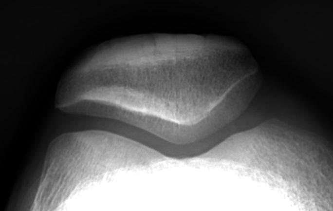
Skyline view (normal). Skyline view of a normal right patella and patellofemoral joint.
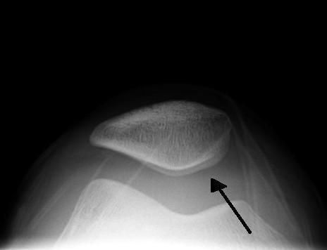
Skyline view (effusion). Skyline view showing a large knee joint effusion (arrow).
CC BY-SA images are shared under the same licence. Practise more films in the clinical image atlas.
Frequently Asked Questions
Waters' or Caldwell — which view for which sinus?▼
Which X-ray views show the acetabulum?▼
How is a small pleural effusion shown on plain X-ray?▼
When is the swimmer's view used?▼
Ready to Start Practicing?
Start Free PracticeNo credit card required