Last verified: 5 October 2026 against BTS pleural procedures 2023, ISO 20696 (urethral catheters) and Wikimedia Commons (licence checked per file).
Quick Answer
French size = 3 × outer diameter in mm. Adults usually need a 12–14 Ch Foley; larger sizes are for clots. Check a Ryle's tube with an aspirate of pH 5.5 or below, or a chest X-ray if there is no aspirate — and never pass it through the nose in a basal skull fracture.
French size and catheter colour codes
French gauge (Fr, also Ch or FG) is the outer diameter in millimetres × 3 (ISO 20696: Fr = 3 × D). So 1 Fr ≈ 0.33 mm, 12 Fr = 4 mm and 18 Fr = 6 mm. It measures the outside of the tube, not the lumen.
| Foley size | Funnel colour (common convention) |
|---|---|
| 6 Fr | Light green |
| 8 Fr | Blue |
| 10 Fr | Black |
| 12 Fr | White |
| 14 Fr | Green |
| 16 Fr | Orange |
| 18 Fr | Red |
| 20 Fr | Yellow |
ISO 20696:2018 sets catheter sizes but its public text does not set funnel colours. The colours below are the convention listed in the MSF medical catalogue specification for ISO 20696 Foley catheters (6–20 Fr only); a children's hospital guideline also refers to an international colour code. Brands do not all follow it — the 18 Fr catheter in one reference photo has a yellow valve, not red — so always read the size printed on the funnel. No authoritative colour code was found for nasogastric tubes, so none is given.
Typical sizes
| Device | Patient | Size | Note |
|---|---|---|---|
| Urethral catheter (Foley) | Adult male | 12–14 Ch | Smallest size that drains; increase gradually, larger sizes only for haematuria with clots (NHS UHBristol catheterisation policy) |
| Urethral catheter (Foley) | Adult female | 12–14 Ch | Female-length catheter may be offered; never put a female-length catheter in a male (NHS UHBristol catheterisation policy) |
| Urethral catheter (Foley) | Adult — by urine | 12/14 Ch clear · 16 Ch light haematuria/small clots · 18 Ch moderate clots or heavy debris | (NHS Borders catheterisation policy) |
| Foley balloon | Adult | 10 ml | 30 ml balloons mainly after prostate surgery (NHS UHBristol catheterisation policy) |
| Suprapubic catheter | Adult | 16 Ch (commonly used) | (NHS UHBristol catheterisation policy) |
| Urinary catheter | Child under 6 months | 6–8 Fr Nelaton (non-balloon) | (Royal Children's Hospital Melbourne guideline) |
| Urinary catheter | Child 1–3 years | 6–10 Fr Foley (1 y: 6–8, preferably 8; 2 y: 8; 3 y: 8–10) | (Royal Children's Hospital Melbourne guideline) |
| Urinary catheter | Child 5–12 years | 10 Fr (5–6 y) · 10–12 Fr (8 y) · 12–14 Fr (12 y) Foley | (Royal Children's Hospital Melbourne guideline) |
| Nasogastric feeding tube (fine bore) | Adult | 6–12 Fr | Wide-bore tubes (Ryle's type) are for gastric drainage or decompression (NHS UHBW nasogastric tube policy) |
| Chest drain — small-bore | Adult | 14F or less (Seldinger, up to 12F, suits most indications) | Pneumothorax and pleural infection, including empyema (BTS Clinical Statement on pleural procedures 2023) |
| Chest drain — large-bore | Adult | Above 14F; 28F gave the best balance in a massive-haemothorax model; above 32F rarely needed | Unstable haemothorax, ventilated or trauma pneumothorax with big air leak, after thoracic surgery (28–32F post-thoracotomy) (BTS Clinical Statement on pleural procedures 2023) |
Ryle's tube: indications, contraindications and position check
Indications
- Gastric aspiration or decompression (wide-bore tube)
- Enteral feeding, fluids and medication (fine-bore 6–12 Fr tube)
- Stomach decompression during surgery, e.g. laparoscopic cholecystectomy
Contraindications
- Basal skull fracture
- Maxillofacial disorders
- Unstable cervical spine injury
- Nasal, pharyngeal or oesophageal obstruction or ulceration; choanal atresia
- Tracheo-oesophageal fistula, oesophageal/pharyngeal pouch, oesophageal stricture
Contraindications to bedside nasal insertion; some are relative. Case reports describe a nasogastric tube entering the cranium through a basal skull fracture.
Length to insert: NEX: tip of nose → earlobe → xiphisternum.
- First line: aspirate 0.5–1 ml and test with CE-marked pH paper — pH 5.5 or below confirms gastric placement
- Readings of 5–5.5 should be checked by a second competent person
- Second line: chest X-ray when no aspirate or pH above 5.5; the film must show both hemidiaphragms and be reported for the tube position
- Feeding into the lung through a misplaced tube is a Never Event — never use the tube before position is confirmed
Urinary
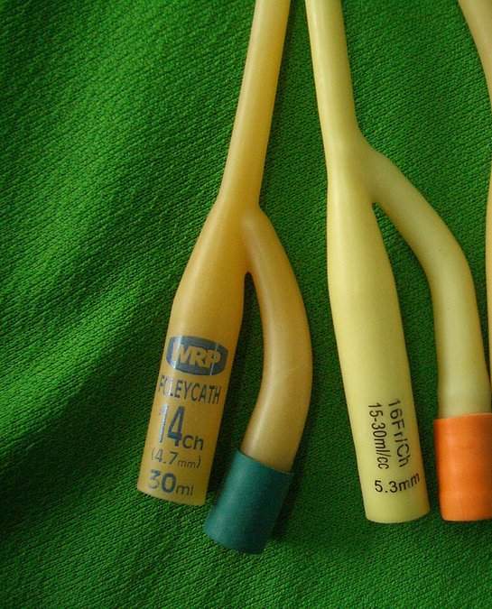
Foley catheter (2-way)
Indwelling urethral catheter for continuous bladder drainage; a balloon inflated with sterile water holds it in the bladder.
Spot it: Two arms at the outer end: a wide drainage funnel and a narrow balloon-inflation arm ending in a valve; the size (Fr/Ch) and balloon volume are printed on the funnel.
Exam angle: Sized in French (Fr = 3 × outer diameter in mm). Use the smallest size that drains: 12–14 Ch for adult men and women. Fill the balloon with 10 ml; 30 ml balloons are mainly for after prostate surgery.
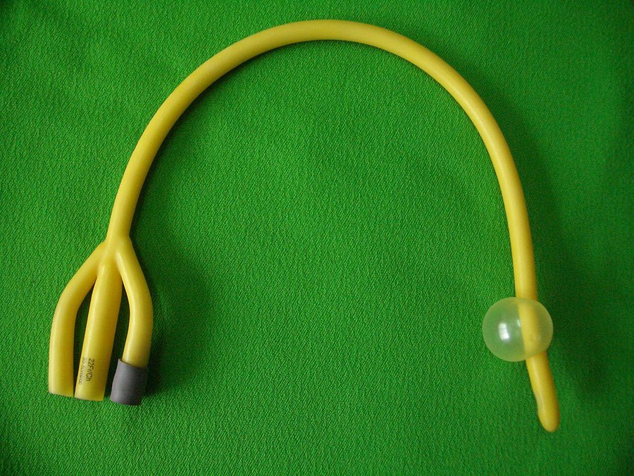
Foley catheter (3-way)
Three-channel Foley catheter used for continuous bladder irrigation: one channel drains, one fills the balloon, one carries irrigation fluid in.
Spot it: Three arms at the outer end (drainage funnel, balloon valve, irrigation inlet).
Exam angle: 3-way = continuous bladder irrigation; it is a larger catheter, so anaesthetic gel is advised; ISO 20696 lists irrigation-lumen flow rates only from 14 Fr upward.
Suprapubic catheter
Catheter passed through the abdominal wall just above the pubic bone into the bladder dome, avoiding the urethra.
Spot it: Tube exits the lower abdomen above the pubic bone instead of the urethra.
Exam angle: Indications include failed urethral catheterisation due to obstruction, urethral trauma, urinary retention and advanced neurological disease; 16 Ch is commonly used to keep a good tract.
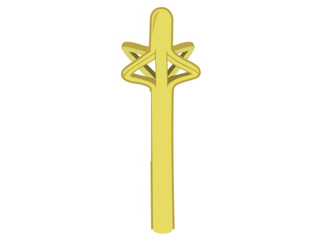
Malecot catheter
Self-retaining drainage catheter, e.g. for nephrostomy, bladder drainage and other post-operative drainage.
Spot it: Tip opens into 'wings' (flower-like struts) that hold it in place — no balloon.
Exam angle: Winged retention tip (vs the mushroom head of de Pezzer); no balloon to inflate. Some papers loosely call it a 'mushroom-tip' catheter.
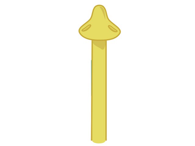
de Pezzer catheter
Self-retaining drainage catheter with a mushroom-shaped head, e.g. for suprapubic drainage after open prostatectomy and for abscess or cavity drainage.
Spot it: Single mushroom-shaped head with drainage holes at the tip — no wings, no balloon.
Exam angle: Mushroom head = de Pezzer; winged tip = Malecot; both are self-retaining without a balloon.
GI
Ryle's tube (nasogastric tube)
Wide-bore nasogastric tube used to aspirate and decompress the stomach (e.g. during surgery); fine-bore 6–12 Fr nasogastric tubes are preferred for feeding.
Spot it: Long, flexible single-lumen tube with length markings, passed through the nose into the stomach.
Exam angle: Measure length nose–earlobe–xiphisternum (NEX). Confirm position before any use: aspirate pH 5.5 or below first, chest X-ray if no aspirate or pH above 5.5. Basal skull fracture is a contraindication to bedside nasal insertion.
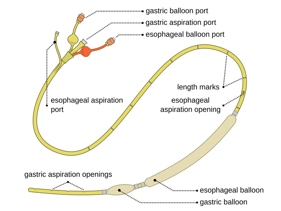
Sengstaken–Blakemore / Minnesota tube
Balloon tamponade for life-threatening bleeding from oesophageal or gastric varices, as a temporary bridge to definitive treatment.
Spot it: Multi-lumen tube with a round gastric balloon, a long oesophageal balloon and several ports; the Minnesota version adds an oesophageal aspiration port (four lumens).
Exam angle: Classic SB tube: gastric balloon, oesophageal balloon, gastric suction port (three lumens, no oesophageal suction). Gastric balloon inflated first; use for up to 24 h — longer risks oesophageal necrosis.
Airway
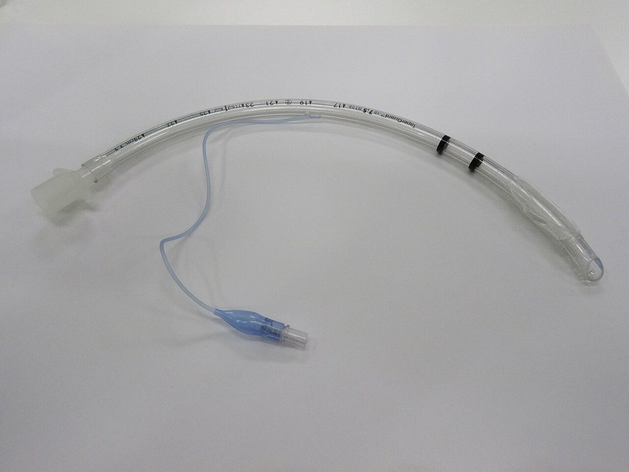
Endotracheal tube (cuffed)
Tube passed through the larynx into the trachea; its inflated cuff seals the trachea to prevent aspiration and allow positive-pressure ventilation.
Spot it: Curved clear tube with a connector at one end, a cuff near the tip and a thin line to a pilot balloon.
Exam angle: Keep cuff pressure 20–30 cm H2O: over-inflation causes mucosal ischaemia and later tracheal stenosis; under-inflation allows microaspiration and VAP. The Murphy eye is a side vent in case the tip is blocked.
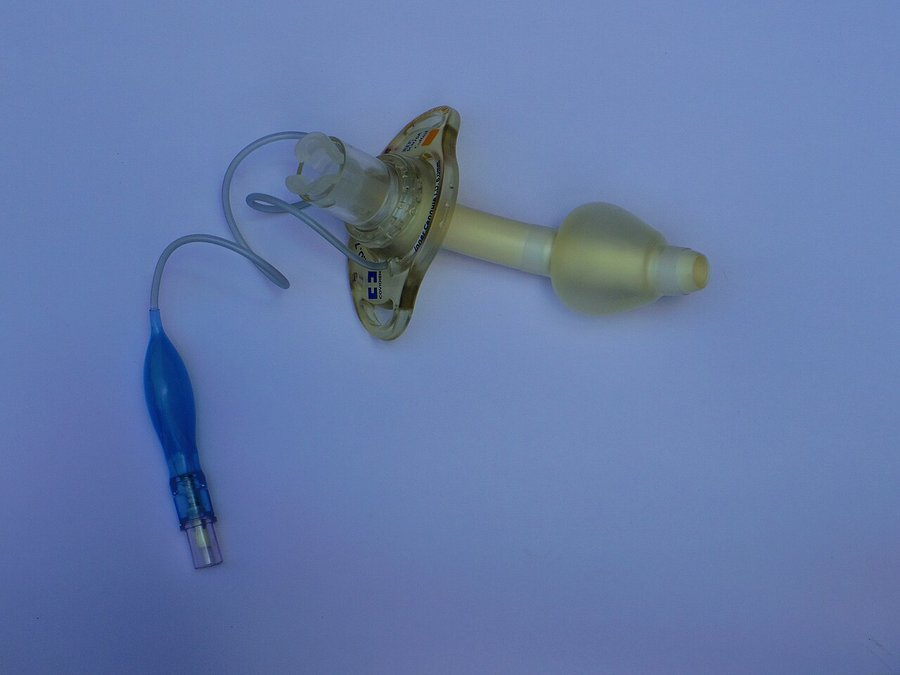
Tracheostomy tube
Tube placed through a surgically created opening in the front of the neck to give temporary or permanent access to the airway for oxygenation and ventilation.
Spot it: Short curved tube with a neck plate (flange); cuffed versions have a pilot balloon.
Exam angle: Parts: flange, outer cannula, inner cannula, obturator. Clean or replace the inner cannula regularly to prevent blockage; secure the flange to prevent displacement.
Chest & drains
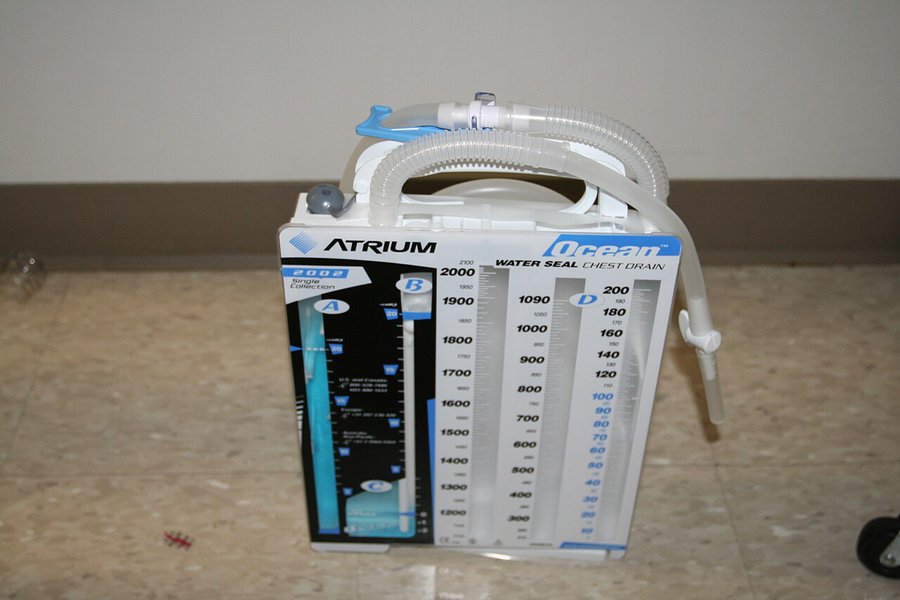
Intercostal chest drain with underwater seal
Drains air, fluid, blood or pus from the pleural space — e.g. pneumothorax failing other treatment, large effusion, pleural infection, traumatic haemothorax.
Spot it: Tube from the chest to a collection bottle where its end sits under water (the water seal); fluid in the tubing swings with breathing.
Exam angle: Bottle always below the insertion site and upright, tube end under water; record swinging and bubbling; never clamp a bubbling drain. Small-bore (14F or less) for most indications; large-bore for haemothorax in an unstable patient.
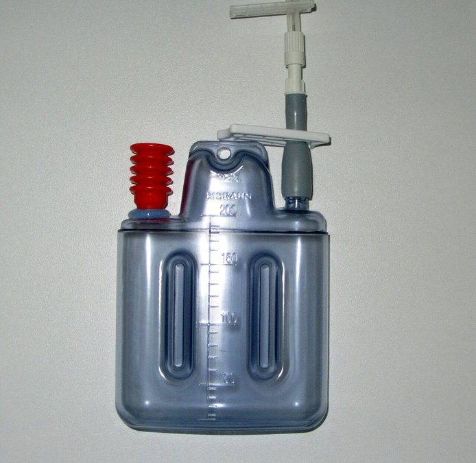
Closed suction drain (Romovac / Redon)
Active surgical drain: the wound tube connects to a sealed collection bottle under negative pressure, e.g. a subcutaneous drain after wound closure.
Spot it: Tube leading to a sealed bottle or bellows that keeps suction on the wound.
Exam angle: Active (negative-pressure, closed) drain, in contrast to passive drains like the Penrose; typically removed once output falls (e.g. below 30 ml/day in one trial).
Penrose drain
Passive surgical drain placed in a wound or body cavity to let fluid escape.
Spot it: Open drain with no suction bottle attached.
Exam angle: Classic example of a passive drain (vs active closed-suction drains).
T-tube (Kehr's tube)
T-shaped tube placed in the common bile duct through the choledochotomy after bile duct exploration, to decompress the biliary tree and reduce bile leakage.
Spot it: T-shaped tube placed into the common bile duct through the choledochotomy.
Exam angle: Used after common bile duct exploration; a T-tube cholangiogram checks for residual stones. Primary duct closure is an alternative that shortens hospital stay.
Vascular
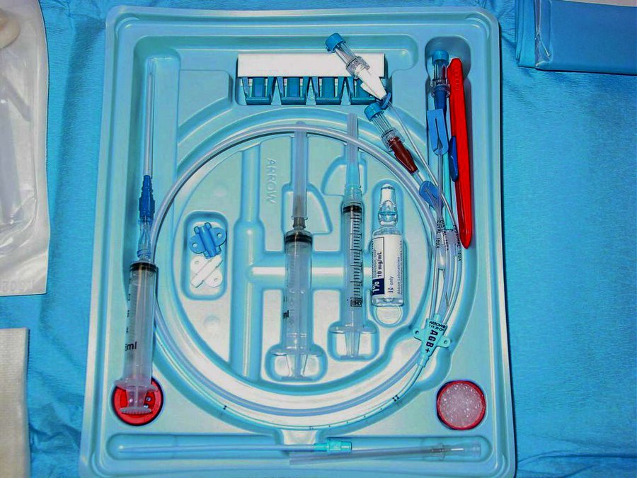
Central venous catheter (CVC)
Catheter placed in a large central vein for rapid fluid infusion, vasoactive drugs, frequent blood sampling, chemotherapy, antibiotics and haemodialysis.
Spot it: Long catheter with several lumens and hubs, supplied as a sterile kit with syringes and an introducer needle.
Exam angle: Tip should lie at the junction of the superior vena cava and right atrium; the carina is the X-ray landmark. Complications: pneumothorax, arterial puncture, arrhythmia, thrombosis, infection, air embolism.
Frequently Asked Questions
What does the French size of a catheter mean?▼
What size Foley catheter is used in an adult?▼
How is the position of a Ryle's (nasogastric) tube confirmed?▼
Why is a nasogastric tube avoided in basal skull fracture?▼
Ready to Start Practicing?
Start Free PracticeNo credit card required