What is the FEV1/FVC ratio?
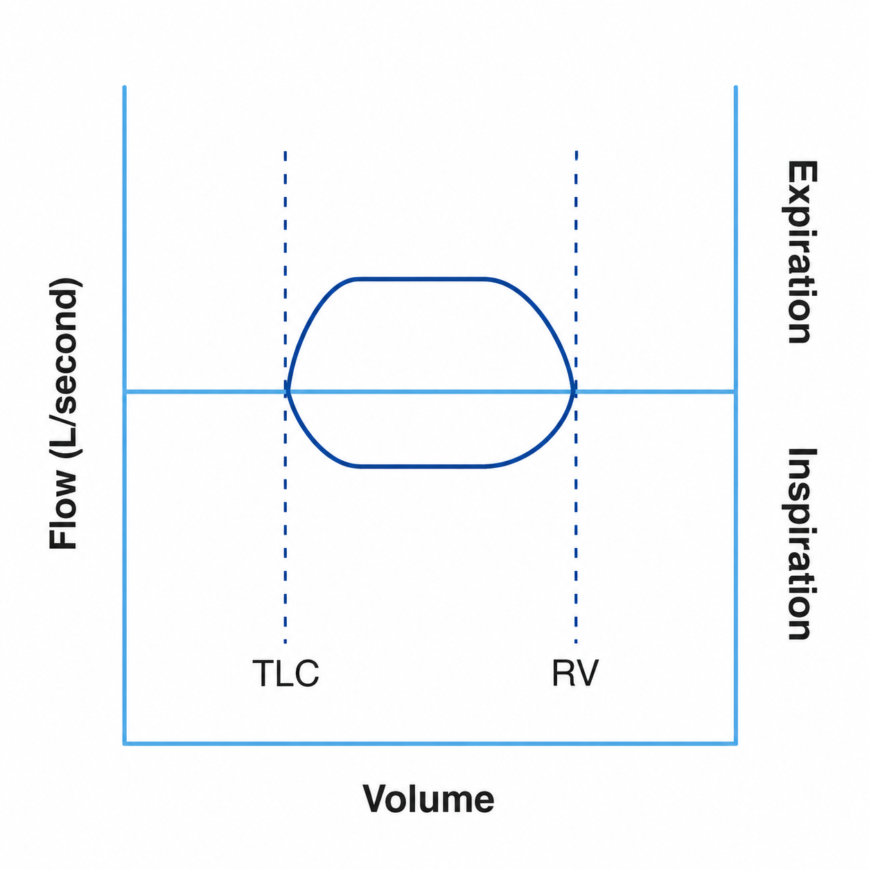
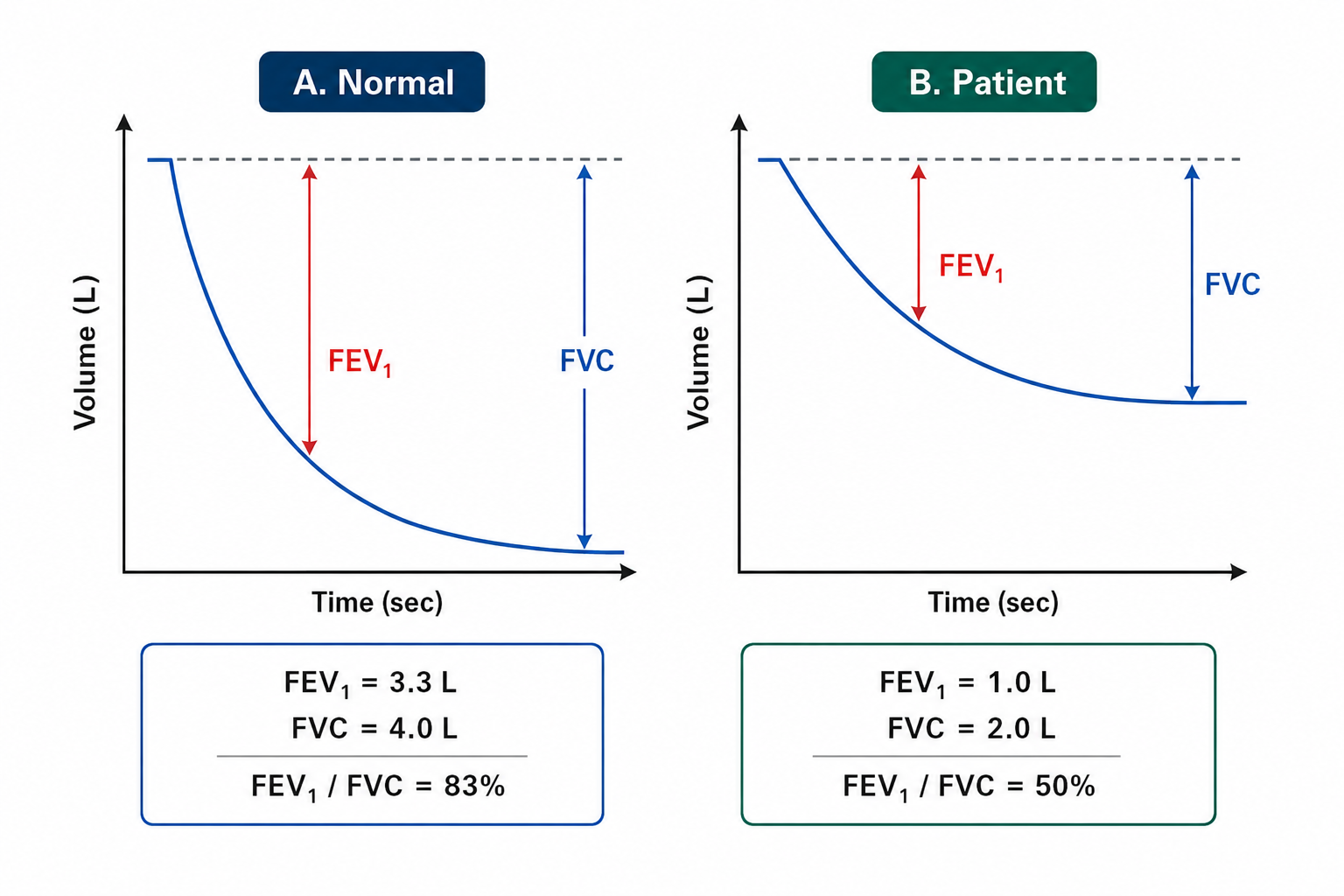
Spirometry asks the patient to breathe in fully to total lung capacity (TLC) and then blow out as hard, fast and completely as possible down to residual volume (RV). Two numbers come out of that single forced breath: the forced vital capacity (FVC), the total volume exhaled, and the forced expiratory volume in 1 second (FEV1), the volume exhaled in the first second. Dividing one by the other gives the FEV1/FVC ratio — the proportion of the vital capacity that leaves the lungs in the first second.
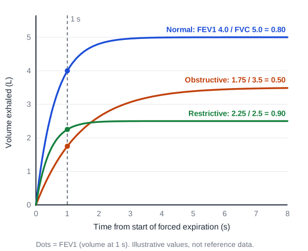
The ratio matters because it separates two very different problems. In obstruction (asthma, COPD, bronchiectasis) the airways narrow or collapse during forced expiration, so air comes out slowly: FEV1 falls much more than FVC and the ratio drops. In restriction (lung fibrosis, chest-wall or neuromuscular disease) the lungs are simply smaller: FEV1 and FVC fall together, so the ratio stays normal or even rises because stiff lungs empty quickly.
FEV1/FVC ratio = FEV1 (L) ÷ FVC (L)
Reported as a decimal (0.78) or a percentage (78%). Some reports also give FEV1 and FVC as % predicted — do not confuse ‘FEV1 % predicted’ with the FEV1/FVC ratio.
| Index | What it measures | Why examiners care |
|---|---|---|
| FVC | Total volume exhaled forcefully after a full breath in | Falls in restriction; may also fall in obstruction because of air trapping |
| FEV1 | Volume exhaled in the first second of the FVC manoeuvre | Falls in both patterns; FEV1 % predicted grades COPD severity |
| FEV1/FVC | Fraction of FVC exhaled in 1 s | The single number that defines obstruction |
| FEF25–75% | Mean flow over the middle half of the FVC | Low values suggest small-airway disease, but it is highly variable |
| PEF | Peak expiratory flow at the start of the blow | Effort-dependent; used for home monitoring of asthma |
How is spirometry performed, and what makes a test acceptable?
The 2019 ATS/ERS standard describes four phases of the forced manoeuvre: a maximal inspiration, a ‘blast’ of expiration, continued complete expiration for up to 15 seconds, and (on devices that measure it) a maximal inspiration back to TLC. Healthy adults usually empty their lungs within about 5–6 seconds; patients with obstruction take longer, which is why a short blow in a COPD patient falsely lowers FVC.
- End of forced expiration is accepted when there is an expiratory plateau (≤0.025 L change in the last 1 second), the expiratory time reaches 15 seconds, or the FVC matches a larger previous FVC within the repeatability limit.
- At least three acceptable manoeuvres are needed before and after a bronchodilator.
- Repeatability (age > 6 years): the two largest FVC values, and the two largest FEV1 values, must each agree within 0.150 L.
- Up to about eight consecutive forced manoeuvres is the usual ceiling, to avoid tiring the patient.
- Values are reported at BTPS (body temperature, ambient pressure, saturated with water vapour).
What is a normal FEV1/FVC ratio — 0.70 or the lower limit of normal?
In healthy adults roughly 70–85% of the FVC is exhaled in the first second, depending on age. The ratio is not a fixed biological constant: it falls with age (and with height), even in lifelong non-smokers, because lungs lose elastic recoil. That creates the classic debate between a fixed cut-off of 0.70 and a lower limit of normal (LLN) from reference equations.
The LLN is the value below which only 5% of a healthy reference population falls — the 5th percentile, equivalent to a z-score of −1.645. The ERS/ATS 2022 interpretive standard defines obstruction as an FEV1/FVC below the LLN and expresses results as z-scores (Global Lung Function Initiative, GLI, equations). GOLD, in contrast, still uses a post-bronchodilator FEV1/FVC < 0.7 to confirm COPD because it is simple and does not depend on reference equations — while acknowledging that it can under-diagnose young adults and over-diagnose the elderly.
| Feature | Fixed ratio < 0.70 | Below LLN (z < −1.645) |
|---|---|---|
| Used by | GOLD for diagnosing COPD (post-bronchodilator) | ERS/ATS 2022 interpretive standard |
| Accounts for age, sex, height | No | Yes — from reference equations such as GLI |
| Older adults | Over-diagnoses obstruction (the LLN drops below 0.70 from middle age — roughly the mid-40s to 50s, depending on sex and reference equation) | Fewer false positives |
| Young adults | Can miss early obstruction (their LLN is above 0.70) | Detects it |
| Practical advantage | Simple; used in the COPD trials behind treatment advice | Statistically sound, but depends on the reference set |
How do you tell obstructive, restrictive and mixed patterns apart?
Read spirometry in a fixed order: (1) the ratio, (2) the FVC, (3) the TLC if lung volumes were measured, then (4) DLCO. A low ratio means obstruction. A normal ratio with a low FVC only suggests restriction — restriction is confirmed by a TLC below the LLN (older texts use < 80% predicted).
| Pattern | FEV1/FVC | FEV1 | FVC | TLC | Classic causes |
|---|---|---|---|---|---|
| Obstructive | ↓ (< 0.70 or < LLN) | ↓ | Normal or ↓ | Normal or ↑ (hyperinflation) | Asthma, COPD, bronchiectasis, cystic fibrosis |
| Restrictive | Normal or ↑ | ↓ | ↓ | ↓ (< LLN) | Interstitial lung disease (IPF, sarcoidosis), neuromuscular weakness, chest-wall disease, severe obesity |
| Mixed | ↓ | ↓ | ↓ | ↓ | Most often COPD combined with congestive heart failure or obesity |
| Pseudorestriction (obstruction with air trapping) | ↓ or borderline | ↓ | ↓ | Normal or ↑ (RV ↑) | COPD, asthma with gas trapping |
| Non-specific pattern / PRISm | Normal | ↓ | ↓ | Normal (or not measured) | Early disease, poor effort, small-airway disease, obesity |
Two newer labels appear in recent guidelines. PRISm (preserved ratio impaired spirometry) describes a post-bronchodilator FEV1/FVC ≥ 0.7 with an FEV1 < 80% predicted; it is common in smokers and linked with higher mortality, and some patients later develop obstruction. The non-specific pattern is a low FEV1 and FVC with a normal ratio and a normal TLC — neither true obstruction nor true restriction.
Severity of an obstructive or restrictive defect has traditionally been graded by FEV1 % predicted. The ERS/ATS 2022 standard instead uses the FEV1 z-score: −1.65 to −2.5 mild, −2.5 to −4.0 moderate, and below −4.0 severe. For COPD specifically, the GOLD grades (below) remain the exam standard.
What is pseudorestriction, and why can a ‘normal’ ratio mislead?
In obstruction, airways close early during forced expiration and trap air, so RV rises and FVC falls. The FVC can fall so much that the spirometry looks like restriction — or the FEV1/FVC ratio drifts up into the ‘normal’ range even though obstruction is present. Pseudorestriction is this picture: a raised RV from air trapping with a normal or slightly increased TLC, producing a reduced FVC.
- Clue on spirometry: low FVC with a scooped (concave) expiratory flow-volume curve and a long expiratory time.
- Clue on lung volumes: TLC normal or high with a raised RV/TLC ratio (air trapping) — true restriction would show a low TLC.
- Useful check: compare FEV1 % predicted with TLC % predicted. A ‘normal’ FEV1 of 95% predicted is relatively low if the TLC is 110% predicted.
What counts as significant bronchodilator reversibility?
Spirometry is repeated after an inhaled short-acting bronchodilator (for example salbutamol), after the wait time set in the laboratory protocol. Large improvements suggest variable airflow limitation — the hallmark of asthma — whereas COPD usually shows only partial reversibility.
| Criterion | Definition | Status |
|---|---|---|
| ATS/ERS 2005 | Increase in FEV1 and/or FVC of ≥ 12% and ≥ 200 mL from the pre-bronchodilator baseline | The classic exam answer; still widely used |
| ERS/ATS 2021–22 | Increase in FEV1 and/or FVC of > 10% of the predicted value | Current interpretive standard; independent of baseline size |
2005 rule: (post-BD FEV1 − pre-BD FEV1) ≥ 200 mL AND ≥ 12% of pre-BD FEV1
The same test can be applied to FVC. The newer rule divides the change by the predicted value, so patients with a very low baseline no longer ‘pass’ on percentage alone.
Worked example (a favourite recall): pre-bronchodilator FEV1 0.9 L and FVC 1.9 L; post-bronchodilator FEV1 1.9 L and FVC 3.9 L. The ratio stays below 0.70 (obstruction), but FEV1 rose by 1.0 L — far more than 200 mL and 12% — so the answer is obstructive disease with significant reversibility.
What do flow-volume loop shapes tell you?
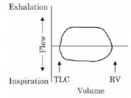
A flow-volume loop plots flow (y-axis, expiration above the line) against volume (x-axis). Its shape often reveals the site of obstruction faster than any number. Diffuse small-airway obstruction gives a scooped, concave expiratory limb; restriction gives a narrow but normally shaped loop; central (large-airway) lesions flatten one or both limbs into a plateau.

| Pattern | Loop appearance | Typical examples |
|---|---|---|
| Normal | Rapid rise to peak expiratory flow, then a near-linear fall; rounded inspiratory limb | — |
| Diffuse obstruction | Reduced peak flow and a scooped (concave) expiratory limb | Asthma, COPD/emphysema |
| Restriction | Small (narrow) loop with preserved or convex shape; flows high for the volume | Pulmonary fibrosis, neuromuscular disease |
| Variable extrathoracic obstruction | Inspiratory limb flattened; expiratory limb near normal | Vocal cord paralysis or dysfunction, laryngeal lesions |
| Variable intrathoracic obstruction | Expiratory limb flattened; inspiratory limb near normal | Tracheomalacia, intrathoracic tracheal tumour |
| Fixed central obstruction | Both limbs flattened — a ‘box’ shape | Post-intubation tracheal stenosis, circumferential tracheal tumour |
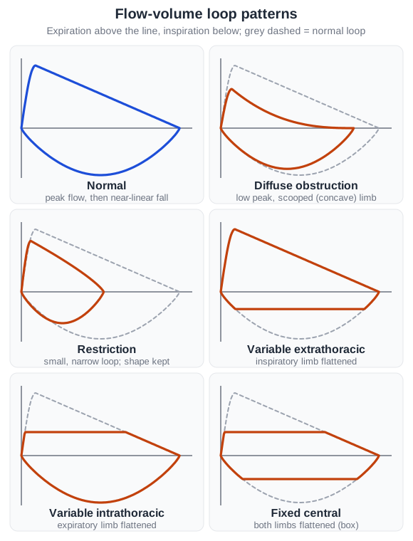
Central airway obstruction often shows on spirometry only when the lumen is severely narrowed, so a normal FEV1 does not exclude it. Ratios such as FEF50%/FIF50% and the Empey index (FEV1/PEF) have been used to support the diagnosis, but the loop shape plus CT or bronchoscopy remain the practical tools.
How does DLCO sharpen the diagnosis after spirometry?
The diffusing capacity for carbon monoxide (DLCO) measures how well gas crosses from alveoli into capillary blood during a breath-hold. It rises with alveolar surface area and capillary haemoglobin, and falls when the membrane is destroyed or thickened, when the pulmonary vascular bed is lost, or when haemoglobin is low.
| Spirometry pattern | DLCO | Think of |
|---|---|---|
| Obstructive | ↓ | Emphysema (alveolar wall destruction) |
| Obstructive | Normal or ↑ | Asthma |
| Restrictive | ↓ | Interstitial lung disease (IPF, sarcoidosis, drug-induced fibrosis) |
| Restrictive | Normal | Extrapulmonary restriction — neuromuscular weakness, chest-wall disease, obesity |
| Normal | ↓ | Pulmonary vascular disease (pulmonary hypertension, chronic thromboembolism) or anaemia |
| Any | ↑ | Alveolar haemorrhage, polycythaemia, increased pulmonary blood flow, asthma |
How does GOLD grade COPD severity with spirometry?
GOLD makes the diagnosis of COPD with a post-bronchodilator FEV1/FVC < 0.7 in the right clinical context, then grades the severity of airflow obstruction using the post-bronchodilator FEV1 as % predicted. Symptoms and exacerbation history (the GOLD ‘ABE’ assessment) are assessed separately and guide treatment.
| GOLD grade | Severity | Post-bronchodilator FEV1 (% predicted) |
|---|---|---|
| GOLD 1 | Mild | ≥ 80% |
| GOLD 2 | Moderate | 50% to < 80% |
| GOLD 3 | Severe | 30% to < 50% |
| GOLD 4 | Very severe | < 30% |
GOLD also names ‘pre-COPD’ — symptoms, structural lesions such as emphysema, or physiological abnormalities with a post-bronchodilator FEV1/FVC ≥ 0.7 — and PRISm. Both groups are at risk of developing airflow obstruction, but not all of them do.
Which spirometry traps keep appearing in NEET PG and INI-CET?
- A low FEV1 is not obstruction — both patterns lower FEV1. Look at the ratio.
- A high ratio (e.g. 0.90) with low FVC in a smoker with clubbing and basal crackles points to pulmonary fibrosis, not COPD.
- Bronchiectasis and cystic fibrosis are obstructive; sarcoidosis, IPF and neuromuscular weakness are restrictive in the typical MCQ.
- Restriction needs TLC. Spirometry alone can only suggest it.
- Reversibility = FEV1 or FVC ≥ 12% and ≥ 200 mL (classic answer); the newer ERS/ATS rule is > 10% of predicted.
- Box-shaped loop = fixed central obstruction — think tracheal stenosis after prolonged intubation.
- Low DLCO + obstruction = emphysema; low DLCO + normal spirometry = pulmonary vascular disease or anaemia.
- GOLD 4 = FEV1 < 30% predicted with post-bronchodilator ratio < 0.7.