Which muscles form the gluteal region?
The buttock is built from three gluteal muscles: gluteus maximus, gluteus medius and gluteus minimus. The gluteus maximus is the most superficial and largest, a thick, fleshy, quadrangular muscle that gives the buttock its shape and slopes across at roughly 45 degrees to its distal insertion. Gluteus medius lies deep to it, and gluteus minimus is the smallest and lies just beneath the gluteus medius (StatPearls).
| Muscle | Position | Main action | Nerve |
|---|---|---|---|
| Gluteus maximus | Superficial, largest | Extends and externally rotates the thigh; chief antigravity muscle in sitting | Inferior gluteal nerve |
| Gluteus medius | Deep to maximus | Abducts the hip; stabilises the pelvis in single-leg stance | Superior gluteal nerve |
| Gluteus minimus | Deepest, smallest | Abducts; anterior fibres medially rotate the thigh | Superior gluteal nerve |
| Tensor fasciae latae | Anterolateral hip | Abductor-mechanism partner; tightens iliotibial tract | Superior gluteal nerve |
What are the attachments and actions of gluteus maximus?
The gluteus maximus arises from the posterior part of the ilium behind the posterior gluteal line, the lateral mass of the sacrum, and by soft-tissue origins from the sacrotuberous ligament and the lumbar fascia. It has two insertions: the superficial fibres into the iliotibial tract, and the deep fibres into the gluteal tuberosity of the femur between adductor magnus and vastus lateralis (StatPearls).
- Main actions: extension and external rotation of the thigh; it extends forcefully only when needed, for example rising from sitting, climbing stairs or hills, and running.
- Chief antigravity muscle during sitting, controlling hip flexion; it also supports the pelvis and trunk.
- Three bursae: trochanteric (between maximus and greater trochanter), ischial (between maximus and ischial tuberosity) and gluteofemoral (between iliotibial tract and vastus lateralis).
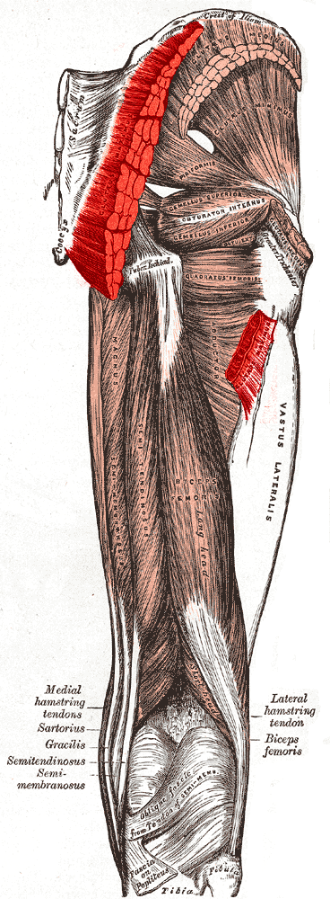
Why is the piriformis the key landmark of the gluteal region?
The piriformis passes through the greater sciatic foramen, nearly fills the notch and divides it into superior and inferior parts. Several gluteal nerves and vessels are named for their relation to it: the superior gluteal nerve and vessels pass above the piriformis, the inferior gluteal nerve and vessels pass below it, and all the other nerves and vessels exit below (StatPearls). The piriformis, obturator internus and the superior and inferior gemelli have tendons that fuse before inserting on the greater trochanter; it is one of the short external rotators of the hip.
| Position | Structures |
|---|---|
| Above piriformis | Superior gluteal nerve, superior gluteal artery and vein |
| Below piriformis | Inferior gluteal nerve and vessels, sciatic nerve, pudendal nerve, and the other nerves and vessels |
The sciatic nerve usually leaves below the piriformis, but variations exist: the whole nerve may pass above or through the muscle, or it may divide high, so that the tibial and common peroneal parts descend separately on either side of the piriformis. Entrapment of the sciatic nerve by the muscle underlies piriformis syndrome, which presents with gluteal pain and referred pain along the sciatic nerve distribution, worse with prolonged sitting, stair-climbing and squatting; it is a clinical diagnosis.

What does the superior gluteal nerve supply?
The superior gluteal nerve arises from the posterior divisions of L4, L5 and S1 of the sacral plexus. It leaves the pelvis through the greater sciatic foramen above the piriformis with the superior gluteal vessels, then runs between gluteus minimus and gluteus medius. It divides into a superior branch (to gluteus medius, occasionally minimus) and an inferior branch (to gluteus minimus, gluteus medius and tensor fasciae latae). On the deep surface of gluteus medius it lies roughly 5 cm above the tip of the greater trochanter, which matters for surgical approaches.
Gluteus medius, gluteus minimus and tensor fasciae latae together form the abductor mechanism, which stabilises the pelvis during the single-leg phase of gait and allows foot clearance on the swing side. The superior gluteal artery, the largest branch of the internal iliac artery, accompanies the nerve and divides into a superficial branch supplying gluteus maximus and a deep branch under gluteus medius.
What do the inferior gluteal nerve and vessels supply?
The inferior gluteal nerve arises from the ventral rami of L5, S1 and S2 and emerges from the inferior border of the piriformis, in contrast to the superior gluteal nerve at its superior border. It supplies gluteus maximus only. The inferior gluteal artery descends along the greater trochanter and accompanies the sciatic nerve, which is clinically important: rupture of the inferior gluteal artery can cause gluteal compartment syndrome and sciatic nerve palsy. It can also form a pseudoaneurysm after intramuscular injection (StatPearls).
| Feature | Superior gluteal nerve | Inferior gluteal nerve |
|---|---|---|
| Roots | L4, L5, S1 (posterior divisions) | L5, S1, S2 (ventral rami as given in StatPearls) |
| Relation to piriformis | Above | Below |
| Supplies | Gluteus medius, minimus, tensor fasciae latae | Gluteus maximus only |
| Injury effect | Trendelenburg gait | Difficulty with extension: rising, stairs |
What is the Trendelenburg sign and which lesion causes it?
Paralysis of the gluteus medius from superior gluteal nerve injury produces the characteristic Trendelenburg gait. The weak muscles are the hip abductors — gluteus medius and minimus — so the pelvis sags on the opposite (normal) side during the stance phase on the affected leg. Severe weakness makes the patient lurch or lean towards the affected side to bring the centre of gravity over the stance leg.
- The examiner stands or sits behind the patient.
- The patient lifts one foot, so that the opposite side of the pelvis is elevated, and holds for at least 30 seconds, then repeats on the other side (modification attributed to Hardcastle).
- In a healthy person the unsupported side stays level or rises slightly.
- If the abductors on the stance side are weak, the pelvis drops on the unsupported side: a positive test.
The test is valid only if the hip is painless and has no abduction or adduction deformity: a painful hip prevents balance, an adductor deformity can give a false negative and an abductor deformity a false positive. Causes of abductor failure include poliomyelitis, L5 radiculopathy, superior gluteal nerve damage, gluteus medius and minimus tendinitis or abscess and total hip arthroplasty; mechanical causes include a short femoral neck and coxa vara. Early osteonecrosis can mask the sign.
Which gluteal region clinical points are tested?
- Trendelenburg gait: superior gluteal nerve, gluteus medius/minimus, L5 radiculopathy, poliomyelitis, hip arthroplasty.
- Piriformis syndrome: gluteal pain with sciatica-like referral; a clinical diagnosis; MRI and nerve studies help exclude other causes.
- Sciatic variation: nerve may pierce or pass above piriformis, giving a basis for entrapment.
- Bursae: trochanteric, ischial and gluteofemoral bursae associated with gluteus maximus; greater trochanteric pain syndrome involves gluteal tendinopathy.
- Injection hazards: pseudoaneurysm of the inferior gluteal artery and sciatic nerve palsy after rupture.
- Sciatic nerve parts: the tibial and common peroneal components leave together or separately near the piriformis.
For practice on how these stems are framed, use the NEET PG PYQ bank and the most repeated topics list.