What is lymphoedema and why does it happen?
Lymphoedema (American spelling lymphedema) is a chronic, progressive condition in which impaired lymphatic drainage lets protein-rich fluid accumulate in the interstitial tissue. When the transport capacity of the lymphatics is exceeded, the persistent oedema triggers inflammation and progressive tissue remodelling — fibrosis, fat deposition and skin change — which is why the swelling hardens over time.
The limbs are involved most often, but the head and neck, trunk and genitalia can also be affected. Patients describe swelling, heaviness, reduced mobility and skin thickening, and they are prone to recurrent infections such as cellulitis. Pain is usually mild unless there is secondary infection.
How is lymphoedema classified — primary versus secondary?
| Feature | Primary | Secondary |
|---|---|---|
| Cause | Congenital or genetic malformation of the lymphatics (hypoplasia, aplasia, hyperplasia) | Acquired injury, obstruction or overload of normal lymphatics |
| Frequency | Rare | Far more common |
| Examples | Congenital, praecox (puberty to early adulthood — Meige disease; about 94% of primary cases), tarda (after age 35) | Filariasis (worldwide), cancer surgery, lymph node dissection, radiation, infection, trauma, malignancy |
| Genes / syndromes | FLT4 (VEGFR-3) and FOXC2; Turner, Noonan, Klippel-Trénaunay-Weber, Hennekam | Not genetic |
By age of onset, primary lymphoedema is congenital (birth or infancy), praecox (the commonest) or tarda. Turner syndrome classically presents with neonatal pedal oedema. Hennekam syndrome is an autosomal recessive lymphatic dysplasia (CCBE1 or FAT4) with intestinal and pulmonary lymphangiectasia.
Among secondary causes, lymphatic filariasis (Wuchereria bancrofti) remains the leading cause in tropical and subtropical regions, whereas in developed countries cancer therapy predominates — typically after lymph node dissection, radiation or tumour infiltration (breast, gynaecological, genitourinary cancers and melanoma carry the highest risk). Sentinel lymph node biopsy is now preferred over full dissection to reduce risk.
What are the ISL stages of lymphoedema and what is Stemmer sign?
| Stage | Name | Key findings |
|---|---|---|
| 0 | Latency / subclinical | Impaired lymph transport but no visible or palpable oedema; limb 'at risk'; may last months to years |
| I | Spontaneously reversible | Soft pitting oedema that subsides with elevation or overnight; minimal or no fibrosis |
| II | Spontaneously irreversible | Oedema persists despite elevation; fibrosis (induration); pitting harder to elicit; hyperkeratosis, papillomatosis |
| III | Lymphostatic elephantiasis | Extensive non-pitting swelling, marked fibrosis, skin thickening, papillomatosis, deep skin folds; recurrent cellulitis |

Stemmer sign — inability to pinch a fold of skin at the base of the second toe or finger — is described as pathognomonic of lymphoedema. Other clues: swelling that begins distally and is asymmetrical, skin changes such as hyperkeratosis and peau d'orange, and a leg that includes the feet (primary lymphoedema often involves the feet).
How is lymphoedema assessed and what mimics it?
Diagnosis is mainly clinical. Objective tools help follow progress: a limb circumference difference of 2 cm or more, or a volume difference of 200 mL or more between limbs, is significant; volume can be estimated by perometry or 3D infrared scanning. Bioimpedance spectroscopy (L-Dex change above about 6.5 to 7.5 units) can detect subclinical lymphoedema.
- Duplex ultrasound — excludes deep vein thrombosis and venous causes.
- Lymphoscintigraphy — the functional gold standard; shows delayed tracer clearance and dermal backflow.
- ICG lymphography — real-time mapping of superficial lymphatics.
- MR lymphangiography — visualises deep lymphatic channels and soft tissue.
| Condition | Distinguishing point |
|---|---|
| Lipoedema | Symmetrical fat deposition in calves and thighs; spares the feet; minimal pitting |
| Chronic venous insufficiency | Stasis oedema with hemosiderin skin change; mixed disease is phlebolymphoedema |
| Deep vein thrombosis | Acute, usually unilateral swelling |
| Systemic causes | Heart failure, nephrotic syndrome, cirrhosis, hypoproteinaemia, drugs (calcium channel blockers), hypothyroidism |
What causes lymphatic filariasis and how is it transmitted?
Lymphatic filariasis (LF) — commonly called elephantiasis — is a neglected tropical disease caused by thread-like nematodes of the filarial family. Three species cause it: *Wuchereria bancrofti* (about 90% of cases), *Brugia malayi* (most of the rest) and *Brugia timori*. Humans are infected when mosquitoes deposit infective larvae on the skin during a blood meal.
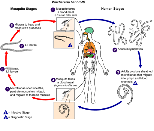
- An infected mosquito bites and deposits infective larvae on the skin; they enter through the bite wound.
- The larvae migrate to the lymphatic vessels and nodes and mature into adult worms that live roughly 6 to 8 years.
- Adults produce millions of microfilariae (immature larvae) that circulate in the blood.
- A mosquito ingests microfilariae with a blood meal; they mature to infective larvae in the mosquito (about 10 to 12 days) and reach the proboscis.
The vector depends on geography: Culex (urban and semi-urban areas), Anopheles (rural areas) and Aedes (Pacific islands). Infection is usually acquired in childhood and damages the lymphatics silently; visible disease appears later in life. India alone accounts for about 40% of the world's lymphatic filariasis cases, and bancroftian filariasis is the second most common cause of infectious disability after leprosy.
What are the clinical stages of filariasis?
Infection passes through asymptomatic, acute and chronic phases. Most infected people have no external signs yet still have lymphatic and kidney damage. About one-third eventually develop clinical disease.
| Phase | Features |
|---|---|
| Asymptomatic (microfilaraemia) | No symptoms; sub-clinical lymphatic damage; source of transmission |
| Acute | Attacks of adenolymphangitis (ADL) — fever, painful lymph nodes and vessels, local swelling; also epididymo-orchitis and funiculitis. Most are secondary bacterial skin infection on damaged lymphatics, some are immune responses to the parasite; they may last weeks and cause lost wages |
| Chronic | Lymphoedema and elephantiasis of limbs, hydrocele (scrotal swelling), breast and genital involvement |
A filarial hydrocele may contain clear fluid, lymph (chylocele) or blood and lymph (haematochylocele). A reaction after treatment can occur because dying worms release antigens and their Wolbachia endosymbionts, triggering acute lymphangitis.
How is lymphatic filariasis diagnosed?
- Night blood smear — W. bancrofti microfilariae show nocturnal periodicity, peaking in blood between 10 PM and 2 AM, matching mosquito biting time. Smears may be stained with Giemsa or Leishman, among others.
- Concentration techniques such as the Knott method improve detection when parasite numbers are low.
- Complete blood count — eosinophilia suggests active filariasis.
- Ultrasound — the filarial dance sign (movement of adult worms within lymphatics) is seen in about 80% of cases.
- Lymphoscintigraphy shows lymphatic abnormality but cannot separate filarial from non-filarial causes.
How are filariasis and lymphoedema treated, and how is filariasis eliminated?
Elimination relies on mass drug administration (MDA) — an annual dose of medicines to the entire at-risk population. The drugs mainly reduce microfilariae in the blood (limited effect on adult worms), which stops transmission to mosquitoes. Two-medicine MDA has interrupted transmission when given annually for at least 4 to 6 years with effective coverage.
| Setting | Regimen |
|---|---|
| Countries without onchocerciasis | DEC 6 mg/kg + albendazole 400 mg |
| Countries with onchocerciasis | Ivermectin 200 mcg/kg + albendazole 400 mg |
| Areas co-endemic with loiasis | Albendazole 400 mg alone, twice per year |
| Without onchocerciasis, where programme conditions allow | Triple drug: ivermectin + DEC + albendazole |
For individual patients, doxycycline (targeting Wolbachia) for 6 weeks improves lymphoedema and hydrocele, particularly early in disease. Morbidity management is essential: hygiene and skin care, exercise, elevation, treatment of ADL episodes, and hydrocelectomy (surgery alleviates most hydroceles).
General lymphoedema care follows complex decongestive therapy (CDT) in two phases: an intensive phase (manual lymph drainage, multilayer compression bandaging, skin care, exercise) and a maintenance phase (compression garments, exercise, self-management). Compression is essential at all stages. Surgery is for refractory disease — lymphovenous anastomosis, vascularised lymph node transfer or liposuction (suction-assisted protein lipectomy) — while radical excision carries high morbidity. The US FDA has not approved any drug for lymphoedema itself.
What are the high-yield exam points?
- Leading cause of secondary lymphoedema worldwide: lymphatic filariasis; commonest species: W. bancrofti (about 90%).
- Primary lymphoedema: praecox (Meige) is the commonest type; genes FLT4 and FOXC2.
- Stemmer sign = cannot pinch skin at base of second toe/finger.
- ISL: 0 latent, I reversible, II irreversible with fibrosis, III elephantiasis.
- Microfilariae show nocturnal periodicity — draw blood 10 PM to 2 AM.
- Filarial dance sign on ultrasound; lymphoscintigraphy is the functional gold standard in lymphoedema.
- Adult worm lifespan 6 to 8 years; MDA must run 4 to 6 years.
- Related parasitic topics: intestinal nematodes; scrotal swellings: undescended testis and scrotal swellings; programmes: national health programmes.