What is otosclerosis?
Otosclerosis is a disorder of bone remodelling in the otic capsule — the dense bone around the inner ear. Normal endochondral bone is replaced by foci of irregular, spongy bone that later hardens abnormally. When a focus involves the stapes footplate and its annular ligament at the oval window, the stapes can no longer vibrate, and sound cannot pass efficiently into the cochlea. The result is conductive hearing loss.
If the foci spread into the cochlea itself (cochlear otosclerosis), sensorineural loss is added, giving a mixed hearing loss — usually affecting the higher frequencies.

Where does otosclerosis start and what causes it?
The first lesion usually appears at the fissula ante fenestram, a small area just in front of the oval window, between the oval window and the cochleariform process. From there it spreads through vascular channels to the anterior footplate and annular ligament. Active, vascular foci appear red; mature foci are sclerotic.
| Factor | What is known |
|---|---|
| Genetics | Mostly autosomal dominant with reduced penetrance (about 40%) and variable expressivity; many loci described |
| Sex | About twice as common in women (2:1), suggesting a role for sex hormones |
| Age | Early adult onset |
| Pregnancy | Appears to worsen hearing loss; the exact relationship is unclear |
| Measles virus | Measles RNA found in stapes footplates; measles vaccination associated with less otosclerosis surgery |
| Ethnicity | Less common in Asian and Black populations than in White populations |
How common is otosclerosis and what does the lesion look like?
Many more people have otosclerotic foci than have symptoms. Clinical otosclerosis affects roughly 0.04–1% of White individuals, yet histological foci may be found in up to 10% of that group. Histological otosclerosis is less frequent in Black (about 1%) and Asian (about 5%) populations. Only foci that reach the stapes footplate or cochlea cause hearing loss.
| Feature | Active (immature) focus | Mature (inactive) focus |
|---|---|---|
| Cells | Abundant osteoclasts and osteoblasts; osteoclasts resorb normal bone, which is replaced by connective tissue | Fewer cells |
| Vascularity | Many marrow and vascular spaces — appears red (basis of the Schwartze sign) | Less vascular |
| Bone | Irregular spongy bone | Hardened, sclerotic bone |
| CT appearance | Lucent (spongiotic) foci; 'halo' around the cochlea in cochlear disease | Sclerotic foci near the oval window |
What are the symptoms and signs of otosclerosis?
- Slowly progressive hearing loss, bilateral in about 70% of patients.
- Tinnitus is very common — about 89% in one surgical series.
- Paracusis Willisii — the patient hears better in a noisy background. It is not pathognomonic, but it indicates a conductive loss.
- Normal tympanic membrane on otoscopy in most patients.
- Schwartze sign — a reddish blush on the cochlear promontory seen through a thin eardrum, due to increased vascularity of an active focus.
What do the tuning fork tests show in otosclerosis?
| Test | Finding | Why |
|---|---|---|
| Rinne | Negative — bone conduction louder than air conduction | Fixed stapes blocks air-conducted sound; becomes reliably negative once the air–bone gap exceeds about 30 dB |
| Weber | Lateralised to the affected ear, or to the worse ear in bilateral disease | Conductive loss masks room noise, so bone-conducted sound seems louder in that ear |
| Gellé | Negative — no change in loudness when canal pressure is raised | With a mobile chain, pressure in the canal reduces bone-conducted hearing; a fixed stapes shows no change |
The Gellé test places a vibrating fork on the mastoid while the air pressure in the ear canal is raised. In normal ears and in sensorineural loss, canal pressure measurably shifts bone-conduction thresholds. When the stapes is fixed, nothing changes — a negative Gellé test. Gellé himself proposed it as a diagnostic criterion for otosclerosis in 1885, and a 2025 surgical series found it negative in 98.7% of ears.
What do audiometry, tympanometry and CT show?
- Pure tone audiometry: air-conduction loss, usually starting in the low frequencies, with an air–bone gap.
- Carhart notch: an apparent dip in the bone-conduction line of about 20–30 dB at 2,000 Hz, caused by loss of the ossicular chain's resonance — not true cochlear damage. It usually disappears after stapedectomy.
- Mixed loss: in advanced or cochlear disease, sensorineural loss is added, mainly at high frequencies.
- Tympanometry: usually normal (type A) in early otosclerosis, which helps distinguish it from ossicular discontinuity. Speech discrimination is also normal early.
- Stapedial reflexes: may still be normal in early disease.
- High-resolution CT temporal bone: foci of spongy or sclerotic bone anterior to the oval window; in cochlear otosclerosis a 'halo sign' of lucency around the cochlea.
How is otosclerosis treated?
| Option | Role |
|---|---|
| Hearing aids | Effective at any stage, alone or with other treatment; used in many patients bilaterally |
| Sodium fluoride | Prescribed to slow progression; efficacy is still debated. Considered when hearing worsens in pregnancy or with a strong family history |
| Bisphosphonates | Antiresorptive; not to be used in pregnancy |
| Stapedotomy / stapedectomy | Definitive surgery for conductive loss; usual threshold is an air–bone gap of about 30 dB |
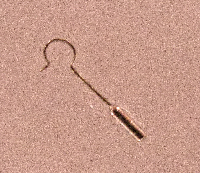
In stapes surgery the fixed stapes superstructure is removed and a prosthesis (piston) is placed from the incus to the oval window. In stapedectomy the footplate is removed; in small-fenestra stapedotomy — now the most common operation — only a small hole is made in the footplate for the piston. Surgery can be done under local or general anaesthesia.

What are the rules and risks of stapes surgery?
- Worse ear first: in bilateral disease the ear with greater loss is operated first.
- Never both ears at the same sitting — to avoid the remote risk of bilateral total sensorineural loss.
- Contraindications: the only hearing ear, active infection of the canal or middle ear, and poor general condition that prevents anaesthesia.
- Main risk: sensorineural hearing loss, up to an anacusic 'dead ear' — higher in revision surgery.
- After surgery: the Carhart notch usually disappears; some patients still need a hearing aid.
How is otosclerosis different from other causes of conductive deafness?
| Condition | Clue |
|---|---|
| Otosclerosis | Young adult, family history, progressive, type A tympanogram, Carhart notch |
| Ossicular discontinuity | Tympanometry helps to separate it from otosclerosis |
| Malleus or incus fixation | Can also produce a Carhart notch; found at surgery |
| Otitis media with effusion | Commonest cause of acquired hearing loss in children; fluid behind the drum |
| Chronic otitis media | Perforation and discharge — see chronic suppurative otitis media |