What is strabismus and how is it described?
Strabismus (squint) means the two eyes are not directed at the same target together — one or both eyes deviate inwards, outwards, up or down. The misalignment may come from a refractive error, abnormal binocular fusion or a neuromuscular problem. It is among the commonest eye problems in children, and it is most often diagnosed before age 6, with a peak at about 3 years.
| Term | Meaning |
|---|---|
| Orthophoria | Perfect alignment even without a stimulus for fusion |
| Heterophoria (phoria) | Latent deviation, held in check by fusion; seen only when fusion is broken |
| Heterotropia (tropia) | Manifest deviation, visible on clinical examination (for example with the corneal light reflex) |
| Eso- / Exo- | Eye turns toward the nose (convergent) / away from the nose (divergent) |
| Hyper- / Hypo- | Eye higher / lower than the fellow eye |
| Incyclo- / Excyclo- | Torsional turn: top of the eye rotates nasally / temporally |
Squint is also classified by onset (infantile if noted at or before 6 months, acquired after that), laterality (unilateral or alternating), constancy (constant or intermittent) and comitance (comitant or incomitant). Alternating fixation in an infant suggests there is no amblyopia; strong fixation with one eye points to amblyopia in the other.
How do paralytic and concomitant squint differ?
This is the most examined distinction. In comitant (concomitant) strabismus the angle of deviation is the same in all positions of gaze; it is typically childhood-onset and linked to refractive or sensory causes. In incomitant strabismus the angle varies with the direction of gaze; it is more often linked to cranial nerve palsy, restrictive disease or orbital pathology.
| Feature | Concomitant | Paralytic / incomitant |
|---|---|---|
| Angle across gaze | Constant in all directions | Varies — greatest in the direction of the paretic muscle |
| Ocular movements | Full in each eye (ductions normal) | Limited movement of the affected eye |
| Primary vs secondary deviation | Equal | Secondary deviation (paretic eye fixating) is always greater than primary |
| Typical onset | Childhood | Acquired, often adult (III, IV, VI nerve palsy) |
| Diplopia | Usually absent (suppression) | Common at onset, especially if previously normal binocular vision |
| Head posture | Not typical | Compensatory head posture to avoid diplopia |
| Typical causes | Uncorrected hypermetropia, accommodative factors, sensory loss | Cranial nerve palsy, thyroid eye disease, orbital fracture, myasthenia |
How are Hirschberg and Krimsky tests done?
Both tests estimate the angle of a manifest squint when the patient cannot cooperate with cover tests — a quick bedside estimate in infants.
| Test | Method | Key points |
|---|---|---|
| Hirschberg | Shine a light and note where the corneal light reflex falls on each cornea | Rough estimate: each 1 mm of reflex displacement = 7° or 14 prism dioptres (pupil 4 mm). Reflex at the pupil margin ≈ 15°; at the limbus ≈ 45° |
| Krimsky | Prisms placed in front of the fixing eye (modified: in front of the deviating eye) until the light reflexes are centred | Measures tropias, not phorias |
Remember that pseudostrabismus can mimic a squint — for example, a low-birth-weight history suggests retinopathy of prematurity, whose ectopic macula can cause it. The corneal light reflex position is what confirms real misalignment.
What do the cover, uncover and alternate cover tests show?
- Cover test — detects heterotropia. Cover the fixating eye and watch the uncovered eye at distance and near. If the uncovered eye moves to take up fixation, a tropia is present (outward movement = esotropia; inward = exotropia).
- Uncover test — detects heterophoria. Cover an eye for 2 to 3 seconds, then watch the same eye as the cover is removed; a phoric eye drifts and then recovers as fusion returns. Done at distance and near.
- Alternate cover test — a dissociation test that reveals the total deviation (tropia plus phoria) when fusion is interrupted.
- Prism cover test — alternate cover test combined with prisms to measure the deviation at near and distance.
Which other tests are used to assess a squint?
| Test | Use |
|---|---|
| Maddox rod | Turns a white light into a red streak; dissociates the eyes to measure phoria |
| Maddox wing | Dissociates the eyes at near (33 cm); measures heterophoria, including cyclophoria |
| Hess chart (Hess / Lees screen) | Plots the field of binocular single vision; used in incomitant squint from palsy or restriction |
| Parks three-step test | Identifies the paretic muscle in vertical diplopia, especially superior oblique palsy |
| Forced duction test | Separates mechanical restriction from paralysis |
| Near point of convergence | Measured with the RAF rule; should be nearer than 10 cm |
| Stereoacuity / fusion tests | Titmus stereo-fly, Lang cards; Worth 4-dot and Bagolini glasses; synoptophore for both |
| AC/A ratio | Convergence per dioptre of accommodation; normal about 3 to 5 : 1 |
| Cycloplegic refraction | 1% cyclopentolate; hypermetropia is the commonest refractive error in squint |
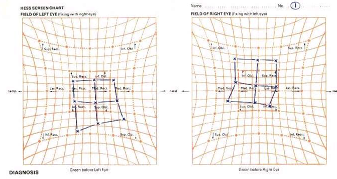

Hess chart interpretation: after a palsy, the ipsilateral antagonist contracts and overacts, and the yoke muscle of the other eye is secondarily inhibited. The eye with the smaller field has the paretic muscle, with the greatest restriction in the direction of that muscle's action; the larger field belongs to the eye with the overacting yoke muscle.
What are the common types of squint and their key features?
| Type | Key features |
|---|---|
| Infantile (congenital) esotropia | Large, stable angle before 6 months; cross-fixation, latent nystagmus, inferior oblique overaction; DVD in 80% by age 3; early muscle surgery (6 months to 2 years) |
| Accommodative esotropia | Age 2 to 5 years; uncorrected hypermetropia, excess accommodative convergence; refractive type has normal AC/A with hyperopia >2 D; treat with full cycloplegic correction; bifocals if high AC/A |
| Intermittent exotropia | Appears with inattention, bright light, fatigue; correct refraction, orthoptic exercises (pencil push-ups), bilateral lateral rectus recession |
| Dissociated vertical deviation (DVD) | Bilateral, asymmetric; an eye drifts up under cover; Bielschowsky phenomenon is specific; surgery: superior rectus recession with Faden |
| Duane retraction syndrome | Globe retraction and narrowed fissure on adduction; anomalous innervation of lateral rectus by 3rd nerve (6th nerve agenesis); more in girls, left eye |
| Brown syndrome | Limited elevation in adduction from superior oblique tendon restriction; forced duction test positive |
| Mobius syndrome | Cannot abduct either eye past midline; bilateral aplasia of 6th and 7th cranial nerves |
| Microtropia | Angle <5° (8 PD); amblyopia, anisometropia, foveal suppression |
- Lateral rectus (VI) palsy — esotropic eye with limited abduction; face turns toward the side of the palsy.
- Oculomotor (III) palsy — ptosis with an abducted, intorted eye and defective accommodation.
- Superior oblique (IV) palsy — limited depression in adduction, vertical and torsional diplopia on down-gaze; a right palsy gives right hypertropia that increases on left gaze and head tilt to the same side. Use the Parks three-step test.
How is squint treated?
The aims are proper alignment, treatment of amblyopia, preserving binocularity and abolishing diplopia. Treatment is a ladder:
- Refractive correction first. Full correction of hypermetropia is the treatment of choice in all esotropias; in intermittent exotropia, full myopic correction often controls the deviation. Executive bifocals are used in convergence excess.
- Amblyopia therapy. Patching the fellow eye for 2 to 6 hours per day is the gold standard, most effective before age 7; atropine 1% penalisation of the fellow eye is equally effective.
- Orthoptic exercises (fusional exercises, pencil push-ups) for intermittent exotropia.
- Prisms for deviations under 20 PD; contraindicated with amblyopia, suppression or anomalous retinal correspondence.
- Miotics (ecothiopate iodide 0.125%) as a short-term option in accommodative esotropia.
- Botulinum toxin A as an adjunct or to test for post-operative diplopia.
- Extraocular muscle surgery only after conservative measures fail: esotropia over 15 PD and exotropia over 20 PD after full spectacle correction; ideal age for infantile squint is before 2 years.
For a related examination, see abnormal pupils and cranial nerves; for lens causes of sensory squint, see cataract types and surgery.