What is the Chapel Hill 2012 classification of vasculitis?
Vasculitis is inflammation of blood vessel walls that leads to end-organ damage. More than 30 distinct vasculitides are described. Primary systemic vasculitis is classified mainly by the International Chapel Hill Consensus Conference (CHCC 2012) nomenclature, which sorts diseases by the size of the vessel predominantly affected. In 2022, ACR/EULAR added new classification criteria for GPA, MPA, EGPA, GCA and Takayasu arteritis.
| Vessel size | Diseases | One-line clue |
|---|---|---|
| Large vessel | Giant cell arteritis (GCA); Takayasu arteritis | Aorta and its major branches; granulomatous |
| Medium vessel | Polyarteritis nodosa (PAN); Kawasaki disease | Stenoses and aneurysms; coronary aneurysm in Kawasaki |
| Small vessel — ANCA-associated | Granulomatosis with polyangiitis (GPA); microscopic polyangiitis (MPA); eosinophilic GPA (EGPA) | Pauci-immune; glomerulonephritis and lung |
| Small vessel — immune complex | IgA vasculitis; cryoglobulinaemic vasculitis | Palpable purpura; IgA or cryoglobulin deposits |
| Variable vessel | Behçet disease | Oral and genital ulcers, uveitis, pathergy |
How do giant cell arteritis and Takayasu arteritis differ?
Both are granulomatous large-vessel vasculitides in which inflammation starts in the vasa vasorum of the adventitia and spreads through the wall. The main discriminator is age: GCA affects people over 50; Takayasu affects mostly women under 50.
| Feature | Giant cell arteritis | Takayasu arteritis |
|---|---|---|
| Age / sex | Over 50, incidence rises with age | Mostly women under 50; female : male about 9 : 1 |
| Population | Predominantly White; the commonest primary vasculitis | More prevalent in South Asian countries |
| HLA | HLA-DRB1*04:01 and *01:01 | HLA-B52 |
| Key symptoms | New headache, visual loss or amaurosis fugax, jaw claudication (about half), fever of unknown origin | Systemic symptoms, limb claudication, absent pulses ('pulseless disease'), bruits, BP difference between arms, hypertension |
| Association | Polymyalgia rheumatica in about one-third | Renovascular hypertension from renal artery stenosis |
| Diagnosis | Raised ESR and CRP; temporal artery biopsy is the gold standard; ultrasound halo sign | No specific blood test; MR angiography preferred, or CT angiography; biopsy rarely done |
| Treatment | High-dose glucocorticoids at once (prednisone 1 mg/kg/day; IV pulse if vision is threatened); tocilizumab spares steroids | Corticosteroids first, with or without immunosuppressants; surgical revascularisation for symptomatic occlusive disease |

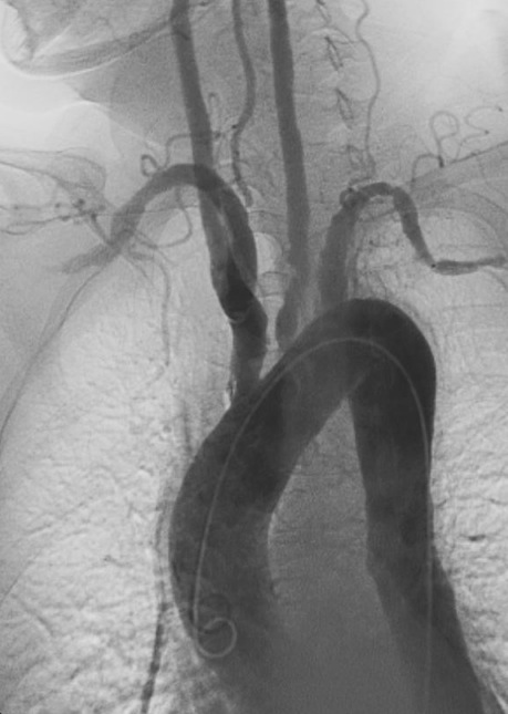
What are the medium-vessel vasculitides — PAN and Kawasaki disease?
Polyarteritis nodosa is a necrotising vasculitis of medium arteries with a well-established link to hepatitis B: immune complexes of viral antigen deposit in the vessel wall. Without hepatitis B it is extremely rare. It shows a slight male predominance and peaks at 40 to 60 years.
- Skin — palpable purpura, livedo reticularis, ulcers, digital ischaemia (mostly lower limbs).
- Kidney — renal artery vasculitis with infarction (non-glomerular disease); malignant hypertension is a key sign.
- Nerve — mononeuritis multiplex with wrist or foot drop.
- Gut and heart — mesenteric ischaemia, myocardial ischaemia; myalgia.
- Diagnosis — no specific blood test; hepatitis B and C serology; arteriography, CT or MR angiography shows stenoses and aneurysmal dilatations.
Kawasaki disease affects medium (and small) arteries in children, mainly under 5 years, with much higher rates in Japan, Korea and Taiwan. The danger is coronary artery aneurysm, so every suspected case needs ECG and echocardiography.
| Requirement | Detail |
|---|---|
| Fever | 5 days or more, plus at least 4 of the 5 features below |
| Eyes | Bilateral painless bulbar conjunctival injection without exudate |
| Mouth | Red mouth and pharynx, strawberry tongue, red cracked lips |
| Rash | Polymorphous exanthem (morbilliform, maculopapular or scarlatiniform) |
| Extremities | Swelling of hands and feet with red palms and soles; periungual peeling 2–3 weeks after fever onset |
| Lymph node | Cervical lymphadenopathy over 1.5 cm |
How do GPA, MPA and EGPA differ?
The three ANCA-associated vasculitides are pauci-immune small-vessel vasculitides: ANCAs prime neutrophils, which release proteinase-3 (PR3) and myeloperoxidase (MPO) and damage the endothelium. ANCA is found in 73–95% of GPA or MPA cases, but up to 10% show the 'opposite' pattern, so ANCA type supports rather than defines the diagnosis.
| Feature | GPA (Wegener) | MPA | EGPA (Churg-Strauss) |
|---|---|---|---|
| ANCA | c-ANCA / anti-PR3 (about 80%); anti-MPO about 15% | p-ANCA / anti-MPO | ANCA in only 30–40%, usually p-ANCA / MPO |
| Granulomas | Yes — necrotising granulomatous inflammation | No granulomas | Yes, extravascular, with tissue eosinophilia |
| Upper airway | Sinusitis, nasal crusting, epistaxis, otitis media, hearing loss, saddle nose | Usually spared | Nasal polyps, chronic sinusitis |
| Lung | Nodules that can cavitate, haemoptysis, diffuse pulmonary haemorrhage | Pulmonary capillaritis, haemorrhage | Asthma (often years before), fleeting infiltrates |
| Kidney | Necrotising crescentic glomerulonephritis | Pauci-immune rapidly progressive GN | Crescentic or necrotising GN, mainly in ANCA-positive patients |
| Blood | Raised ESR/CRP | Raised ESR/CRP | Eosinophilia of 1000/mm³ or more (or over 10%), high IgE |
| Other | Higher relapse risk with PR3-ANCA | More common in Asian populations | Cardiac involvement; mononeuritis multiplex |
GPA triad: upper respiratory tract + lower respiratory tract + kidney. A patient with recurrent 'sinusitis', a lung infiltrate, palpable purpura and haematuria should be tested for ANCA. MPA is distinguished from GPA by the absence of granulomatous inflammation in the upper respiratory tract and by pulmonary capillaritis.
What is IgA vasculitis (Henoch-Schönlein purpura)?
IgA vasculitis is the commonest vasculitis of childhood, affecting about 20 per 100,000 children per year, and is rare in adults. It is an immune-complex small-vessel vasculitis: galactose-deficient IgA1 and anti-IgA1 antibodies form complexes that deposit in vessel walls. It is commoner from September to April and often follows group A streptococcus, parainfluenza or parvovirus B19 infection.
| Mandatory | Plus at least one of |
|---|---|
| Palpable purpura or petechiae without thrombocytopenia, mainly on the lower limbs | Abdominal pain |
| Arthralgia or arthritis | |
| Renal involvement — proteinuria, haematuria or red cell casts | |
| Histology: leukocytoclastic vasculitis or proliferative glomerulonephritis with predominant IgA deposits |

What are cryoglobulinaemic vasculitis and Behçet disease?
| Type | Composition | Typical link |
|---|---|---|
| I | Monoclonal immunoglobulin (usually IgM) | Lymphoproliferative disease |
| II (mixed) | Polyclonal IgG + monoclonal IgM with rheumatoid factor activity | Vasculitis; hepatitis C |
| III (mixed) | Polyclonal IgG + polyclonal IgM with rheumatoid factor activity | Vasculitis; hepatitis C, connective tissue disease |
Chronic hepatitis C accounts for 70–90% of mixed cryoglobulinaemic vasculitis. Features: palpable purpura, arthralgia, neuropathy and membranoproliferative glomerulonephritis; complement (especially C4) is low. Blood must be collected and kept at 37 °C or the cryoglobulins precipitate and the test is falsely negative.
Behçet disease is a variable-vessel vasculitis strongly linked to HLA-B51 and commonest along the ancient Silk Road. It causes recurrent painful oral aphthae, genital ulcers, uveitis (often panuveitis), erythema nodosum, vascular thrombosis and pathergy (a sterile pustule at a needle-prick site). In the ICBD score, oral ulcers, genital ulcers and eye lesions score 2 points each; 4 or more confirms the diagnosis.
Which clue points to which vasculitis?
Any vasculitis can begin with constitutional symptoms — fever, weight loss, malaise and fatigue — so the hardest step is thinking of it at all. StatPearls advises suspecting vasculitis when systemic symptoms combine with dysfunction of one or more organs, or when a 'pneumonia' or 'sinusitis' keeps relapsing and then a purpuric rash or haematuria appears. Use the organ pattern and the patient's age to narrow the list.
| Clue in the question | Think of |
|---|---|
| Over 50, new headache, jaw claudication, raised ESR | Giant cell arteritis |
| Young woman, unequal arm BP, absent radial pulse | Takayasu arteritis |
| Hepatitis B, mononeuritis multiplex, renal infarcts, aneurysms on angiography | Polyarteritis nodosa |
| Child under 5, fever 5 days, strawberry tongue, peeling fingers | Kawasaki disease |
| Sinusitis + lung nodules + glomerulonephritis, c-ANCA | Granulomatosis with polyangiitis |
| Pulmonary-renal syndrome, p-ANCA, no granulomas | Microscopic polyangiitis |
| Late-onset asthma, eosinophilia, neuropathy | Eosinophilic granulomatosis with polyangiitis |
| Child, purpura on buttocks and legs, abdominal pain, haematuria | IgA vasculitis |
| Hepatitis C, purpura, low C4, MPGN | Cryoglobulinaemic vasculitis |
| Oral and genital ulcers, uveitis, pathergy | Behçet disease |
What are the common exam traps in vasculitis?
- Kawasaki is medium-vessel (not small) and is the classic cause of coronary aneurysm in children.
- PAN kidney disease is arterial (renal artery vasculitis, infarcts, malignant hypertension) or non-glomerular — unlike the glomerulonephritis of the ANCA vasculitides.
- MPA has no granulomas; GPA and EGPA do.
- EGPA is ANCA-positive in only 30–40% — a negative ANCA does not exclude it.
- IgA vasculitis needs purpura without thrombocytopenia — low platelets point elsewhere.
- Cryoglobulin sample must be kept warm (37 °C).
- Takayasu diagnosis is by imaging; GCA diagnosis is by temporal artery biopsy (or ultrasound halo sign).