What are chromosomal syndromes and how do they arise?
A chromosomal syndrome is a recognisable pattern of malformations caused by a change in the number or structure of chromosomes. The exam favourites are numerical (aneuploidy) disorders: a trisomy (2n + 1) such as Down syndrome, or a monosomy (2n − 1) such as Turner syndrome (45,X). Within three years of 1959, numerical abnormalities had been identified in Turner, Down and Klinefelter syndromes.
The usual mechanism is nondisjunction — replicated chromosomes fail to separate in one of the two meiotic divisions. For autosomal trisomies this happens predominantly in meiosis I of oocyte formation, which is why risk climbs with advanced maternal age: StatPearls notes that trisomy 21 rates increase nearly exponentially after a maternal age of 35. Sex chromosome aneuploidies behave differently — paternal nondisjunction accounts for at least half of them.
- Nondisjunction (meiotic) — full trisomy in every cell; the commonest route for trisomies 21, 18 and 13.
- Robertsonian translocation — two acrocentric chromosomes fuse at their centromeres, losing both short arms. A balanced carrier is healthy but can produce an unbalanced (trisomic) child; carrier frequency is about 1 in 1000.
- Mosaicism — a post-zygotic error leaves two cell lines (normal and abnormal); the phenotype is usually milder and more variable.

What causes Down syndrome and what are its features?
Down syndrome (trisomy 21) is the most common viable autosomal trisomy and the leading genetic cause of intellectual disability. Live-birth incidence is roughly 1 in 700 to 1 in 800 (StatPearls range: 1 in 319 to 1 in 1000 across populations). About 50% to 75% of affected fetuses are lost through miscarriage. Chromosome 21 is the smallest and least gene-dense autosome — one reason trisomy 21 survives better than other trisomies.
| Type | Share of cases | Mechanism | Exam point |
|---|---|---|---|
| Nondisjunction (free trisomy 21) | About 95% | Meiotic failure of chromosome 21 to separate | Linked to maternal age; karyotype 47,XX or XY,+21 |
| Robertsonian translocation | About 3–4% | Usually long arms of 14 and 21 fused — der(14;21) | 46 chromosomes on count; check parents' karyotype |
| Mosaic | About 1–2% | Post-zygotic nondisjunction — two cell lines | Often milder phenotype |
Features: flat facies, prominent epicanthic folds, flat occiput, Brushfield spots in the iris, single transverse palmar crease, clinodactyly and hypotonia — present in nearly all infants and the cause of delayed motor milestones. Nearly all have mild to moderate intellectual disability.
| System | Association | Number to remember |
|---|---|---|
| Heart | Congenital heart disease; commonest is AVSD (endocardial cushion defect), then VSD | CHD in up to 50%; AVSD about 40% of these; VSD about 32% |
| Gut | Duodenal atresia/stenosis, annular pancreas, imperforate anus, Hirschsprung disease | 2% of Down syndrome have Hirschsprung; 12% of Hirschsprung patients have Down syndrome |
| Blood | Transient myeloproliferative disorder (TAM) in the newborn; AMKL (GATA1), ALL | TMD in about 10% of neonates; 20–30% of them later develop AMKL before age 4 |
| Brain | Early-onset Alzheimer disease — APP gene sits on chromosome 21 | 50–70% have dementia by age 60 |
| Endocrine | Congenital and acquired hypothyroidism; subclinical hypothyroidism common | About half show subclinical hypothyroidism |
| Spine | Ligamentous laxity → atlantoaxial instability | Clinical surveillance; no routine X-ray screening |
| Gut (immune) | Celiac disease | About 5% |

What screening does a child with Down syndrome need?
The American Academy of Pediatrics' 2022 health-supervision guidance (summarised in StatPearls) sets out a schedule that is commonly paraphrased in questions:
- Echocardiogram for every newborn with Down syndrome, regardless of prenatal imaging.
- Thyroid: review newborn screen, then TSH at 6 and 12 months and yearly thereafter (every 6 months if antithyroid antibodies are present).
- Hearing: ear-specific testing in the first month, repeated at 6 months, then yearly.
- Eyes: ophthalmology review within the first 6 months (strabismus, cataract, nasolacrimal duct obstruction, refractive error).
- Blood: CBC with differential and iron studies from age 1, yearly.
- Atlantoaxial instability: history and examination for myelopathy at each visit; the AAP no longer recommends routine neck X-rays in asymptomatic children.
What are the features of Edwards syndrome (trisomy 18)?
Edwards syndrome (trisomy 18) is the second most common autosomal trisomy, first reported by Edwards in 1960. Complete trisomy accounts for about 94% of cases, usually from maternal nondisjunction, mostly in meiosis II; mosaic (under 5%) and partial (2%) forms make up the rest. Live-birth prevalence ranges from 1 in 3600 to 1 in 10,000, with a female-to-male ratio of 3:2 because male fetuses are lost more often.
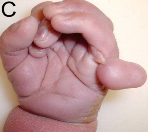
- Hands: clenched fists with overriding (overlapping) fingers with hypoplastic nails.
- Feet: rocker-bottom feet with prominent calcanei.
- Head and face: microcephaly, prominent occiput, micrognathia, low-set malformed ears.
- Chest: short sternum.
- Heart: defects in about 90% — VSD, ASD, PDA, polyvalvular disease.
- Other: omphalocele, horseshoe kidney, severe growth restriction and profound developmental delay; increased risk of Wilms tumour and hepatoblastoma.
- Antenatal clues: growth restriction, polyhydramnios, choroid plexus cysts, clenched hands, omphalocele, single umbilical artery.
Survival: among live-born infants, 60–75% survive the first week, 20–40% the first month and 10–19% the first year; intensive care including cardiac surgery can raise 1-year survival to 30–50%. Current neonatal resuscitation guidance no longer advocates automatically withholding active management.
What are the features of Patau syndrome (trisomy 13)?
Patau syndrome (trisomy 13) is the third most common autosomal trisomy, about 1 in 10,000 to 20,000 live births, with antenatal mortality above 95%. Complete trisomy is commonest (about 80%); maternal meiotic nondisjunction accounts for about 91% of cases. Partial trisomy usually comes from a Robertsonian translocation, typically t(13;14). The defects reflect failed prechordal mesoderm fusion, so they cluster in the midline.
- Holoprosencephaly spectrum (about 74%) — the forebrain fails to divide into two hemispheres. Trisomy 13 is the most common cause of holoprosencephaly.
- Midline face: cleft lip and/or palate (about 45%), hypotelorism, microphthalmia or anophthalmia, cyclopia in severe forms.
- Postaxial polydactyly (about 63–67%).
- Cutis aplasia (punched-out scalp defect).
- Heart: congenital heart disease in most (ASD, VSD, PDA, hypoplastic left heart).
- Other: omphalocele, renal anomalies, cryptorchidism; postnatal mortality about 50% in the first month and up to 90% in the first year.
What are the features of Turner syndrome (45,X)?
Turner syndrome is a phenotypic female with one intact X and complete or partial loss of the second sex chromosome. It occurs in about 1 in 2000 to 2500 live female births, is the most common sex chromosome abnormality in females and the most common genetic cause of primary amenorrhoea. More than 99% of 45,X fetuses miscarry, usually before 28 weeks. Pure 45,X accounts for about 45% of cases; the rest are mosaics (45,X/46,XX; 45,X/46,XY) or structural changes such as isochromosome Xq or ring X.
| Stage | Typical findings |
|---|---|
| Fetus | Increased nuchal translucency, cystic hygroma, non-immune hydrops, left-sided heart lesions such as coarctation, horseshoe kidney |
| Newborn | Lymphoedema of hands and feet, webbed neck, low posterior hairline, nail dysplasia, high-arched palate |
| Childhood | Short stature (SHOX haploinsufficiency), shield chest with widely spaced nipples, cubitus valgus, short 4th metacarpals, Madelung deformity |
| Adolescence | Delayed puberty / primary amenorrhoea from premature ovarian insufficiency; raised FSH |
- Ovaries: replaced by fibrous tissue — streak gonads with few or no follicles.
- Heart: congenital heart disease in about 50% — bicuspid aortic valve and coarctation of the aorta; lifelong risk of aortic dilatation and dissection.
- Kidney: horseshoe kidney, collecting-system malformations, malrotation.
- Brain: intelligence usually normal, with visuospatial and executive weaknesses.
- Autoimmune: hypothyroidism, celiac disease, inflammatory bowel disease.
- Y material (in 45,X/46,XY mosaics) → risk of gonadoblastoma; prophylactic gonadectomy is decided individually.
Treatment: growth hormone improves final height even though GH deficiency is not the cause; estrogen replacement begins if breast development has not started by 11–12 years, starting low and titrating, preferably with 17β-estradiol, then cyclic estrogen–progesterone until about the age of natural menopause.
What are the features and hormone levels in Klinefelter syndrome?
Klinefelter syndrome is a phenotypic male with two or more X chromosomes; 47,XXY accounts for more than 90%, with mosaic 46,XY/47,XXY and 48,XXXY or 49,XXXXY variants making up the rest. It affects 1 in 500 to 1 in 1000 males, yet up to two-thirds are never diagnosed — the average age at diagnosis is about 30 years, often during an infertility work-up (about 3% of men evaluated for infertility have it).
- Tall stature with long limbs and a low upper-to-lower segment ratio (extra SHOX dosage).
- Small, firm testes — rarely above 4 mL — from hyalinisation and fibrosis of the seminiferous tubules; azoospermia and infertility.
- Gynaecomastia, sparse facial and body hair, incomplete puberty.
- Mean full-scale IQ about 85–90 with language and expressive speech difficulties.
- Risks: male breast cancer, extragonadal germ cell tumours, non-Hodgkin lymphoma, osteoporosis, type 2 diabetes and venous thromboembolism.
| Hormone | Level | Why |
|---|---|---|
| FSH | Raised (more than LH) | Loss of Sertoli cell feedback |
| LH | Raised | Low testosterone feedback |
| Testosterone | Low or low-normal | Leydig cell failure; high SHBG lowers free testosterone further |
| Estradiol | High-normal or raised; estradiol-to-testosterone ratio raised | Explains gynaecomastia |
| Inhibin B | Usually undetectable in adults | Sertoli cell dysfunction |
| AMH | Low in adults | Sertoli cell dysfunction |
How do the five chromosomal syndromes compare at a glance?
| Syndrome | Karyotype | Frequency | Hallmark features | Typical heart lesion |
|---|---|---|---|---|
| Down | 47,+21 (95% nondisjunction) | About 1 in 700–800 births | Hypotonia, Brushfield spots, single palmar crease, duodenal atresia, early Alzheimer | AVSD |
| Edwards | 47,+18 | 1 in 3600–10,000 live births | Clenched hands with overlapping fingers, rocker-bottom feet, prominent occiput, short sternum | VSD/ASD/PDA (heart defects in about 90%) |
| Patau | 47,+13 | 1 in 10,000–20,000 live births | Holoprosencephaly, cleft lip/palate, postaxial polydactyly, microphthalmia, cutis aplasia | ASD/VSD/PDA |
| Turner | 45,X (about 45%) or mosaic | 1 in 2000–2500 female births | Short stature, webbed neck, lymphoedema, streak ovaries, primary amenorrhoea | Bicuspid aortic valve, coarctation |
| Klinefelter | 47,XXY (over 90%) | 1 in 500–1000 male births | Tall, small firm testes, gynaecomastia, azoospermia, high FSH | Not characteristic |
Which prenatal screening markers point to each trisomy?
Prenatal screening combines ultrasound with maternal serum analytes. First-trimester screening (11–14 weeks) uses nuchal translucency plus PAPP-A and free β-hCG; a nuchal translucency above 3 mm is abnormal. The second-trimester triple screen measures β-hCG, maternal serum AFP and unconjugated estriol; the quadruple screen adds inhibin A. Cell-free DNA (cfDNA/NIPT) gives the highest detection rate, and the American College of Medical Genetics and Genomics recommends it as the preferred first-line screen for trisomies 13, 18 and 21 in singleton pregnancies.
| Trisomy | AFP | Unconjugated estriol | β-hCG | Inhibin A | PAPP-A |
|---|---|---|---|---|---|
| 21 (Down) | Low | Low | High | High | Low |
| 18 (Edwards) | Low | Low | Low | Normal | Low |
| 13 (Patau) | Normal | Normal | Normal | Normal | Low |
- Ultrasound soft markers for Down syndrome (11–24 weeks): increased nuchal fold, small or absent nasal bone, large ventricles.
- Increased nuchal translucency is not specific — it also occurs in trisomies 13 and 18 and in Turner syndrome; cystic hygroma points strongly to Turner.
- Diagnostic tests: chorionic villus sampling (typically 11–13 weeks) or amniocentesis (typically 15–18 weeks) with karyotype or chromosomal microarray. Screening results alone never confirm a diagnosis.
How are chromosomal syndromes asked in NEET PG and INI-CET?
- Image of a newborn's hand or foot — clenched fist with overlapping fingers or rocker-bottom foot → trisomy 18.
- Midline facial cleft + polydactyly + holoprosencephaly on scan → trisomy 13.
- Most common heart defect in Down syndrome → AVSD (endocardial cushion defect).
- Quadruple test pattern — high hCG and inhibin A with low AFP and estriol → Down syndrome.
- Girl with short stature, webbed neck and primary amenorrhoea → Turner; heart lesion → bicuspid aortic valve / coarctation; ovary → streak gonad.
- Tall man with small firm testes, gynaecomastia and azoospermia → Klinefelter; hormones → high FSH and LH, low testosterone.
- Translocation Down syndrome → next step is parental karyotyping.