What are the craniovertebral joints?
The craniovertebral junction is where the skull meets the upper cervical spine. Its joints are the atlanto-occipital joint (occiput and C1, the atlas) and the atlantoaxial joint (C1 and C2, the axis). The atlas and axis are unique among the cervical vertebrae, and together they form the most flexible segment of the whole spine.
Together with their ligaments and the suboccipital muscles they stabilise the head, support its weight in the neutral position, protect the spinal cord from compression, and allow the vertebral artery to pass to the brain. Most of the characteristic head movements come from just these two joints.
What is special about the atlas (C1) and axis (C2)?
The atlas (C1) is a ring with no vertebral body and no spinous process. It articulates with the occiput above and the axis below through condyles on its lateral masses. The axis (C2) has the dens (odontoid process) projecting upward from the body, plus a bifid spinous process and transverse foramina. The dens is the remnant of the C1 body that fused with C2.

| Feature | Atlas (C1) | Axis (C2) |
|---|---|---|
| Body | Absent | Present, with the dens projecting upward |
| Spinous process | Absent | Bifid |
| Transverse foramen | Present; groove for the vertebral artery on the posterior arch | Present (bilateral) |
| Articulates with | Occiput above, axis below | Atlas above, C3 below |
| Special structure | Anterior and posterior arches; transverse ligament between the lateral masses | Dens and superior articular facets |
What is the atlanto-occipital joint?
The atlanto-occipital joint joins the occiput to the atlas. It is the main contributor to flexion and extension of the head, giving about half of the neck's total range in these movements. It is reinforced by the anterior and posterior atlanto-occipital membranes, which StatPearls lists among the external ligaments of the cervical spine.

Clinically, the atlanto-occipital joint is also named as a possible origin of cervicogenic headache, together with the upper cervical joints. The pain is typically ipsilateral pericranial muscle pain, with the C1 to C3 nerve roots thought to be the source. Failure of the occiput-atlas relationship is the basis of atlanto-occipital dislocation, covered below.
What is the atlantoaxial joint and how does it move?
The atlantoaxial joint is a uniaxial pivot joint that permits rotation only. It is really three synovial joints: one median joint (the dens against the anterior arch of the atlas, held back by the transverse ligament) and two lateral gliding joints between the lateral masses. Its articular capsule connects the lateral masses of the atlas with the back of the axis, and the joint is supplied mainly by branches of the second cervical spinal nerve.
| Feature | Atlanto-occipital | Atlantoaxial |
|---|---|---|
| Bones | Occiput and C1 | C1 and C2 (median and two lateral joints) |
| Main movement | Flexion and extension (about 50% of neck range) | Rotation (about 50% of cervical rotation) |
| Type | Synovial | Median pivot; lateral gliding |
| Nerve supply | Upper cervical nerves (C1 to C3 roots implicated in cervicogenic headache) | Mainly branches of C2 |
| Disc | None | None |
Which ligaments stabilise the craniovertebral junction?
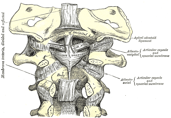
Because the atlantoaxial joint is the most mobile part of the cervical spine, it depends on ligaments rather than discs. StatPearls names the main stabilisers as the transverse (cruciform) ligament and the alar ligaments; the full craniovertebral set is the cruciform ligament, tectorial membrane, alar ligaments and apical ligament of the dens.
| Ligament | Role / exam point |
|---|---|
| Transverse ligament (cruciform ligament of the atlas) | Runs behind the dens between the lateral masses of C1 and prevents anterior subluxation of the atlas on the axis |
| Alar ligaments | Paired stabilisers; the tip of the dens is attached at their insertion (type I dens fracture) |
| Tectorial membrane | Listed among the internal ligaments of the upper cervical spine |
| Apical ligament of the dens | Part of the craniovertebral ligament set |
| Atlanto-occipital membranes | External ligaments covering the atlanto-occipital joint (anterior and posterior) |
The suboccipital muscles — rectus capitis posterior major and minor and obliquus capitis superior and inferior — act as postural stabilisers and initiate craniocervical movement, preventing posterior shift of the occiput on C1. The vertebral artery leaves the axis transverse foramen, passes slightly laterally to the atlas transverse foramen and enters the cranium; the two vertebral arteries join as the basilar artery.
How do the atlas and axis develop, and what are the variants?
The axis has one ossification centre for the body, two for the neural arches and one for the dens. The dens-body junction is a cartilaginous plate that does not fuse until about 4 to 6 years; this can be mistaken for a fracture on a child's X-ray. Development is complete by about 25 years.
- Os odontoideum — a controversial congenital or traumatic variant in which a hypoplastic dens has a separate ossicle at its upper margin; can be orthotopic or dystopic.
- Persistent ossiculum terminale — failure of the secondary ossification centre at the top of the dens to fuse; usually stable and found incidentally.
- Incomplete formation of the dens or transverse ligament — predisposes to instability and injury.
What injuries and instability affect the craniovertebral junction?
| Type | Site | Comment |
|---|---|---|
| Type I | Avulsion of the dens tip at the alar ligament insertion | Rare and usually stable |
| Type II | Base of the dens | Unstable; nonunion risk if age over 50, displacement over 6 mm or comminution |
| Type III | Extends into the axis body | Larger fracture surface |
- Jefferson fracture — fracture of the atlas, usually of the anterior and posterior arches after axial loading (classically diving headfirst into shallow water); the transverse ligament may rupture, and the transverse and alar ligaments can also tear without a fracture.
- Atlanto-occipital dissociation — usually high-energy trauma; high mortality because of the brainstem and vessels. On imaging a basion-dens distance over 10 mm is highly likely abnormal.
- Non-traumatic atlantoaxial subluxation — from ligamentous laxity in rheumatoid arthritis, trisomy 21, Morquio syndrome, Marfan syndrome and Grisel syndrome (nasopharyngeal torticollis).
- Rotatory subluxation — more common in children, who have more joint and ligament hypermobility.
Dens fractures with nonunion risk are treated by internal or external fixation; a halo vest gives rigid immobilisation and restricts C1-C2 flexion by about 75%, with a lower nonunion risk in patients under 50 with minimal displacement and no comminution. Atlas, axis and ligament injuries are best evaluated with CT and MRI.
Which imaging measurements matter at the craniovertebral junction?
In basilar invagination with atlantoaxial instability, the dens and atlas move upward or are unstable, causing cervicomedullary compression and progressive myelopathy. Surgeons assess the atlantodental interval (ADI), the clivo-axial angle, Chamberlain's line and McRae's line; in one 2026 series of three patients, ADI was 6.2 to 9.0 mm and the clivo-axial angle 125 to 132 degrees.
| Measurement | Use |
|---|---|
| Atlantodental interval (ADI) | Atlantodental interval; raised in atlantoaxial instability (6.2 to 9.0 mm in the 2026 case series) |
| Basion-dens distance | Over 10 mm is highly likely atlanto-occipital dissociation |
| Chamberlain's line / McRae's line | Reference lines to quantify basilar invagination |
| Clivo-axial angle | Angle used with the lines above to quantify craniovertebral deformity |