What are gallstones and how do they form?
Gallstones (cholelithiasis) form when substances in bile exceed their solubility. Supersaturated bile throws out tiny crystals that are trapped in gallbladder mucus as sludge, and these grow into stones. Bile stasis (a sluggish gallbladder) gives the crystals time to grow.
| Type | Made of | Typical setting |
|---|---|---|
| Cholesterol (most common; about 75% in Western countries) | Cholesterol crystals | Obesity, diabetes, dyslipidaemia, oestrogen, rapid weight loss |
| Black pigment | Calcium bilirubinate | Haemolysis (for example sickle cell disease), Crohn disease, cirrhosis |
| Brown pigment | Calcium bilirubinate with calcium salts and cholesterol | Bacterial or parasitic infection and strictures; can form inside the bile ducts |
- Risk factors: female sex, obesity, age in the 40s, pregnancy, family history, rapid weight loss, haemolysis, diabetes. Women of reproductive age or on oestrogen pills have about a 2-fold higher risk; progesterone in pregnancy slows emptying.
- Classic profile: 'fat, forty, fertile and flatulent'.
- Only about 10% of gallstones contain enough calcium to show on a plain X-ray.
- Most gallstones are silent: about 20% cause symptoms over 20 years, so asymptomatic stones are not removed routinely.

How do biliary colic, acute cholecystitis and cholangitis differ?
The three conditions are best seen as steps along one path: a stone briefly blocks the cystic duct (colic), stays there and inflames the gallbladder (cholecystitis), or passes into the common bile duct and the stagnant bile gets infected (cholangitis).
| Feature | Biliary colic | Acute cholecystitis | Acute cholangitis |
|---|---|---|---|
| Cause | Gallbladder contracting against a stone in the cystic duct | Persistent cystic duct obstruction → inflammation | Common bile duct obstruction + infection |
| Pain | RUQ/epigastric, after fatty meals, settles in hours | Constant RUQ pain, usually more than 6 hours | RUQ pain |
| Fever | No | Often | Yes, with rigors |
| Jaundice | No | Usually no | Yes |
| Sign | Tenderness only | Murphy sign; sometimes a tender RUQ mass | Charcot triad; Reynolds pentad if severe |
| Blood tests | Usually normal | Raised WBC, mild LFT changes | Raised WBC, cholestatic LFTs, raised bilirubin |
- Murphy sign: inspiratory arrest while the right upper quadrant is palpated. The sonographic Murphy sign is maximal tenderness when the probe presses on the gallbladder.
- Charcot triad: fever, right upper quadrant pain and jaundice. It is very specific (about 96%) but insensitive (about 26%).
- Reynolds pentad: Charcot triad + altered mental status + shock (sepsis) — suppurative cholangitis.
- About 95% of acute cholecystitis is calculous. Acalculous cholecystitis occurs in critically ill patients and those on long-term TPN.
- Cholangitis organisms are mainly gut Gram-negatives: *E. coli* (25–50%), Klebsiella, Enterococcus and Enterobacter.
How are gallstones and acute cholecystitis diagnosed?
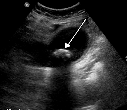
- Ultrasound is the first-choice test. It detects stones as small as 2 mm (echogenic focus with posterior acoustic shadowing), sludge and polyps.
- Ultrasound signs of acute cholecystitis: gallbladder wall thicker than 3 mm, pericholecystic fluid, a stone impacted in the neck and a sonographic Murphy sign.
- HIDA scan (cholescintigraphy) when ultrasound is unclear: non-filling of the gallbladder means cystic duct obstruction and supports acute cholecystitis. With CCK, an ejection fraction below 35% suggests biliary dyskinesia.
- CT often comes first in the emergency department but is less sensitive for stones.
- Common bile duct stones: a dilated duct on ultrasound raises suspicion; MRCP is non-invasive, while ERCP can both diagnose and remove stones.
What are the Tokyo Guidelines 2018 for acute cholecystitis and cholangitis?
The Tokyo Guidelines 2018 (TG18) kept the TG13 diagnostic criteria and severity grading for acute cholecystitis without change, because validation studies showed the grades predicted mortality, conversion to open surgery and length of stay.
| Element | What it includes |
|---|---|
| A. Local signs of inflammation | Murphy sign, or RUQ mass, pain or tenderness |
| B. Systemic signs of inflammation | Fever, raised CRP or raised WBC |
| C. Imaging | Findings typical of acute cholecystitis |
| Suspected diagnosis | One item in A + one item in B |
| Definite diagnosis | A + B + C |
| Grade | Criteria |
|---|---|
| Grade III (severe) | Dysfunction of any one organ system: hypotension needing dopamine ≥5 µg/kg/min or any noradrenaline; reduced consciousness; PaO2/FiO2 < 300; oliguria or creatinine > 2.0 mg/dL; PT-INR > 1.5; platelets < 100,000/mm3 |
| Grade II (moderate) | Any one of: WBC > 18,000/mm3; palpable tender RUQ mass; symptoms > 72 hours; marked local inflammation (gangrenous or emphysematous cholecystitis, pericholecystic or hepatic abscess, biliary peritonitis) |
| Grade I (mild) | Does not meet Grade II or III — a healthy patient with mild local inflammation |
| Grade | Criteria |
|---|---|
| Grade III (severe) | Organ dysfunction in any one system (same organ list as for cholecystitis) |
| Grade II (moderate) | Any two of: WBC > 12,000 or < 4,000/mm3; fever ≥ 39 °C; age ≥ 75 years; total bilirubin ≥ 5 mg/dL; low albumin |
| Grade I (mild) | Meets neither Grade II nor Grade III at diagnosis |
How are acute cholecystitis and cholangitis managed?
- Biliary colic: analgesia and a low-fat diet; recurrent attacks → elective laparoscopic cholecystectomy.
- Acute cholecystitis: IV fluids, analgesia and antibiotics against Gram-negative rods and anaerobes, then early laparoscopic cholecystectomy during the same admission in fit patients. Early surgery within 72 hours of symptom onset carried a 30-day morbidity of 6.6% and mortality of 1.1% in one large study.
- Grade III or unfit patients: percutaneous cholecystostomy (gallbladder drainage) as a bridge, followed by delayed cholecystectomy, or as definitive care in the very frail.
- Acute cholangitis: fluids, early antibiotics and biliary drainage (ERCP preferred; percutaneous or surgical if needed). In TG18 data, early or urgent drainage lowered 30-day mortality in Grade II cholangitis.
- Gallstone pancreatitis: treat the pancreatitis; cholecystectomy follows once the patient recovers.
- Ursodeoxycholic acid dissolves fewer than half of stones and does not treat the cause — it is not routine therapy.
What is Calot's triangle and the critical view of safety?
The hepatocystic triangle (commonly called Calot triangle) is the key landmark in cholecystectomy. Its boundaries are the cystic duct (on the right), the common hepatic duct (on the left) and the inferior surface of the liver (above). It contains the cystic artery and the cystic lymph node of Lund (often called Calot's node).
- Clear all fat and fibrous tissue from the hepatocystic triangle.
- See only two structures entering the gallbladder — the cystic duct and the cystic artery.
- Separate the lower third of the gallbladder from the liver bed to expose the cystic plate.
These three steps make up the critical view of safety (CVS). Only after it is achieved are the duct and artery clipped and divided — the aim is to avoid bile duct injury, one of the most serious complications. The cystic artery arises from the right hepatic artery in about 90% of people.

What are Mirizzi syndrome and gallstone ileus?
Mirizzi syndrome is obstructive jaundice from a stone impacted in Hartmann's pouch or the cystic duct that compresses the common hepatic duct from outside. Over time it can erode into the duct and form a cholecystobiliary fistula.
| Type | Description |
|---|---|
| I | External compression, no fistula (IA cystic duct present; IB cystic duct obliterated) |
| II | Fistula involving less than one-third of the common hepatic duct circumference |
| III | Fistula involving one-third to two-thirds |
| IV | Fistula involving more than two-thirds |
Treatment is cholecystectomy; advanced cases may need a partial (subtotal) cholecystectomy or open surgery. Gallbladder cancer was found in 5–28% of operated cases, usually only on histology.
Gallstone ileus is a mechanical small-bowel obstruction from a large stone (usually over 2 cm) that enters the bowel through a biliary–enteric fistula, most often cholecystoduodenal. The stone usually lodges at the terminal ileum / ileocaecal valve. It causes 1–4% of mechanical bowel obstructions overall but up to 25% in older patients, and affects women about 3.5 times as often as men, mostly over 60.
