What are the intrinsic muscles of the hand?
Hand muscles are split into extrinsic muscles, whose bellies lie in the forearm and send long tendons into the hand, and intrinsic muscles, which begin and end within the hand. The intrinsic muscles give fine motor control and contribute a large share of pinch and grip strength — weakness of the intrinsics costs roughly 60–80% of pinch and grip strength (StatPearls — Intrinsic Hand Deformity).
- Thenar group (ball of the thumb): abductor pollicis brevis, flexor pollicis brevis, opponens pollicis.
- Adductor pollicis: lies next to the thenar group but is not part of the thenar eminence and has a different nerve.
- Hypothenar group (ball of the little finger): abductor digiti minimi, flexor digiti minimi brevis, opponens digiti minimi — plus the small subcutaneous palmaris brevis.
- Lumbricals: four slender muscles arising from the flexor digitorum profundus (FDP) tendons.
- Interossei: dorsal (abductors) and palmar (adductors), between the metacarpals.
Only two nerves supply them: the median nerve (enters through the carpal tunnel) and the ulnar nerve (enters through Guyon's canal). The radial nerve supplies no intrinsic hand muscle — its role in the hand is the forearm extensors and sensation over the dorsum.

Which muscles make up the thenar and hypothenar eminences?
| Muscle | Main attachments | Action | Nerve |
|---|---|---|---|
| Abductor pollicis brevis | Flexor retinaculum + scaphoid tubercle → lateral base of thumb proximal phalanx | Abducts thumb | Median (recurrent branch) |
| Flexor pollicis brevis | Superficial head: flexor retinaculum; deep head: trapezium tubercle → base of thumb proximal phalanx | Flexes thumb | Superficial head: median; deep head: ulnar |
| Opponens pollicis | Flexor retinaculum + trapezium → lateral side of 1st metacarpal | Opposition | Median (recurrent branch) |
| Adductor pollicis | 2nd and 3rd metacarpals + capitate → medial base of thumb proximal phalanx | Adducts thumb | Ulnar (deep branch) |
| Abductor digiti minimi | Pisiform → medial base of little-finger proximal phalanx | Abducts little finger | Ulnar (deep branch) |
| Flexor digiti minimi brevis | Flexor retinaculum + hook of hamate → medial base of proximal phalanx | Flexes little finger at MCP | Ulnar (deep branch) |
| Opponens digiti minimi | Flexor retinaculum + hamate → medial side of 5th metacarpal | Opposes little finger | Ulnar (deep branch) |
| Palmaris brevis | Subcutaneous, over the hypothenar eminence | Wrinkles hypothenar skin | Ulnar (superficial branch) |
The thenar muscles (APB, FPB, opponens) receive the recurrent motor branch of the median nerve, given off just distal to the carpal tunnel. This branch lies superficially near the middle of the thenar mass and carries no sensory fibres, so a knife or glass laceration can paralyse opposition while sensation stays normal — easy to miss in the emergency department (StatPearls — Median Nerve).
What do the lumbricals and interossei do?
Both groups insert into the extensor expansion (dorsal hood), so they share one combined action: flex the metacarpophalangeal (MCP) joints and extend the interphalangeal (IP) joints — the 'writing' or 'L' position of the fingers. This is the key to understanding claw hand: lose them and the MCP joints hyperextend while the IP joints flex.
| Muscle | Origin | Special action | Nerve |
|---|---|---|---|
| 1st and 2nd lumbricals | Radial side of FDP tendons to index and middle fingers (unipennate) | MCP flexion + IP extension | Median |
| 3rd and 4th lumbricals | Adjacent sides of the medial FDP tendons (bipennate) | MCP flexion + IP extension | Ulnar (deep branch) |
| Dorsal interossei (4) | Adjacent sides of two metacarpals | Abduct index, middle and ring fingers from the axis of the middle finger | Ulnar (deep branch) |
| Palmar interossei (3) | Palmar surface of one metacarpal | Adduct index, ring and little fingers towards the middle finger | Ulnar (deep branch) |
Some texts count a fourth palmar interosseous to the thumb; StatPearls lists 4 dorsal and 3 palmar in its intrinsic-muscle chapter. Either way, every interosseous is supplied by the deep branch of the ulnar nerve — which is why finger-spreading and finger-gripping tests examine the ulnar nerve.
Which intrinsic muscles does the median nerve supply — the LOAF rule?
| Nerve and branch | Muscles supplied |
|---|---|
| Median — recurrent motor branch | Abductor pollicis brevis, opponens pollicis, superficial head of flexor pollicis brevis |
| Median — in the palm | 1st and 2nd lumbricals |
| Ulnar — superficial branch | Palmaris brevis (plus sensory digital nerves) |
| Ulnar — deep branch | Hypothenar muscles, 3rd and 4th lumbricals, all interossei, adductor pollicis, deep head of flexor pollicis brevis |
The ulnar nerve carries C8 and T1 fibres, and the intrinsic muscles as a whole depend on the C8–T1 segments. That is why a lower trunk (C8–T1) brachial plexus injury — Klumpke palsy — paralyses every intrinsic muscle at once, producing a 'total claw hand' with palmar wasting, unlike an isolated ulnar lesion that spares the LOAF muscles.
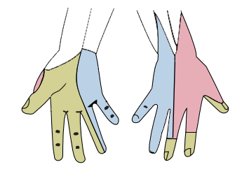
What is the sensory supply of the hand?
| Nerve | Palmar side | Dorsal side | Spared in |
|---|---|---|---|
| Median | Lateral 3½ digits (thumb, index, middle, radial half of ring); palm and thenar base via the palmar cutaneous branch | Distal parts (beyond PIP) of index, middle and radial half of ring, including nail beds | Thenar-base sensation is spared in carpal tunnel syndrome (palmar cutaneous branch passes superficial to the retinaculum) |
| Ulnar | Medial 1½ digits and hypothenar skin | Medial dorsum of hand, little finger and ulnar half of ring (dorsal cutaneous branch) | Dorsal sensation is spared in Guyon canal lesions (dorsal branch leaves in the forearm) |
| Radial (superficial branch) | None | Radial dorsum of the hand, dorsal thumb and proximal phalanges of index and middle fingers | Lost in wrist drop at the spiral groove; not in posterior interosseous lesions |
Two branches localise lesions neatly. The palmar cutaneous branch of the median nerve arises proximal to the wrist and runs over the flexor retinaculum, so carpal tunnel syndrome spares sensation over the thenar eminence. The dorsal cutaneous branch of the ulnar nerve leaves the main trunk in the forearm, so wrist (Guyon canal) lesions spare the dorsal ulnar hand while elbow (cubital tunnel) lesions involve it.
Why does ulnar nerve injury cause a claw hand?
Claw hand is an intrinsic-minus posture: the paralysed interossei and lumbricals can no longer flex the MCP joints or extend the IP joints, so the intact extrinsic muscles win — the long extensors hyperextend the MCP joints and FDP/FDS flex the IP joints. In a pure ulnar lesion the ring and little fingers claw most, because the median-supplied 1st and 2nd lumbricals still hold the index and middle fingers (StatPearls — Claw Hand).
- Froment sign — pinching paper between thumb and index finger, the thumb IP joint flexes because flexor pollicis longus (median) substitutes for a weak adductor pollicis (ulnar).
- Jeanne sign — reciprocal hyperextension of the thumb MCP joint during the same pinch.
- Wartenberg sign — the little finger rests abducted: the third palmar interosseous is weak and the radial-supplied extensor digiti minimi pulls unopposed.
- Duchenne sign — clawing of the ring and little fingers (MCP hyperextension, PIP flexion).
- Egawa (Pitres-Testut) sign — cannot abduct the middle finger side to side with the palm flat (2nd and 3rd dorsal interossei).
- Masse sign — flattened palmar metacarpal arch from hypothenar and interosseous wasting.
| Site | Motor | Sensory |
|---|---|---|
| Elbow (cubital tunnel) — commonest site | FCU and ulnar FDP weak + all ulnar intrinsics; milder claw | Palmar and dorsal ulnar 1½ digits |
| Guyon canal, zone I (before bifurcation) | All ulnar intrinsics | Palmar ulnar 1½ digits; dorsum spared |
| Zone II (deep branch) | Pure motor — interossei, ulnar lumbricals, adductor pollicis | None |
| Zone III (superficial branch) | None (palmaris brevis only) | Pure sensory — palmar ring/little fingers |
After carpal tunnel syndrome, ulnar entrapment is the second commonest compressive neuropathy of the upper limb, and the cubital tunnel is its commonest site. Leprosy is an important systemic cause of ulnar claw hand in Indian practice.

What is ape hand and the hand of benediction?
Ape hand follows loss of the thenar muscles — classically a low median lesion (wrist laceration, long-standing carpal tunnel syndrome) or injury to the recurrent motor branch. The thenar eminence flattens and the thumb lies adducted, in the plane of the palm, unable to abduct or oppose (StatPearls — Median Nerve Injury).
Hand of benediction (papal benediction sign) is taught in Indian exams as the sign of a high (elbow or above) median lesion: when the patient tries to make a fist, the index and middle fingers stay extended because FDS and the lateral half of FDP are paralysed, while the ring and little fingers flex through the ulnar-supplied FDP. Flexor pollicis longus is also lost, so the thumb tip cannot flex.
| Lesion | Lost | Sensation | Sign |
|---|---|---|---|
| High median (above elbow) | Pronators, FCR, FDS, lateral FDP, FPL + thenar + lateral lumbricals | Lost — lateral 3½ digits | Benediction on fist; pointing index; ape thumb |
| Anterior interosseous nerve | FPL, lateral half of FDP, pronator quadratus | Normal — purely motor | Cannot make an 'O' — flattened pinch |
| Low median (wrist / carpal tunnel) | Thenar muscles + 1st/2nd lumbricals | Lost — lateral 3½ digits; thenar base spared in CTS | Ape hand, thenar wasting |
| Recurrent motor branch | APB, opponens, superficial FPB | Normal | Loss of opposition only |
How does a radial nerve injury affect the hand?
The radial nerve supplies no intrinsic muscle, but it supplies the wrist and finger extensors, so its injury produces wrist drop. The spiral groove of the humerus is the most common site — mid-shaft humerus fracture or prolonged compression ('Saturday night palsy'). Triceps strength is typically preserved at this level; brachioradialis and the extensors are weak, and sensation is lost over the dorsum of the hand, especially the first web space (StatPearls — Wrist Drop).
| Level | Typical cause | Motor loss | Sensory loss |
|---|---|---|---|
| Axilla | Crutch palsy, shoulder dislocation | Triceps + wrist and finger extensors | Posterior arm, forearm, dorsum of hand |
| Spiral groove | Humeral shaft fracture, Saturday night palsy | Wrist drop; triceps spared | Dorsum of hand, first web space |
| Posterior interosseous nerve | Radial tunnel / arcade of Frohse, elbow trauma | Finger and thumb drop; wrist extension partly kept (ECRL is supplied by the radial nerve proper) | None — purely motor |
| Superficial radial nerve (Wartenberg syndrome) | Tight wristbands, handcuffs, casts | None | Radial dorsum of the hand |
What passes through the carpal tunnel and Guyon's canal?
| Feature | Carpal tunnel | Guyon's canal (ulnar tunnel) |
|---|---|---|
| Boundaries | Roof: transverse carpal ligament (flexor retinaculum); floor and walls: carpal bones | Medial: pisiform; lateral: hook of hamate; roof: volar carpal ligament and palmaris brevis; floor: transverse carpal ligament |
| Contents | Median nerve + 9 flexor tendons (4 FDS, 4 FDP, FPL) | Ulnar nerve + ulnar artery (with venae comitantes, fat) |
| Not inside | Palmar cutaneous branch of median; ulnar nerve and artery; palmaris longus | Dorsal cutaneous branch of ulnar nerve |
| Syndrome | Carpal tunnel syndrome — the most common entrapment neuropathy overall | Guyon canal (ulnar tunnel) syndrome — ganglion, hook of hamate fracture, repetitive hypothenar pressure |
| Sensory pattern | Lateral 3½ digits; thenar base spared | Palmar ulnar 1½ digits; dorsal hand spared |
In carpal tunnel syndrome, numbness and paresthesia start in the thumb, index, middle and radial half of the ring finger, later come weak abduction and opposition and thenar atrophy. Phalen test: both wrists held fully flexed back-to-back for one minute reproduces the symptoms. Tinel test: tapping over the carpal tunnel — StatPearls quotes a sensitivity of 50% and specificity of 77%. Surgical release divides the transverse carpal ligament; the commonest complication is a neuroma of the palmar cutaneous branch (StatPearls — Carpal Tunnel Syndrome).

How are hand muscles and nerves asked in NEET PG and INI-CET?
- Nerve supply one-liners — adductor pollicis (ulnar, deep branch), deep head of FPB (ulnar), palmaris brevis (ulnar, superficial branch), 1st/2nd lumbricals (median).
- Action questions — abduction of fingers (dorsal interossei), adduction (palmar interossei), MCP flexion with IP extension (lumbricals and interossei).
- Clinical signs — Froment (adductor pollicis vs FPL), Wartenberg (third palmar interosseous), Egawa (dorsal interossei), inability to make an 'O' in anterior interosseous nerve palsy.
- Image-based — photograph of a claw, ape thumb, benediction posture or wrist drop: name the nerve and the level.
- Paradox questions — ulnar paradox (distal lesion claws more); Klumpke palsy = total claw.
- Tunnels — contents of the carpal tunnel, boundaries of Guyon's canal, which sensory branch is spared.
For more patterns across years, see most repeated topics and solve recalled papers on the NEET PG PYQ and INI-CET PYQ pages. Related upper-limb topics: Erb's palsy and the brachial plexus and Volkmann's ischaemic contracture.