What is idiopathic intracranial hypertension?
Idiopathic intracranial hypertension (IIH), also called pseudotumor cerebri (PTC), is a neurological disorder of raised intracranial pressure (ICP) with no identifiable structural cause. The brain imaging is normal apart from soft signs of high pressure, and the CSF composition is normal — only the pressure is raised. Because the optic nerve head is swollen by the pressure, the disease carries a real risk of permanent vision loss if it is missed.
The classic patient is an overweight woman of childbearing age. The condition also occurs in women of all ages, in men and in children of both sexes, but the textbook stem almost always describes a young obese woman with a headache and blurred vision.
Who gets IIH — epidemiology and risk factors?
IIH most often affects women aged 20 to 44 years, and about 90% of post-pubertal cases are female. The strongest association is with raised body mass index: among women whose weight is 20% or more above ideal body weight, the annual incidence reaches 19.3 per 100,000; across all women aged 15 to 44 it is 3.5 per 100,000; in the general population it is 0.9 per 100,000.
Before puberty (generally under 12 years) boys and girls are affected at similar rates and obesity is a weaker risk factor. After puberty the female excess and the link with obesity become obvious.
| Factor | Point to remember |
|---|---|
| Female sex, age 20–44 | About 90% of post-pubertal cases are female |
| Obesity / recent weight gain | The main modifiable risk factor; incidence rises with BMI |
| Vitamin A and retinoids | Drug-induced IIH is linked to vitamin A and its metabolites |
| Other drugs (secondary PTC) | Tetracyclines, lithium, steroids, progestins, estrogens, testosterone, growth hormone |
| Venous outflow problems | Cerebral venous sinus thrombosis, jugular compression, SVC syndrome, dural AV fistula cause secondary PTC |
What causes the raised pressure?
The mechanism is not fully understood. The pressure rises because of dysregulation of CSF volume — increased CSF production, decreased CSF absorption, or both.
- Increased production — CSF is made by the choroid plexus. Obesity is linked to upregulation of aquaporin 1 (AQP1), a water channel involved in CSF production. Acetazolamide reduces AQP1 expression, which fits its benefit.
- Reduced absorption — CSF drains through the arachnoid granulations into the venous sinuses. Venous sinus stenosis (common in IIH) raises venous pressure and reduces the gradient needed for absorption.
- Glymphatic dysfunction — the glymphatic system, described in 2012 as another CSF clearance pathway, may also contribute.
- Hormonal factors — altered 11β-HSD1 (cortisol metabolism) activity and the role of GLP-1 receptors in the choroid plexus are being studied; small randomised trials of an 11β-HSD1 inhibitor and a GLP-1 agonist lowered ICP.
What are the symptoms and signs of IIH?
| Feature | Frequency / detail |
|---|---|
| Headache | Most frequent complaint — up to 98%; often daily, worse in the morning or with Valsalva |
| Transient visual obscurations | Up to 70%; seconds-long loss of vision in one or both eyes |
| Pulsatile tinnitus | Up to 60%; whooshing in time with the pulse, abolished by ipsilateral jugular compression |
| Photopsia | Up to 54%; flashes of light |
| Persistent vision loss | Up to 32%, especially in untreated patients |
| Horizontal diplopia | From abducens (VI) nerve palsy causing esotropia |
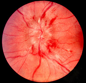
On examination the defining sign is papilledema, together with obesity. Visual field defects are more common than loss of acuity, so perimetry is more sensitive than acuity testing. The commonest field changes are enlargement of the physiological blind spot and inferonasal field defects.
What are the diagnostic criteria for IIH?
The modified Dandy criteria remain the most widely used. All of the following must be present:
- Signs and symptoms of increased intracranial pressure.
- No localising neurological findings on examination.
- No deformity, displacement or obstruction of the ventricular system and otherwise normal neurodiagnostic studies, except raised CSF pressure.
- An awake and alert mental status.
- No other cause of raised intracranial pressure.
The 2013 revised criteria widened the definition to include patients without papilledema and those with secondary PTC. A definite diagnosis needs: papilledema; a normal neurological examination apart from cranial nerve abnormalities; imaging with no hydrocephalus, meningeal enhancement or mass lesion; normal CSF composition; and a raised lumbar puncture opening pressure.
| Group | Opening pressure required |
|---|---|
| Adults | 25 cm H2O or more |
| Children (1–18 years) | 28 cm H2O or more |
| Children who are neither sedated nor obese | 25 cm H2O or more is enough |
Without papilledema, the diagnosis can still be made if the other criteria are met plus an abducens palsy. With neither papilledema nor abducens palsy, at least 3 imaging signs are needed: empty sella, flattening of the posterior globe, distension of the perioptic subarachnoid space (with or without optic nerve tortuosity) and transverse sinus stenosis.
Which investigations are done, and what do they show?
- MRI brain with MR venography — the preferred imaging to exclude mass, hydrocephalus and venous sinus thrombosis. The brain parenchyma and ventricles look normal in IIH. CT is the alternative if MRI cannot be done, with lower sensitivity and specificity.
- Supportive MRI signs — transverse sinus stenosis, posterior scleral flattening, perioptic subarachnoid space distension, empty sella, enlarged Meckel's cave and vertical tortuosity of the optic nerve. Findings beyond empty sella, optic nerve sheath distension and smooth non-flow-related venous sinus stenosis should prompt a search for another diagnosis.
- Lumbar puncture — measures the opening pressure and gives CSF for analysis.
- Ophthalmoscopy — papilledema graded with the Frisén scale from 0 (normal disc) to 5 (severe disc oedema obscuring all retinal vessels). Greater disc swelling generally corresponds to greater visual impairment.
- Visual acuity and automated perimetry — perimetry detects loss earlier than acuity and is used to monitor treatment.
- Complete blood count — to exclude anaemia and lymphoproliferative disorders as causes of papilledema.
How is IIH treated?
Treatment aims to protect vision and relieve headache. It is stepped according to visual function and response.
| Step | What | Key points |
|---|---|---|
| 1. Weight loss | Sustained loss of 5–10% of body weight | Lowers ICP and is linked to remission; weight regain raises the risk of recurrence |
| 2. Acetazolamide | Carbonic anhydrase inhibitor, cornerstone of medical therapy | Reduces CSF secretion (via AQP1 and Na+/K+ ATPase); IIH Treatment Trial used up to 4 g/day with a low-sodium diet and improved perimetry, papilledema and opening pressure. Side effects: paraesthesia, dysgeusia, fatigue |
| 3. Topiramate | Second-line | Lowers ICP, helps headache and also promotes weight loss |
| 4. Other diuretics | Furosemide, spironolactone | Used if acetazolamide or topiramate not tolerated; weaker evidence |
| 5. Surgery / endovascular | For progressive symptoms or worsening vision despite medical therapy | CSF diversion (ventriculoperitoneal or lumboperitoneal shunt), transverse sinus stenting, optic nerve sheath fenestration |
- Optic nerve sheath fenestration (ONSF) — an opening in the optic nerve sheath relieves pressure around the nerve; it consistently improves acuity, fields and papilledema but headache relief is less reliable.
- Transverse sinus stenting — reserved for stenosis with a trans-stenotic pressure gradient of 8 cm H2O or more, confirmed by venography and manometry. It avoids open surgery and the repeated revisions that shunts need.
- Shunts — ventriculoperitoneal (catheter usually through the foramen of Monroe) or lumboperitoneal (from the thecal sac).
- Fulminant IIH — acute, rapidly progressive visual loss; a neuro-ophthalmic emergency needing urgent, aggressive intervention.
What are the differentials and common exam traps?
- Mass lesion or hydrocephalus — excluded by imaging; in IIH the ventricles are normal or small.
- Cerebral venous sinus thrombosis — the key secondary PTC to exclude with MR venography. See Virchow's triad.
- Meningitis / chronic meningitis — abnormal CSF composition; IIH CSF is normal.
- Drug-induced — vitamin A, retinoids, tetracyclines, lithium, steroids and sex-hormone preparations.
- Other causes of disc swelling — papilledema means disc oedema from raised ICP.
For related neuro-ophthalmology revision see visual pathway lesions and cranial nerves. Acetazolamide belongs with the carbonic anhydrase inhibitors in diuretics.