What is Kussmaul breathing?
Kussmaul breathing (Kussmaul respiration) is a pattern of deep, laboured breathing — large tidal volumes, usually with a raised rate — seen in severe metabolic acidosis. It is named after the 19th-century German physician Adolf Kussmaul, who described it in patients with diabetic coma. The textbook setting is diabetic ketoacidosis (DKA), where it often comes with a fruity (acetone) smell on the breath, dehydration and abdominal pain.
What sets it apart from ordinary tachypnoea is depth. Patients with metabolic acidosis increase alveolar ventilation first by taking bigger breaths and only later by breathing faster. This hyperpnoea is the most efficient way to clear CO₂ because each large breath wastes proportionally less air on anatomical dead space. The breathing is regular — there are no pauses or apnoeas, which separates it from Cheyne-Stokes and Biot breathing.

Clinically the change is easy to miss until the acidosis is marked: the increase in minute ventilation may only become obvious once the pH has fallen very low. That is why unexplained deep breathing in a drowsy diabetic, a patient with renal failure or a suspected poisoning should prompt an immediate blood gas.
Why does metabolic acidosis cause deep, rapid breathing?
- An acid accumulates (e.g. β-hydroxybutyrate and acetoacetate in DKA) and consumes bicarbonate, so HCO₃⁻ and pH fall.
- Peripheral chemoreceptors in the carotid and aortic bodies sense the rise in arterial H⁺; central chemoreceptors near the ventral surface of the medulla respond to pH and PaCO₂ changes in brain interstitial fluid.
- The medullary respiratory centre increases ventilation — mainly tidal volume at first.
- PaCO₂ falls. Because pH depends on the HCO₃⁻/PaCO₂ ratio, lowering PaCO₂ pulls the pH back towards normal.
- Respiratory compensation starts within minutes and is complete in about 12–24 hours, but it never fully normalises the pH.
Where the acid comes from in DKA: insulin deficiency with excess counter-regulatory hormones (glucagon, catecholamines, cortisol, growth hormone) releases free fatty acids from adipose tissue, and the liver oxidises them into the ketoacids β-hydroxybutyrate and acetoacetate. Their H⁺ is buffered by bicarbonate, leaving unmeasured ketoanions — a high anion gap acidosis. Some acetoacetate breaks down to acetone, which is exhaled and gives the fruity breath that often accompanies the deep breathing.
pH = 6.1 + log [HCO₃⁻ ÷ (0.03 × PaCO₂)]
Henderson–Hasselbalch equation: when HCO₃⁻ falls, only a proportional fall in PaCO₂ can protect the pH — which is exactly what Kussmaul breathing achieves.
How does Winter's formula tell you if compensation is adequate?
Expected PaCO₂ (mmHg) = (1.5 × HCO₃⁻) + 8 ± 2
Winter's formula — valid for primary metabolic acidosis. Compare the measured PaCO₂ with the expected range.
| Measured PaCO₂ | Interpretation |
|---|---|
| Within the expected range | Appropriate respiratory compensation (simple metabolic acidosis) |
| Higher than expected | Concomitant respiratory acidosis — ventilation is not keeping up (exhaustion, sedation, lung disease, neuromuscular weakness) |
| Lower than expected | Concomitant respiratory alkalosis — an extra drive to breathe (e.g. sepsis, early salicylate poisoning, anxiety) |
| HCO₃⁻ | Expected PaCO₂ | Measured PaCO₂ | Conclusion |
|---|---|---|---|
| 12 mEq/L | 24–28 mmHg | 26 | Appropriately compensated metabolic acidosis |
| 10 mEq/L | 21–25 mmHg | 30 | Metabolic acidosis + respiratory acidosis (relative hypoventilation) |
| 11 mEq/L | 22.5–26.5 mmHg | 31 | Primary metabolic acidosis with inadequate compensation |
| 18 mEq/L | 33–37 mmHg | 35 | Appropriate compensation |
A PaCO₂ that is normal (say 40 mmHg) in a patient with bicarbonate of 10 is not reassuring — it is far above the expected ~23 and signals a patient who can no longer breathe hard enough — a concomitant respiratory acidosis and a warning of impending respiratory failure. Winter's formula is also step 4 of the stepwise ABG approach explained on the anion gap page.
How do recalled NEET PG ABG questions use Winter's formula?
Examiners like to hide a second disorder behind an obvious metabolic acidosis. The way to catch it is always the same: identify the primary process from pH and HCO₃⁻, then compare the measured PaCO₂ with Winter's prediction.
- pH 7.15, HCO₃⁻ 10, PaCO₂ 30. Primary metabolic acidosis. Expected PaCO₂ = 1.5 × 10 + 8 = 23 ± 2 (21–25). Measured 30 is too high → a superimposed respiratory acidosis from relative hypoventilation (reduced CO₂ washout).
- Glucose 345 mg/dL, pH 7.17, HCO₃⁻ 11, PaCO₂ 31, tachypnoea. The primary disturbance is metabolic acidosis (DKA picture). Expected PaCO₂ 22.5–26.5, so compensation is incomplete; ‘fully compensated’ is wrong because the pH is still well below 7.35.
- Type 1 diabetic, confused, Kussmaul breathing, BP 70/50, glucose 450, ketones 4+. Before any further test, the first step is an isotonic saline bolus to treat shock; insulin comes after fluids are running (and after checking potassium).
- HCO₃⁻ 6, PaCO₂ 13, anion gap 20. Expected PaCO₂ 17 ± 2 (15–19). A measured 13 is below the range → metabolic acidosis with an additional respiratory alkalosis.
Which conditions cause Kussmaul breathing?
Any severe metabolic acidosis can drive Kussmaul breathing. Very low pH values (below about 7.1–7.2) are most often due to a high anion gap acidosis, and diabetic ketoacidosis is the commonest single cause.
| Cause | Supporting clues |
|---|---|
| Diabetic ketoacidosis | Type 1 diabetes, hyperglycaemia, ketonaemia, fruity breath, vomiting, abdominal pain |
| Alcoholic or starvation ketoacidosis | Binge drinking with vomiting and poor food intake; prolonged fasting |
| Lactic acidosis | Shock, sepsis, bowel or limb ischaemia, metformin |
| Uraemia (advanced kidney failure) | High urea and creatinine, anaemia, oliguria |
| Toxic alcohols | Methanol (visual loss after illicit liquor), ethylene glycol (oxalate crystals, kidney injury) |
| Salicylate poisoning | Early hyperventilation with respiratory alkalosis, then metabolic acidosis |
| Severe normal anion gap acidosis | Profuse diarrhoea, renal tubular acidosis |
How is Kussmaul breathing different from Cheyne-Stokes, Biot and apneustic breathing?
Breathing patterns are a favourite image- and graph-based MCQ. Learn them by rhythm (regular or not), depth and where the lesion or problem lies.
| Pattern | What you see | Typical cause / lesion |
|---|---|---|
| Kussmaul | Deep, regular, usually rapid; no pauses | Metabolic acidosis — DKA, uraemia, lactic acidosis, toxic alcohols |
| Cheyne-Stokes | Smooth crescendo–decrescendo waxing and waning of depth, then an apnoea (about 10 s or longer); cycle about a minute | Heart failure (prolonged circulation time, unstable CO₂ feedback), bilateral hemispheric damage, metabolic encephalopathy, stroke; also at high altitude |
| Biot (ataxic) | Irregular clusters of breaths that start and stop abruptly, separated by variable apnoeas; rate and depth vary | Dorsomedial medulla damage — a warning sign of impending respiratory arrest |
| Apneustic | Prolonged pause at full inspiration (about 2–3 s) before expiration | Low pontine lesions, e.g. basilar artery occlusion |
| Central neurogenic hyperventilation | Sustained rapid, deep breathing without acidosis | Pontomesencephalic (upper brainstem) lesions |
| Agonal gasps | Occasional slow gasping breaths | Lower medullary damage; terminal pattern |
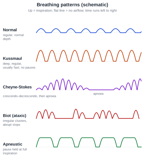
Cheyne-Stokes and Biot can look alike on a monitor. The difference is that Cheyne-Stokes depth rises and falls gradually and regularly, whereas Biot breathing switches on and off abruptly and irregularly. In heart failure, Cheyne-Stokes breathing during sleep is a form of central sleep apnoea.
What is Kussmaul's sign, and why is it a different thing?
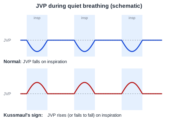
Kussmaul's sign is a paradoxical rise — or failure to fall — of the jugular venous pressure (JVP) during inspiration. Normally inspiration lowers intrathoracic pressure, draws blood into the right heart and the JVP falls (by at least about 3 mmHg). When the right heart cannot accept the extra venous return, the pressure backs up into the neck veins instead. Same surname, completely different system: Kussmaul breathing is about acid-base; Kussmaul's sign is about right-heart filling.
| Cause | Why the JVP cannot fall |
|---|---|
| Constrictive pericarditis (classic) | A rigid, often calcified pericardium cannot expand; right atrial pressure is high and inspiratory pleural pressure is poorly transmitted to the heart |
| Restrictive cardiomyopathy | A stiff myocardium limits right ventricular filling |
| Right ventricular infarction | A failing, non-compliant right ventricle (often with inferior MI) |
| Tricuspid stenosis | Obstruction to right ventricular inflow |
| Massive pulmonary embolism, advanced heart failure | Acute or chronic right ventricular pressure overload |
Constriction vs restrictive cardiomyopathy is the harder INI-CET distinction, because both can show Kussmaul's sign, a steep y descent and the ventricular ‘square root’ (dip-and-plateau) pressure tracing. What favours constriction is ventricular interdependence: the stiff pericardium fixes the total cardiac volume, so inspiration fills the right ventricle at the expense of the left (the septum shifts leftwards) and expiration does the opposite. Pericardial thickening or calcification on imaging, a pericardial knock, and a history of TB, cardiac surgery or radiation also point to constriction; restriction is a disease of the myocardium itself.
| Feature | Constrictive pericarditis | Restrictive cardiomyopathy | Cardiac tamponade |
|---|---|---|---|
| Kussmaul's sign | Classic | Can occur | Not the typical sign |
| JVP y descent | Steep, prominent (Friedreich's sign) | Steep | Blunted or absent |
| Pulsus paradoxus | About one-third of patients | — | Characteristic (> 10 mmHg) |
| Ventricular pressure trace | Dip-and-plateau (square root) | Dip-and-plateau | — |
| Pericardium | Thickened, often calcified | Normal | Fluid-filled sac |
Where tuberculosis is common — as in India and the rest of South and Southeast Asia — TB is a dominant cause of constrictive pericarditis, so a stem with past TB, a raised JVP that rises on inspiration, right-heart failure and a calcified pericardial rim on X-ray points straight to constriction.
How do you avoid mixing up the Kussmaul eponyms and look-alike signs?
| Sign | Definition | System | Think of |
|---|---|---|---|
| Kussmaul breathing | Deep, regular, rapid respiration | Respiratory / acid-base | DKA, uraemia, lactic acidosis |
| Kussmaul's sign | JVP rises (or fails to fall) on inspiration | Cardiovascular | Constrictive pericarditis, restrictive cardiomyopathy, RV infarction |
| Pulsus paradoxus | Systolic BP falls > 10 mmHg on inspiration | Cardiovascular | Cardiac tamponade, severe asthma/COPD, massive PE, tension pneumothorax |
| Friedreich's sign | Sharp, deep y descent of the JVP | Cardiovascular | Constrictive pericarditis |
| Cheyne-Stokes respiration | Crescendo–decrescendo breathing with apnoea | Respiratory control | Heart failure, bilateral cerebral lesions |
How is Kussmaul breathing tested in DKA questions?
Most NEET PG and INI-CET stems that mention Kussmaul breathing are really DKA questions. The deep breathing is the clue; the question then tests biochemistry, acid-base or first-line management.
- Ketones: DKA produces β-hydroxybutyrate and acetoacetate. The nitroprusside test (urine dipstick, Rothera's) detects acetoacetate and acetone but not β-hydroxybutyrate, the main ketone in DKA — so it can underestimate severity.
- Acid-base: a high anion gap metabolic acidosis with appropriate respiratory compensation (check with Winter's formula). During treatment it often becomes a normal-gap, hyperchloraemic acidosis as ketoanions are excreted and saline is given.
- First step in a shocked patient: restore circulating volume with isotonic (0.9%) saline or a balanced crystalloid before anything else; in adults without cardiac compromise the classic starting rate is 15–20 mL/kg/h (about 1–1.5 L in the first hour), and the 2024 consensus suggests 500–1000 mL/h for the first 2–4 hours.
- Potassium first if low: if serum K⁺ is below 3.5 mmol/L, replace potassium and delay insulin until it is above 3.5, to avoid life-threatening arrhythmias and respiratory muscle weakness (2024 ADA/EASD/JBDS/AACE/DTS consensus; the older ADA 2009 guidance, and many exam keys, use 3.3).
- Bicarbonate therapy in DKA is controversial and not routine.