How are leukaemias classified?
A leukaemia is a clonal malignancy of haematopoietic cells that floods the marrow and spills into the blood. The working classification uses two axes: the lineage of the malignant cell (myeloid or lymphoid) and the pace of the disease (acute, with immature blasts that fail to mature, or chronic, with mature-looking but dysfunctional cells).
| Type | Typical age | Cell | Signature finding |
|---|---|---|---|
| ALL | Children — peak 2–10 years | Lymphoblast (B or T) | TdT+, CNS and testicular spread; commonest childhood cancer |
| AML | Adults — commonest acute leukaemia of adults | Myeloblast | Auer rods, DIC (especially APL) |
| CML | Adults | Mature granulocytes | Philadelphia chromosome t(9;22), BCR::ABL1, massive splenomegaly |
| CLL | Elderly — average 70 years | Mature B lymphocyte | Smudge cells, CD5+ CD23+; commonest adult leukaemia in the West |
ALL accounts for up to 80% of childhood leukaemia but only about 20% of adult cases. In adults, AML is the commonest acute leukaemia, while CLL is the commonest leukaemia overall in Western populations (25–30% of leukaemias in the United States).
What are the key features of acute myeloid leukaemia (AML)?
AML is a clonal proliferation of myeloblasts. Risk factors include prior chemotherapy (secondary AML often appears 5–7 years after exposure), progression of a myeloproliferative or myelodysplastic disorder, radiation, tobacco smoke and benzene. Patients present with pallor, bruising and hepatosplenomegaly from marrow failure; lymphadenopathy is rare.

- Auer rods — red-staining, needle-like bodies in the cytoplasm of myeloblasts — mark a myeloid blast.
- Myeloid markers: myeloperoxidase (MPO), CD13, CD33, CD117 (with CD34 and HLA-DR on immature cells). MPO is absent in pure lymphoid blasts.
- DIC is common (mucosal bleeding, purpura, oozing from IV sites), and schistocytes may appear on the smear.
- Tumour lysis at presentation or with therapy: high LDH, uric acid, potassium and phosphate.
- Leukostasis with very high counts causes visual and neurological symptoms and respiratory distress.
| Abnormality | Fusion / gene | Risk |
|---|---|---|
| t(8;21)(q22;q22) | RUNX1::RUNX1T1 (AML-ETO); about 12% of AML | Favourable |
| inv(16) / t(16;16) | Core-binding factor | Favourable |
| t(15;17) | PML::RARA — APL | Favourable (with ATRA-based therapy) |
| t(6;9), ASXL1 or U2AF1 mutations | — | Adverse |
| FLT3-ITD | Add quizartinib to 7+3 induction (QUANTUM trial) | Targetable |
| IDH1/IDH2 mutations | 15–20% of AML | Targetable |
Treatment: fit patients up to about 70 years receive '7+3' induction — continuous cytarabine for 7 days plus an anthracycline on days 1–3 — followed by high-dose cytarabine (HiDAC) consolidation. Older or unfit patients usually receive a hypomethylating agent (azacitidine or decitabine) with the BCL-2 inhibitor venetoclax.
Why is acute promyelocytic leukaemia (APL) a medical emergency?
APL is the AML subtype defined by t(15;17), which fuses the PML gene on chromosome 15 with the retinoic acid receptor alpha (RARA) gene on chromosome 17. The PML::RARA protein blocks maturation at the promyelocyte stage. Its smear feature is abundant cytoplasmic Auer rods — clumps of azurophilic granules elongated like needles.
- ATRA pushes the arrested promyelocytes on to mature granulocytes — differentiation therapy, not cytotoxic killing.
- Differentiation syndrome can appear from 48 hours to about 3 weeks after starting ATRA.
- Standard-risk APL: ATRA plus arsenic trioxide (ATO), with no need for stem-cell transplant.
- High-risk (Sanz) APL: WBC above 10,000/μL; anthracycline is added to ATRA and ATO once the diagnosis is confirmed.
What are the key features of acute lymphoblastic leukaemia (ALL)?
ALL is a malignancy of B or T lymphoblasts. It is the most common malignancy of childhood, with a peak at 2–10 years. Risk is higher with Down syndrome (trisomy 21), neurofibromatosis type 1, Bloom syndrome and ataxia-telangiectasia. Survival has improved dramatically, with five-year overall survival now above 85%.
- Presentation: fever, fatigue, bleeding and bone pain from marrow infiltration; hepatosplenomegaly.
- CNS involvement is common — cranial nerve palsies and meningeal signs — so every regimen includes intrathecal CNS prophylaxis.
- Other sites: testicular enlargement and a mediastinal mass (typical of T-ALL).
- Treatment runs in phases: induction, consolidation and maintenance, with CNS-directed therapy throughout.
| Feature | Detail | Significance |
|---|---|---|
| TdT | Nuclear enzyme expressed by most ALL cases | Marks a lymphoblast; TdT-negative is the rule in mature B (Burkitt/L3) leukaemia |
| CD10 (CALLA) | Defines 'common' B-ALL (EGIL B-II) | Commonest childhood subtype |
| B-lineage markers | CD19 with CD79a, cytoplasmic CD22 | B-ALL |
| T-lineage marker | Cytoplasmic or surface CD3 | T-ALL |
| t(12;21) (ETV6::RUNX1) | Good-prognosis aberration in B-ALL | Favourable |
| High hyperdiploidy | Extra chromosomes | Favourable / intermediate |
| t(9;22) (Ph+ ALL, BCR::ABL1) | Philadelphia-positive ALL | Poor — outlook improved by tyrosine kinase inhibitors |
| t(4;11) / KMT2A (MLL) at 11q23 | MLL rearrangement | Poor |
What defines chronic myeloid leukaemia (CML) and how is it treated?
CML is a myeloproliferative neoplasm defined by the BCR::ABL1 fusion. In 90–95% of patients it arises from the reciprocal translocation t(9;22)(q34;q11.2) that produces the shortened chromosome 22 called the Philadelphia (Ph) chromosome. ABL1 (chromosome 9) encodes a non-receptor tyrosine kinase; joined to BCR (breakpoint cluster region, chromosome 22), it becomes constitutively active. The usual fusion protein is p210.
| Phase | Blasts | Other features |
|---|---|---|
| Chronic (most patients) | < 2% in blood (usually < 5% in marrow) | Leukocytosis with granulocytes at all stages of maturation, basophilia and eosinophilia; splenomegaly |
| Accelerated | 10–19% | Increasing fibrosis, dysplasia |
| Blast | ≥ 20% or extramedullary blast mass | Usually myeloid blasts; lymphoid blast crisis also occurs |
- Presentation: anaemia and splenomegaly (early satiety, left upper quadrant fullness); basophilia can release histamine and cause peptic ulcers.
- Diagnosis: FISH or RT-PCR for BCR::ABL1 on peripheral blood — marrow biopsy is not required for diagnosis (it shows a 100% cellular marrow).
- First-line tyrosine kinase inhibitors: imatinib 400 mg daily (first generation), or second-generation dasatinib, nilotinib, bosutinib (preferred for intermediate or high-risk scores).
- Ponatinib (third generation) is for patients failing several TKIs and for the T315I resistance mutation.
- Allogeneic stem-cell transplant for TKI-resistant or advanced-phase disease.
What are the hallmarks of chronic lymphocytic leukaemia (CLL)?
CLL/SLL is a monoclonal proliferation of morphologically mature but immunologically dysfunctional B lymphocytes. It is the most common adult leukaemia in Western populations, with an average age at diagnosis of about 70 years, and it arises from a precursor state called monoclonal B-cell lymphocytosis.

- Smudge (basket) cells on the smear — fragile lymphocytes disrupted when the slide is spread; StatPearls calls them pathognomonic.
- Immunophenotype: co-expression of CD5, CD19 and CD23, with low (dim) surface immunoglobulin. CD5 is normally a T-cell antigen.
- Lymphadenopathy in 50–90% (cervical, supraclavicular, axillary); skin is the commonest non-lymphoid site.
- Autoimmunity: autoimmune haemolytic anaemia (Coombs-positive) and immune thrombocytopenia.
- Hypogammaglobulinaemia → recurrent infections.
How is hairy cell leukaemia recognised?
Hairy cell leukaemia (HCL) is a rare mature B-cell neoplasm (about 2% of leukaemias) with a male-to-female ratio of 4:1. Most cases carry the BRAF V600E mutation, which drives the RAS-RAF-MAPK pathway. The cells have abundant pale blue cytoplasm with fine circumferential hair-like projections.
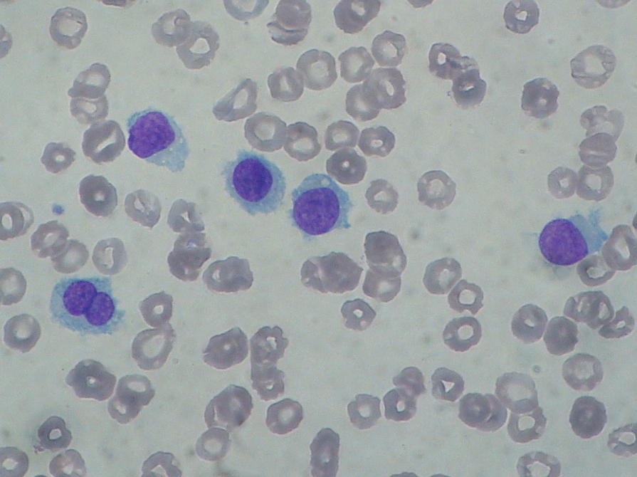
- Pancytopenia with monocytopenia and splenomegaly, usually without lymphadenopathy.
- 'Dry tap' — bone marrow aspiration frequently fails (not in the HCL variant).
- Flow cytometry: CD11c, CD25, CD103, CD123 plus B-cell markers (CD19, CD20, CD22). The variant (HCL-v) is CD25- and CD123-negative and lacks BRAF mutation and monocytopenia.
- Immunohistochemistry: annexin A1 is the most sensitive and specific marker; tartrate-resistant acid phosphatase (TRAP) is a classic routine marker; BRAF V600E can be detected by molecular testing or a mutation-specific antibody.
- Treatment: purine analogues cladribine (first-line; one 5–7-day course gives complete response in about 90%) or pentostatin; vemurafenib (BRAF inhibitor) for refractory disease.
Which markers and translocations should be memorised?
| Leukaemia | Marker profile | Genetics | Treatment pearl |
|---|---|---|---|
| AML | MPO+, CD13, CD33, CD117; Auer rods | t(8;21), inv(16) favourable | 7+3 (cytarabine + anthracycline) |
| APL (M3) | Abundant Auer rods | t(15;17) PML::RARA | ATRA + arsenic trioxide |
| B-ALL | TdT+, CD19, CD79a, CD10 (common ALL) | t(12;21) good; t(9;22), 11q23 poor | Phased chemo + intrathecal CNS prophylaxis |
| T-ALL | TdT+, CD3 (CD10 in about 25%) | — | Mediastinal mass |
| CML | Granulocytes at all stages, basophilia | t(9;22) BCR::ABL1 | Imatinib; ponatinib for T315I |
| CLL | CD5, CD19, CD23; smudge cells | — | AIHA, hypogammaglobulinaemia |
| Hairy cell | CD11c, CD25, CD103, CD123; annexin A1, TRAP | BRAF V600E | Cladribine |