How are the veins of the lower limb organised?
Lower-limb veins form superficial, deep and perforating systems. The defining anatomical boundary is the muscular deep fascia. Superficial veins drain cutaneous and subcutaneous tissues above it; deep veins drain muscles beneath it. Perforating veins cross the fascia to connect the systems. Communicating veins connect veins within the same system, so communicating and perforating are not interchangeable anatomical terms.
Venous return is directed towards the heart and ordinarily from superficial to deep through perforators. Valves limit reflux, while muscular pumps propel blood. The superficial veins are visually prominent, but effective return depends heavily on the deep system and muscle pumps. This distinction matters when planning treatment: a superficial vein may be abnormal, yet the deep outflow must still be assessed.
| System | Relationship to fascia | Examples or role |
|---|---|---|
| Superficial | Above muscular deep fascia | Great and small saphenous veins and tributaries |
| Deep | Beneath muscular fascia | Tibial, fibular, popliteal and femoral veins |
| Perforating | Crosses muscular fascia | Connects superficial and deep systems |
| Communicating | Within one system | Connects superficial to superficial or deep to deep |
Compare the upper limb. The superficial veins of the upper limb arise from the dorsal venous arch and drain mainly through the cephalic vein (radial side) and the basilic vein (medial side). In the cubital fossa they are joined by the median cubital vein, the usual site for venepuncture.
What is the course of the great saphenous vein?
The great saphenous vein, also called the long saphenous vein in older descriptions, arises from the medial end of the dorsal venous arch of the foot. It passes anterior to the medial malleolus, ascends on the medial leg, passes posteromedial to the knee and continues up the medial thigh. It crosses the deep fascia at the saphenous opening to join the common femoral vein.
The important landmarks are the malleolus, medial knee, medial thigh and saphenofemoral junction. A question showing a vein in front of the medial malleolus is identifying the great saphenous, not the small saphenous. The terminal junction is in the groin, rather than the popliteal fossa. Visualise the entire route instead of memorising isolated endpoints.
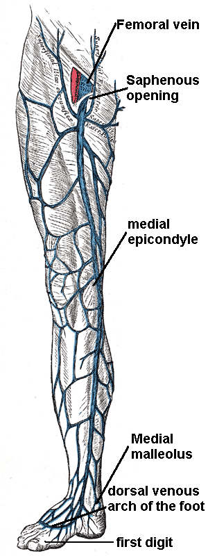
The saphenous nerve is closely related to the vein in the calf, which explains the risk of nerve injury during procedures extending into that region. Near the groin, superficial epigastric, superficial circumflex iliac and superficial external pudendal drainage commonly joins the saphenous system. Tributaries and accessory veins matter because venous disease is not always confined to the main trunk.
How does the small saphenous vein differ from the great saphenous?
The small saphenous vein, also called the short saphenous vein, begins from the lateral side of the dorsal venous network. It passes posterior to the lateral malleolus and ascends the posterior calf. Its usual termination is in the popliteal vein, although its terminal anatomy is variable. A cranial extension can communicate with more proximal veins.
| Feature | Great saphenous | Small saphenous |
|---|---|---|
| Foot origin | Medial dorsal venous arch | Lateral dorsal venous network |
| Ankle landmark | Anterior to medial malleolus | Posterior to lateral malleolus |
| Main route | Medial leg and thigh | Posterior calf |
| Usual deep junction | Common femoral vein | Popliteal vein |
| Important nerve relation | Saphenous nerve in calf | Sural nerve |
| Junction name | Saphenofemoral | Saphenopopliteal |
The sural nerve is an important neighbour of the small saphenous vein. The nerve relationship is a practical reason to map and plan interventions rather than applying a single fixed anatomical picture. Do not assume that every small saphenous terminates at the same level in the popliteal fossa; duplex mapping identifies the actual junction and any extension in the patient.
The great and small saphenous systems can communicate through intersaphenous channels. A proximal extension of the small saphenous is often referred to as the vein of Giacomini. This explains why superficial drainage and reflux patterns may not fit a diagram showing two completely isolated trunks. For the basic exam comparison, however, the ankle position and usual termination remain the most useful discriminators.
What do perforators and the deep venous system contribute?
The deep veins of the calf include anterior tibial, posterior tibial and fibular veins accompanying their arteries, together with muscular veins and sinuses. These drain towards the popliteal and femoral systems. The calf muscle pump empties the deep veins during contraction; relaxation creates conditions that favour inflow from the superficial system through competent perforators.
Perforators cross the deep fascia. Competent valves normally favour flow from superficial to deep. Incompetence allows abnormal transmission of pressure and reflux, contributing to ambulatory venous hypertension. A visible superficial varix may therefore reflect a problem at a junction, a perforator or within a more complex network rather than a single isolated valve.
Older examination terminology includes Cockett perforators for posterior tibial perforators and Boyd perforators among the paratibial group. Modern anatomical descriptions prefer location-based names. The posterior tibial perforators often connect the posterior arch tributary with posterior tibial veins rather than directly joining the main great saphenous trunk. That relationship matters when interpreting anatomy questions and treatment failures.
- Distinguish crossing the fascia from communicating within a single compartment.
- Describe the source and destination when naming a perforator.
- Map deep-vein patency as well as superficial reflux before intervention.
- Avoid memorising one universal termination level for the small saphenous.
- Use location-based anatomy when an old eponym is ambiguous.
Why do varicose veins develop and what symptoms result?
Varicose veins are dilated, tortuous subcutaneous veins. StatPearls uses a diameter of at least 3 mm for the clinical definition. They are a manifestation of chronic venous disease, commonly involving saphenous trunks or tributaries. The appearance alone does not identify the precise site or mechanism of reflux.
Venous wall changes and valve incompetence can interact. Once valve leaflets fail to coapt, blood can reflux and venous pressure remains abnormally elevated during activity. Pregnancy, obesity and prolonged standing can add to the haemodynamic burden. Previous thrombosis may leave valve damage or outflow obstruction, producing secondary chronic venous disease.

Symptoms include aching, heaviness, discomfort, swelling and itching. Chronic venous hypertension may produce ankle pigmentation, eczema, lipodermatosclerosis and ulceration. Symptoms must be assessed alongside the examination, because the amount of visible tortuosity does not reliably convey the full burden of venous disease.
How do you interpret the Brodie–Trendelenburg test?
The classic bedside test attempts to distinguish filling through proximal superficial reflux from filling despite proximal occlusion. With the patient supine, elevate the leg to empty the superficial veins. Occlude the great saphenous near the saphenofemoral junction using pressure or an appropriately placed superficial tourniquet, then ask the patient to stand. Observe filling while occlusion is maintained and after it is released.
Filling despite maintained proximal occlusion suggests an alternative reflux route, classically incompetent perforators. If the veins remain relatively empty during occlusion but fill rapidly from above on release, proximal saphenofemoral/superficial reflux is suggested. Both observations can occur when more than one component is incompetent. The interpretation depends on what was actually occluded and when filling appeared.
| Observation | Classical interpretation | Key caution |
|---|---|---|
| Rapid filling while proximal occlusion is maintained | Reflux through other connections, especially incompetent perforators | Not a precise anatomical map |
| Rapid filling after releasing proximal occlusion | Proximal superficial/saphenofemoral reflux | Assess before and after release separately |
| Abnormal filling in both phases | Combined incompetence may be present | Multiple reflux pathways can coexist |
Bedside tests are useful examination concepts, but they do not replace duplex ultrasound. Technique, multiple reflux pathways and deep-system disease can complicate the result. Avoid calling every positive test isolated deep-vein obstruction, and do not confuse this venous test with the Trendelenburg sign used to assess hip abductor function.
How are chronic venous disease severity and reflux assessed?
The CEAP system describes clinical findings, aetiology, anatomical distribution and pathophysiology. The clinical categories progress from no visible signs to visible veins, oedema, skin changes and ulcer disease. It is a description of chronic venous disease rather than a substitute for investigating the cause of a swollen leg.
| Category | Clinical finding |
|---|---|
| C0 | No visible or palpable venous disease |
| C1 | Telangiectasias or reticular veins |
| C2 | Varicose veins |
| C3 | Oedema |
| C4a | Pigmentation or eczema |
| C4b | Lipodermatosclerosis |
| C5 | Healed venous ulcer |
| C6 | Active venous ulcer |
NICE recommends duplex ultrasound to confirm varicose veins, define the extent of truncal reflux and plan treatment. The scan examines anatomy, flow and compressibility as appropriate. It addresses questions that bedside tests cannot settle reliably, including the distribution of reflux and the actual saphenopopliteal junction anatomy.
Assess the patient standing when examining visible veins, and inspect the ankle and lower leg for chronic changes. Review symptoms, previous thrombosis, prior interventions and bleeding. Sudden unilateral swelling requires assessment for an acute cause rather than automatic attribution to ordinary varicosities. Distal perfusion and the ulcer appearance also matter before choosing compression.
What is the treatment sequence and when is referral urgent?
For confirmed varicose veins with truncal reflux, NICE recommends endothermal ablation, followed by ultrasound-guided foam sclerotherapy if ablation is unsuitable, then surgery if foam is unsuitable. This is a guideline-specific hierarchy, so name the source when using it in a management answer. Anatomy, suitability and patient preference remain part of the decision.
Compression can help symptoms in selected situations, but NICE does not recommend routine compression hosiery as definitive treatment when an intervention is suitable. During pregnancy, compression can be considered for symptom relief, while intervention is generally deferred except in exceptional circumstances. This connects normal pregnancy venous effects with the management of symptomatic varicosities.
Bleeding varicose veins require immediate vascular referral. Symptomatic primary or recurrent veins, venous skin changes, suspected venous incompetence with superficial thrombosis, and active or healed venous ulcers also warrant referral. A painful hard vein suggests superficial thrombosis; an acutely swollen limb raises a separate concern for deep thrombosis.
- Match the symptomatic distribution to the duplex findings.
- Assess deep outflow rather than treating a visible superficial vessel in isolation.
- Keep nerve relations in mind: saphenous with great saphenous, sural with small saphenous.
- Distinguish a historical bedside test question from a current imaging or treatment question.