What are neural tube defects and why do they occur?
Neural tube defects (NTDs) are the most common severe malformations of the central nervous system, second only to cardiovascular anomalies as a cause of congenital morbidity and mortality. The nervous system is ectodermal in origin: the neural plate folds dorsally under the influence of the underlying notochord, and its edges fuse to form the neural tube. This process, neurulation, begins as early as 3 to 4 weeks after conception — often before the woman knows she is pregnant. Failure to complete it produces an NTD.
Closure is not simultaneous along the whole tube. It ends at two openings, the rostral (cranial) neuropore and the caudal neuropore. Failure at the rostral end gives anencephaly; failure at the caudal end gives spina bifida. The overall incidence quoted in StatPearls is about 1 to 2 per 1000 births.

What are the types of neural tube defect?
| Defect | What is wrong | Key points |
|---|---|---|
| Anencephaly | Rostral neuropore fails to close; brain and cranial vault grossly malformed, hindbrain relatively normal | Highly lethal; diagnosed easily on antenatal ultrasound; polyhydramnios is common |
| Encephalocele | Herniation of brain tissue (with or without meninges) through a skull defect | Associated with communicating hydrocephalus; occipital type is part of Meckel-Gruber syndrome |
| Spina bifida occulta | Vertebral arch defect; cord, meninges and overlying skin remain intact, no herniation | No herniation of cord or meninges; the overlying skin is intact |
| Meningocele | Herniation of meninges only (CSF-filled sac) | Sac contains meninges and CSF but no neural tissue |
| Myelomeningocele | Herniation of meninges and neural tissue | Sac contains neural tissue; associated with hydrocephalus and Chiari II malformation |
| Myeloschisis | Exposed neural tissue with no skin or meninges covering it | Open defect with CSF leakage |
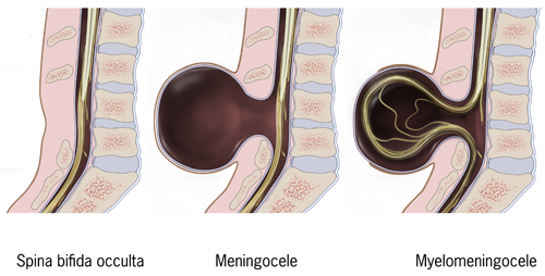
Spina bifida cystica is the umbrella term for meningocele and myelomeningocele — the forms with a visible cystic swelling. In open spina bifida neural tissue is exposed and CSF leaks, which explains the later problems: hydrocephalus, Chiari II malformation and meningitis from ascending infection. About one-third of NTD cases also carry other congenital anomalies such as cleft palate, undescended testis, omphalocele or talipes.
What causes neural tube defects — and which drugs are classic risk factors?
NTDs are multifactorial: genetic susceptibility plus environmental and nutritional insults. The single most important modifiable factor is folate status. Folic acid is converted to tetrahydrofolate by dihydrofolate reductase and, as 5-methyltetrahydrofolate, supplies one-carbon units for purine and pyrimidine (DNA and RNA) synthesis. Without it, cell proliferation during neurulation fails.
| Category | Examples | Mechanism or comment |
|---|---|---|
| Folate antagonists | Valproate, carbamazepine, phenytoin; methotrexate (DHFR inhibitor) | Inhibit folate activity or dihydrofolate reductase; women on antiseizure drugs need higher-dose folic acid |
| Genetic folate pathway | MTHFR C677T polymorphism; defects of folate receptors | Lower enzyme activity and serum folate; NTD can occur even with adequate intake |
| Vitamin B12 / homocysteine | B12 deficiency | Raised homocysteine; one hypothesis is homocysteinylation of folate receptors producing autoantibodies |
| Maternal metabolic | Diabetes (gestational diabetes raises CNS malformation risk 2 to 10 fold), obesity | Hyperglycaemia is teratogenic to the embryo before about 7 weeks |
| Physical and infective | Maternal hyperthermia, hypervitaminosis A, rubella, toxoplasmosis, CMV, radiation, amniotic bands | Antipyretic use for fever may reduce the risk |
| Social | Low socioeconomic status, no folic acid supplementation | Higher rates reported in South-East Asia and other low-resource settings |
NTDs also occur more often in twins than in singletons, and overall prevalence differs by geography and ethnicity, so family history and local epidemiology matter when counselling.
How much folic acid prevents neural tube defects?
Periconceptional folic acid, often with dietary fortification, prevents 50 to 70% of NTDs; StatPearls quotes a 71% reduction from multivitamin folic acid supplementation. The USPSTF (2023 reaffirmation, grade A) recommends that all persons planning or capable of pregnancy take a daily supplement containing 0.4 to 0.8 mg (400 to 800 micrograms) of folic acid.
| Woman | Dose | Timing |
|---|---|---|
| Any woman who could become pregnant | 0.4 to 0.8 mg per day | Start before conception and continue through early pregnancy |
| Previous child with an NTD, or positive family history | 4 mg per day | From 1 month before conception through the first 3 months of pregnancy (to prevent recurrence) |
| Taking antiseizure drugs | Higher dose; keep antiepileptic doses as low as practicable | Higher folate offsets drug antagonism (StatPearls) |
Because neurulation is complete by about 4 weeks — before most pregnancies are recognised — supplementation has to start before conception. StatPearls notes that red-cell folate takes roughly 20 weeks to reach protective levels, so ideally women should start 5 to 6 months before conception. Methionine and inositol have been proposed for folate-resistant cases.
How are neural tube defects diagnosed before birth?
Ultrasound is the investigation of choice for prenatal diagnosis; it localises the site and size of the defect and the affected vertebral level. MRI is used when ultrasound is equivocal. Maternal serum alpha-fetoprotein (MSAFP) is the classic biochemical screen.
| Test | Finding | Comment |
|---|---|---|
| Maternal serum AFP | Raised; > 2.5 MoM strongly suggests an open defect | Done at 15 to 18 weeks; less used now that non-invasive prenatal testing is common |
| Amniotic fluid AFP + acetylcholinesterase (AChE) | Both raised in open defects | Used when screening is abnormal or uncertain |
| Ultrasound — 'lemon' sign | Bifrontal concavity (scalloping) of the skull | Sign of Chiari II; reported in up to 98% of open spinal dysraphism before 24 weeks |
| Ultrasound — 'banana' sign | Cerebellum wrapped around the brainstem | Another sensitive sign of open spina bifida |
| Chromosomal microarray | Associated genetic abnormality | For syndromic cases or multiple anomalies |
AFP is made by the fetal yolk sac, liver and gastrointestinal tract. It is high in fetal plasma and amniotic fluid early on, and it reaches maternal serum by crossing the placenta as gestation advances. In an open defect, fetal AFP leaks into amniotic fluid and then into the maternal circulation, so maternal and amniotic fluid levels rise.
What complications and associated conditions go with spina bifida and anencephaly?
- Polyhydramnios — typical of anencephaly: with no functioning swallowing mechanism, amniotic fluid accumulates. StatPearls cites polyhydramnios in about 27% of anencephalic pregnancies in one series, and in about 50% of patients overall by the second and third trimesters.
- Arnold-Chiari (Chiari II) malformation — downward herniation of cerebellum into the foramen magnum because the cord is tethered by the spina bifida; fetal repair stops the CSF leak that drives it.
- Hydrocephalus — brain malformation and CSF-flow obstruction; encephalocele is associated with the communicating type.
- Meningitis — continuous CSF leak gives easy access to the meninges for oropharyngeal organisms (streptococci, enterococci).
- Neurogenic bladder and bowel, leg weakness, late ambulation, cognitive impairment — the extent depends on the level and span of the lesion.
- Stillbirth and preterm labour — serious complications of NTDs.
- Latex sensitisation — children need repeated exposure to catheters and procedures; non-latex gloves are advised.
| Condition | Why it matters |
|---|---|
| Meckel-Gruber syndrome | Autosomal recessive ciliopathy: occipital encephalocele plus polycystic kidneys and polydactyly; look for renal and respiratory involvement |
| Iniencephaly | Rare lethal defect with retroflexion of the head and cervical spine, often with spina bifida, cardiac and renal anomalies |
| Tethered cord, lipomyelomeningocele | Closed defects causing progressive neurological deficit; a skin-covered hump may mimic an NTD |
How are neural tube defects managed — including fetal surgery?
- Counselling after prenatal diagnosis: expected prognosis, options, referral to neurosurgery, urology and rehabilitation.
- Delivery and immediate care: sterile saline-soaked dressing over the sac, broad-spectrum antibiotics, non-latex gloves and aseptic handling to protect against infection.
- Postnatal closure of spina bifida within 72 hours of birth.
- Hydrocephalus: ventriculoperitoneal (VP) shunt, which drains CSF from the ventricle to the peritoneal cavity. Endoscopic third ventriculostomy with choroid plexus cauterisation is increasingly used in many centres, sometimes to reduce shunt dependence.
- Long-term follow-up for bladder, bowel, orthopaedic and developmental needs.
Prenatal (in-utero) repair of myelomeningocele was tested in the Management of Myelomeningocele Study (MOMS, 2011). Open fetal surgery reduced the need for shunting and improved motor function compared with postnatal repair, and recent data also suggest better bladder outcomes. It carries significant maternal and obstetric risks — preterm delivery, uterine dehiscence (more with open surgery) and, with the percutaneous fetoscopic approach, preterm birth and placental rupture. Myelomeningocele is the only non-lethal condition for which prenatal surgery is routinely considered.
How are neural tube defects tested in NEET PG and INI-CET?
- Embryology — NTDs are ectodermal; failure of the rostral neuropore = anencephaly, caudal neuropore = spina bifida; neurulation at 3 to 4 weeks.
- OBG screening — raised MSAFP and amniotic fluid AChE; the lemon and banana signs; ultrasound as investigation of choice.
- Preventive medicine / OBG — folic acid 0.4 mg vs 4 mg and the timing before conception.
- Pharmacology — valproate, carbamazepine, phenytoin and methotrexate as folate antagonists.
- Pediatrics / Surgery — meningocele vs myelomeningocele, hydrocephalus, VP shunt, closure within 72 hours.