How are polyhydramnios and oligohydramnios defined on ultrasound?
Amniotic fluid volume is judged on ultrasound by two methods. The single deepest vertical pocket (SDP, also called MVP) is the deepest clear fluid pocket, free of cord and fetal parts and at least 1 cm wide. The amniotic fluid index (AFI) is the sum of the deepest vertical pockets in the four abdominal quadrants. The probe is held perpendicular to the floor, in the sagittal plane parallel to the fetal spine.
| Category | Single deepest pocket (SDP) | Amniotic fluid index (AFI) |
|---|---|---|
| Normal | 2 cm or more and under 8 cm | More than 5 cm and under 24 cm |
| Oligohydramnios | Under 2 cm | 5 cm or less |
| Polyhydramnios (any) | 8 cm or more | 24 cm or more |
| Mild polyhydramnios | 8 to 11.9 cm | 24 to under 30 cm |
| Moderate polyhydramnios | 12 to 15.9 cm | 30 to under 35 cm |
| Severe polyhydramnios | 16 cm or more | 35 cm or more |
Which method? AFI can be used only in singleton pregnancy, because four quadrants cannot be assigned to each of two fetuses. The SDP works in singletons and multiples. SMFM, ACOG and AIUM prefer SDP for diagnosing oligohydramnios, as it leads to fewer obstetric interventions without worse perinatal outcomes. SDP tends to overestimate volume, so SDP alone can over-diagnose polyhydramnios. Both measures correlate poorly with true volume (dye dilution is the gold standard) but abnormal values still predict adverse outcomes.
- Colour Doppler helps to exclude cord and fetal parts from a pocket but lowers AFI and SDP by about 20%, and may overcall oligohydramnios without changing outcomes.
- Frequency: polyhydramnios occurs in 1–2% of pregnancies; about 65–70% is mild, 20% moderate and under 15% severe.
How is amniotic fluid made and cleared?
Fluid volume is a balance of production versus removal. In the first trimester it comes from fetal lung secretions and transport of maternal plasma across the membranes. From 14–16 weeks, fetal urine becomes the main source. Estimated fetal urine output is about 4.2 mL/h at 20 weeks, 22.7 mL/h at 30 weeks and 52.2 mL/h at 40 weeks (roughly 100, 545 and 1,250 mL/day). Larger fetuses make more urine; growth-restricted fetuses make less.
Removal is mainly by fetal swallowing (about 500–1,000 mL/day near term) and absorption through the gut. A second route is intramembranous absorption across the fetal surface of the placenta (about 200–500 mL/day), driven by the fact that amniotic fluid is hypotonic to fetal plasma. Swallowing rises as volume rises, which helps self-regulation.
| Problem in | Effect on fluid | Classic examples |
|---|---|---|
| Fetal urine output increased | Polyhydramnios | Maternal diabetes (fetal osmotic diuresis), macrosomia, high-output cardiac states, Bartter syndrome, recipient twin in TTTS |
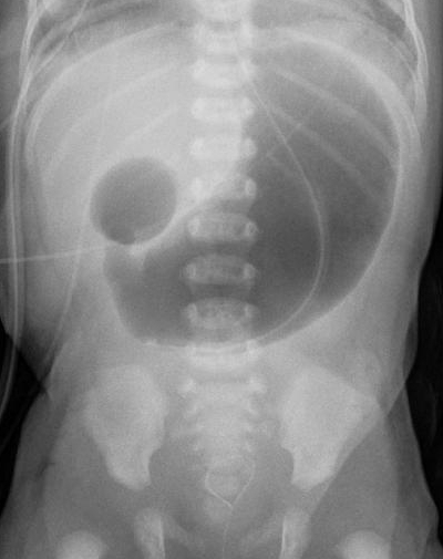
| Fetal swallowing or gut absorption reduced | Polyhydramnios | Oesophageal or duodenal atresia, tracheo-oesophageal fistula, CNS lesions, neuromuscular disease, cleft palate, neck mass |
| Fetal urine output reduced | Oligohydramnios | Renal agenesis or dysplasia, bladder outlet obstruction (posterior urethral valves), growth restriction, ACE inhibitors, ARBs, NSAIDs, donor twin in TTTS |
| Fluid leaks out | Oligohydramnios | Pre-labour rupture of membranes (PROM) |
What causes polyhydramnios?
In 50–60% of patients no cause is found (idiopathic), and that label is a diagnosis of exclusion made only after a work-up. Among pathological causes, gestational diabetes is among the commonest, and chromosomal abnormalities are also relatively common. Rarely, transplacental infection is responsible.
| Mechanism | Examples |
|---|---|
| Idiopathic | 50–60% of cases; mostly mild; excellent prognosis |
| Maternal | Diabetes (pre-gestational or gestational) |
| Impaired swallowing | CNS lesions, myotonic dystrophy, arthrogryposis syndromes, cleft lip or palate, micrognathia, obstructive neck masses |
| Impaired GI absorption | Duodenal atresia, tracheo-oesophageal fistula, oesophageal atresia, thoracic mass, diaphragmatic hernia |
| Excess urine (high-output states) | Severe fetal anaemia (alloimmunisation, parvovirus B19, alpha-thalassaemia), fetal tachyarrhythmia (SVT), sacrococcygeal teratoma, chorioangioma (arteriovenous shunting) |
| Renal | Bartter syndrome |
| Genetic | Trisomies 21, 18 and 13; Noonan, Prader-Willi, Beckwith-Wiedemann syndromes |
| Twins | Recipient twin in twin-twin transfusion syndrome (see below) |
| Infection | Congenital infections such as syphilis, rubella, CMV, toxoplasmosis, parvovirus B19 |
How do you recognise and work up polyhydramnios?
Most patients are asymptomatic and the finding is incidental, or arises from a symphysis–fundal height more than expected (3 cm above gestational age in weeks, or rapid uterine enlargement). Moderate-to-severe cases cause dyspnoea (the diaphragm is pushed up), lower-limb oedema and a grossly distended abdomen with shallow breathing.
- Look for the cause: glucose screening (rescreen if more than 1 month since the first test), anomaly scan, alloimmunisation and syphilis screening, serology for congenital infection if risk factors are present.
- Targeted ultrasound: growth (macrosomia or restriction), hydrops, face and palate, neck masses, heart, stomach bubble (absent or small in oesophageal atresia), hands and feet (arthrogryposis), kidneys, spine and pelvis (sacrococcygeal teratoma), placenta (chorioangioma), movements. MCA peak systolic velocity is used to screen for fetal anaemia, especially in hydrops.
- Genetics: offer amniocentesis with karyotype; microarray or whole-genome sequencing if severe polyhydramnios or an anomaly is found. About 5.5% of moderate or severe polyhydramnios carry a clinically significant genetic anomaly.
- Monochorionic twins: screen for twin-twin transfusion syndrome every 2 weeks from 16 weeks to delivery.
The differential diagnosis for a uterus large for dates includes macrosomia, multiple gestation, maternal fluid overload (cirrhosis, heart failure, end-stage renal disease, pre-eclampsia), placental tumour and concealed abruption. Breathlessness in these women also needs pulmonary embolism, pre-eclampsia and cardiac disease excluded.
What are the complications of polyhydramnios?
Perinatal and maternal morbidity rise with severity. Perinatal mortality in one series of 7,562 high-risk pregnancies was 1.97 per 1,000 with normal fluid and 4.12 per 1,000 with polyhydramnios. The overdistended uterus explains most obstetric complications.
| Complication | Reason |
|---|---|
| Preterm labour and preterm PROM | Uterine overdistension |
| Malpresentation | Excess fluid; confirm presentation on admission |
| Cord prolapse after rupture of membranes | Excess fluid; may present as a pulsating purplish mass in the cervical canal or vagina |
| Placental abruption | Risk rises after membrane rupture in an overdistended uterus |
| Postpartum haemorrhage | Uterine atony after chronic overdistension |
| Macrosomia, shoulder dystocia, caesarean birth | Macrosomia in 15–30% even of mild idiopathic cases; diabetes |
| Transient tachypnoea of the newborn | Retained fetal lung fluid; neonatal support should be available |
| Low 5-minute Apgar, perinatal death | Underlying anomalies; severe and rapidly progressive disease is an independent risk factor |
How is polyhydramnios managed?
Management depends on severity and the underlying cause (for example laser for TTTS). Mild idiopathic polyhydramnios rarely needs treatment, and about 37% of patients in one study resolved spontaneously, all mild.
| Measure | Detail |
|---|---|
| Antepartum surveillance | Not needed for mild idiopathic disease. For moderate to severe disease start at 32–34 weeks (biophysical profile or modified BPP). Repeat growth scans if symptoms worsen. |
| Amnioreduction | Only for symptomatic severe polyhydramnios. Ultrasound-guided transabdominal needle drainage, typically 1.5–3 L. Fluid often reaccumulates; more than one procedure in 42–46%. Risks within 48 h: preterm labour, PPROM, infection, abruption. |
| Indomethacin | Reduces fetal urine and uterine irritability. Not recommended solely to reduce fluid. A 48-hour course is reasonable for preterm contractions before 32 weeks; contraindicated at 32 weeks or more (periventricular leukomalacia, severe IVH, NEC); use nifedipine or terbutaline as tocolytics instead. |
| TTTS | Selective fetoscopic laser photocoagulation is recommended for stage II–IV before 26 weeks; amnioreduction was the historical mainstay. |
| Delivery | Severe cases deliver at a tertiary centre. Mild idiopathic: spontaneous labour at term; induction not before 39 weeks without another indication. Confirm presentation on admission; consider external cephalic version if non-cephalic. |
| Intrapartum | Continuous electronic fetal monitoring; be ready for shoulder dystocia, postpartum haemorrhage (atony), cord prolapse and neonatal transient tachypnoea. |
What causes oligohydramnios?
Causes fall into four groups: maternal (uteroplacental insufficiency, drugs), fetal (urinary tract anomalies, growth restriction, chromosomal), placental/membrane (PROM, abruption, TTTS) and idiopathic. Rupture of membranes is the commonest obstetric cause; preterm PROM alone accounts for over 37% of oligohydramnios diagnosed in the second and third trimesters. In the third trimester about half of cases are idiopathic, and these carry better outcomes.
| Group | Examples |
|---|---|
| Rupture of membranes | PROM and preterm PROM; confirm with speculum exam, fern test, nitrazine paper or amniotic protein point-of-care tests |
| Fetal urinary anomalies | Renal agenesis, dysplastic kidneys, posterior urethral valves (bladder outlet obstruction), obstructive uropathy; absent bladder suggests renal agenesis, thick-walled enlarged bladder suggests outlet obstruction |
| Uteroplacental insufficiency | Fetal growth restriction, chronic hypertension, pre-eclampsia, thrombophilia, chronic abruption, diabetic microvascular disease |
| Drugs | ACE inhibitors, ARBs, NSAIDs (reduce fetal renal blood flow), cocaine |
| Post-term pregnancy | Physiological fall in fluid late in gestation |
| Chromosomal / infection | Aneuploidy; TORCH and parvovirus B19 (less common, usually with other findings) |
| Twins | Donor twin in TTTS |

What are the complications of oligohydramnios, and what is Potter sequence?
Severe oligohydramnios or anhydramnios in the second trimester can cause intrauterine death or previable and periviable birth. Without fluid the fetal chest and limbs are compressed: lungs cannot expand and limbs cannot move. Pulmonary hypoplasia is the most important predictor of fetal mortality in second-trimester oligohydramnios, and is worst when oligohydramnios begins before 26 weeks, when the terminal lung sacs are developing.
- Potter sequence: pulmonary hypoplasia, limb contractures and flattened facies from fetal compression (classically with bilateral renal agenesis).
- Late oligohydramnios (third trimester, isolated): cord compression, abnormal fetal heart rate patterns, caesarean birth, meconium aspiration syndrome and NICU admission.
- Prognosis: worse with underlying genitourinary malformation, chromosomal anomaly or growth restriction. In one series of 131 fetuses with renal-cause oligohydramnios, 35% chose termination, 8% had intrauterine death, 27% died after birth and 30% survived.
How is oligohydramnios managed?
First establish the cause: confirm membrane status, then do a targeted scan for fetal growth and kidneys and bladder; genetic testing if anomalies are present (chromosomal microarray preferred over karyotype); infection serology only if clinically suspected. Anyone with fundal height at least 3 cm smaller than expected, known renal or gut anomaly, aneuploidy markers or risk of uteroplacental insufficiency needs an amniotic fluid scan.
| Measure | Detail |
|---|---|
| Antepartum surveillance (isolated oligohydramnios) | ACOG: start once or twice weekly at diagnosis until delivery; non-stress test plus fluid assessment (modified BPP) or full BPP. Growth scans every 3–4 weeks. Umbilical artery Doppler not needed if isolated with normal kidneys and well-being, but assessed in growth restriction. |
| Maternal hydration | Raises fluid: oral 2 L increases AFI by a mean of 3.8–5 cm, IV 2 L hypotonic fluid by 2.8–3.8 cm. A single episode wears off within about 24 hours, so a reasonable regime is 1.5–2 L of hypotonic fluid daily for at least 2 weeks. |
| Vesicoamniotic shunt | For suspected lower urinary tract obstruction at 16–28 weeks without life-limiting anomalies, in referral centres only. |
| Amnioinfusion | Mixed results; serial amnioinfusion for mid-trimester PPROM has not improved perinatal mortality; for LUTO only in an experimental setting. Prophylactic intrapartum amnioinfusion reduced fetal heart-rate abnormalities, caesarean births and low 5-minute Apgar in older studies. |
| Timing of delivery (isolated) | ACOG: 36 0/7 to 37 6/7 weeks, or at diagnosis if 38 weeks or more. With a cause present, time delivery by that condition (PPROM, abruption, growth restriction). |
| Intrapartum | Continuous fetal monitoring is advised in growth restriction and induction, which is the usual route for oligohydramnios. |
What is twin-twin transfusion syndrome (the oligo-poly sequence)?
TTTS affects 8–10% of monochorionic twin pregnancies. Vascular anastomoses within the shared placenta shunt blood from the donor to the recipient. The donor becomes hypovolaemic, with little urine and oligohydramnios (the 'stuck twin'); the recipient becomes overloaded, with polyuria, polyhydramnios, hydrops and high-output cardiac failure. Fluid discordance, not just growth discordance, is the hallmark. The Quintero staging system defines stage I as the oligo-poly sequence (donor pocket under 2 cm, recipient pocket over 8 cm) with a visible donor bladder and normal Dopplers; stage II adds a donor bladder not seen over 60 minutes; stage III abnormal Dopplers; stage IV ascites or hydrops; stage V demise of one or both twins.
Screen monochorionic twins by ultrasound every 2 weeks from 16 weeks to delivery. Selective fetoscopic laser photocoagulation of the anastomoses treats the cause and is recommended for stage II–IV disease, typically performed at 16–26 weeks. Asymptomatic stage I is usually managed expectantly with at least weekly surveillance, since about 75% remain stable or regress. See twin pregnancy and chorionicity for why only monochorionic twins are at risk.
What are the common exam traps?
- High fluid = high output or low swallow; low fluid = low output or leak. Gut atresia, CNS and neuromuscular disease raise fluid; renal agenesis and PROM lower it.
- Renal agenesis gives oligohydramnios, not polyhydramnios. Oesophageal and duodenal atresia give polyhydramnios.
- ACE inhibitors, ARBs and NSAIDs are drug causes of oligohydramnios; indomethacin (an NSAID) is used to treat polyhydramnios but only below 32 weeks.
- AFI cannot be used in twins; use the single deepest pocket.
- Amnioreduction is for symptomatic severe polyhydramnios only; fluid usually returns.
- Oligohydramnios before 26 weeks gives pulmonary hypoplasia; PROM is the commonest obstetric cause.