What are superficial fungal infections and who causes them?
Superficial mycoses are fungal infections confined to the outermost keratinised layers — the stratum corneum, hair shaft and nail. Dermatophytes digest keratin and belong to three genera: Trichophyton, Microsporum and *Epidermophyton*. The disease caused by them is called tinea (ringworm) and is named by body site. Not every superficial mycosis is a dermatophytosis: tinea (pityriasis) versicolor is caused by the yeast Malassezia, and piedra by Piedraia hortae or Trichosporon.
By natural habitat, dermatophytes are anthropophilic (human to human; for example T. rubrum), zoophilic (animals; for example Microsporum canis) or geophilic (soil). Transmission is by direct contact or fomites such as hats, hairbrushes, towels and communal floors. Predisposing factors are heat, sweating, humidity, occlusive clothing, diabetes and immunosuppression.
How are dermatophytes identified — KOH, Wood's lamp and culture?
- KOH mount — scrape scales from the leading edge of the lesion, add 10–20% potassium hydroxide (with gentle heating, or 20–40% DMSO to clear keratin without heat) and look for hyaline, septate, branching hyphae without constrictions.
- Wood's lamp — useful in tinea capitis and versicolor (see the table); T. tonsurans does not fluoresce.
- Culture on Sabouraud's dextrose agar to identify the species in refractory, recurrent or atypical disease.
- Histology with PAS or methenamine silver stain highlights fungal filaments when biopsy is done.
| Condition | Fluorescence |
|---|---|
| Tinea capitis — M. canis, M. audouinii | Green to yellow-green |
| Tinea capitis — T. schoenleinii (favus) | Blue |
| Tinea capitis — T. tonsurans | None — usually does not fluoresce |
| Tinea (pityriasis) versicolor | Gold-yellow, yellow-green or coppery-orange; present in fewer than 50% of patients |
| Erythrasma (groin differential, bacterial) | Coral red |
What are tinea corporis and tinea cruris?
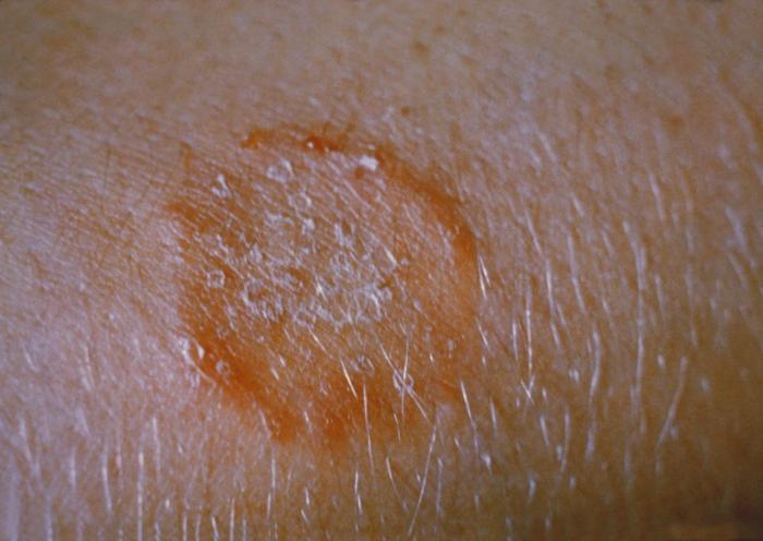
Tinea corporis affects the glabrous (non-hairy) skin of the trunk and limbs. It produces annular, scaly plaques with central clearing and an active, erythematous, sometimes raised border, and is itchy. Trichophyton rubrum accounts for about 80–90% of pathogenic strains. In immunocompromised patients it can be confluent and widespread, and a deep follicular form, Majocchi granuloma, invades the dermis.
Tinea cruris ('jock itch') involves the groin, upper inner thigh, perineum and perianal skin. Sweating, maceration and alkaline pH favour it; it is commoner in adolescent and adult males, and foot-to-groin autoinfection from tinea pedis is important. T. rubrum is the commonest cause, with rising T. mentagrophytes in some regions. The lesion is an erythematous scaly annular plaque with a raised leading edge and central clearing.
| Condition | Distinguishing features |
|---|---|
| Tinea cruris | Raised active margin, central clearing, KOH positive; dermatophyte |
| Candidal intertrigo | Often in women; satellite lesions and erythema without central clearing; may involve scrotum and penis |
| Erythrasma | No active border; coral-red fluorescence under Wood's lamp |
| Psoriasis / seborrhoeic dermatitis | Lesions elsewhere (scalp, extensors, flexures); no response to antifungals |
- Topical is first-line for localised disease: allylamines (terbinafine, butenafine, naftifine) or azoles (clotrimazole, miconazole, ketoconazole) once or twice daily for 2–4 weeks; luliconazole is used once daily for one week.
- Nystatin is ineffective against dermatophytes.
- Oral therapy for extensive, recurrent or failed topical treatment: terbinafine 250 mg daily for 2 weeks, itraconazole 100 mg daily for 2 weeks or 200 mg daily for 1 week (with food), fluconazole 150–200 mg weekly, or griseofulvin 500–1000 mg daily for 2–4 weeks.
- Address predisposing factors: sweating, tight clothing, shared towels, treating associated tinea pedis.
What is tinea capitis and why does it need oral treatment?
Tinea capitis is dermatophyte infection of the scalp hair, mostly caused by Microsporum and Trichophyton. It occurs primarily in children aged 3–14 years and may involve eyelashes and eyebrows. Transmission is from humans, animals or fomites such as hats and hairbrushes, and asymptomatic carriers sustain spread. The fungus invades the hair follicle's outer root sheath and then the hair shaft.
| Pattern | Features |
|---|---|
| Non-inflammatory (grey patch / scaling) | Scaly patch with broken hairs; usually no scarring |
| Black-dot | Hairs break at the scalp surface leaving black dots; the classic presentation |
| Kerion | Painful boggy inflammatory nodule with pus and lymphadenopathy; can cause scarring alopecia |
| Favus | Boggy, inflammatory, crusted type (T. schoenleinii) |
Differentials are bacterial folliculitis, dissecting cellulitis of the scalp, seborrhoeic dermatitis, pustular psoriasis, secondary syphilis and scalp abscess. Most oral antifungals can raise liver enzymes, so monitor liver function during prolonged courses.
What are the clinical types of tinea pedis (athlete's foot)?
Tinea pedis is acquired by walking barefoot in communal areas such as locker rooms, showers and swimming pools. Causes are T. rubrum, T. mentagrophytes/T. interdigitale and Epidermophyton floccosum. Diabetes and occlusive footwear increase risk, and untreated disease can lead to cellulitis and, in diabetics, osteomyelitis.
| Type | Features |
|---|---|
| Interdigital (intertriginous) | Commonest; itchy peeling and maceration in the toe clefts |
| Chronic hyperkeratotic (moccasin) | Diffuse or patchy scaling of the sole, medial and lateral borders of the foot |
| Vesiculobullous | Tense vesicles or bullae on the sole and instep, often due to T. mentagrophytes; burning and itching |
Treatment is topical antifungal plus foot hygiene. Terbinafine and amorolfine act faster than clotrimazole, a keratolytic such as salicylic acid helps hyperkeratotic disease, and tolnaftate powder used after swimming or showering reduces recurrence in community settings. Differentials include psoriasis, erythrasma, candidal infection, dyshidrotic eczema, pustular psoriasis and contact dermatitis.
What is tinea (pityriasis) versicolor and how is it treated?
Tinea versicolor is a common, benign superficial infection by the lipid-dependent yeast *Malassezia* (formerly Pityrosporum), part of the normal skin flora. M. globosa is the most prevalent species, with M. furfur and M. sympodialis also implicated. The disease appears when the yeast converts to its mycelial form. It is not a dermatophyte infection.
- Lesions — multiple well-defined round-to-oval macules with fine scale, either hyper- or hypopigmented (hence 'versicolor'), mainly on the upper trunk, neck and proximal limbs; usually asymptomatic or mildly itchy.
- Epidemiology — most common in warm, humid regions (up to about 50% prevalence in tropical countries) and in adolescents and young adults with active sebaceous glands.
- KOH — short hyphae and round yeast cells: 'spaghetti and meatballs'. Wood's lamp — gold-yellow, yellow-green or coppery-orange fluorescence in fewer than half.

| Route | Options |
|---|---|
| Topical (first-line) | Ketoconazole 2% shampoo left on for 5 minutes, 1–3 days (about 80% mycological cure); selenium sulfide 2.25–2.5% for 10 minutes daily for 1 week; terbinafine 1%, ciclopirox 1% or zinc pyrithione |
| Oral (extensive or recurrent) | Itraconazole 200 mg daily for 5–7 days or fluconazole 300 mg weekly for 2 weeks |
| Ineffective | Oral terbinafine and oral griseofulvin do not work for Malassezia |
What is piedra — black versus white?
Piedra is a superficial infection of the hair shaft producing nodules stuck to the hair. Black piedra is caused by *Piedraia hortae, is common in the tropics, and presents as firm, gritty, brown-black nodules (under about 1 mm) on scalp hair, especially in people with long hair and poor scalp hygiene. White piedra is caused by **Trichosporon* species and forms soft, whitish, loosely adherent** material on hair shafts. KOH shows packed brown hyphae with asci in black piedra, and septate hyphae and arthroconidia in white piedra.
- Treatment of black piedra — complete shaving of the hair, or weekly washing with 2% ketoconazole or 2% miconazole shampoo where shaving is not acceptable; oral terbinafine 250 mg daily for 6 weeks has been used in resistant cases.
- Prevention — avoid sharing combs, clips and hairbands.
- Piedra is asymptomatic apart from the unsightly nodules; trichoscopy, KOH and culture on Sabouraud's agar separate it from head lice nits and hair casts.
Which differences should you memorise for the exam?
| Disease | Organism | Key exam fact |
|---|---|---|
| Tinea corporis | T. rubrum (80–90%) | Annular scaly plaque, central clearing; topical or oral terbinafine |
| Tinea cruris | T. rubrum, T. mentagrophytes | Groin and thigh; separate from candida and erythrasma |
| Tinea capitis | Trichophyton, Microsporum | Children 3–14 years; oral griseofulvin 4–8 weeks; kerion needs steroid |
| Tinea pedis | T. rubrum, T. interdigitale | Interdigital, moccasin, vesiculobullous types |
| Tinea versicolor | Malassezia (yeast) | Not a dermatophyte; spaghetti and meatballs; ketoconazole shampoo; terbinafine ineffective orally |
| Black piedra | Piedraia hortae | Gritty black hair nodules; shaving or azole shampoo |
Related reading: stains and culture media in microbiology, papulosquamous disorders, pigmentary disorders and vaginal candidiasis.