Why do stains and culture media matter so much in exams?
Almost every bacteriology question ends with a lab step: which stain shows it? or which medium grows it? A stain is the fastest bedside clue — a smear can be read in minutes. Culture is slower but confirms the organism and allows antibiotic testing. Questions pair one organism with one classic stain and one classic medium, so learning them as pairs pays off.
The Gram stain is usually the first test performed on a clinical sample. When an organism does not stain well with Gram (mycobacteria, which have a waxy, lipid-rich wall), the acid-fast (Ziehl-Neelsen) stain takes over. Special stains — India ink, Albert, Giemsa, PAS, silver — answer narrower questions about capsules, granules, parasites and fungi.
What are the steps of the Gram stain and why does it work?
- Primary stain — crystal violet: all bacteria take up the violet dye.
- Mordant — Gram's iodine (10 to 60 seconds): forms a crystal violet-iodine complex that is harder to wash out.
- Decolouriser — alcohol or an ethanol-acetone mix: the critical, timed step.
- Counterstain — basic fuchsin or safranin: colours the decolourised (gram-negative) cells pink-red. Basic fuchsin stains gram-negatives more intensely, and some anaerobes stain poorly with safranin.
Why it works: gram-positive bacteria have a thick peptidoglycan wall; the solvent dehydrates it, closes the pores and traps the violet-iodine complex — they stay purple. Gram-negative bacteria have a thin peptidoglycan layer and an outer membrane rich in lipid; the solvent dissolves the lipid, the dye escapes, and the counterstain makes them pink.
A classic clinical use: a urethral Gram stain in a man showing neutrophils with intracellular gram-negative diplococci is considered diagnostic of gonorrhoea (specificity around 99%). The same smear from the pharynx or rectum is not recommended, because harmless Neisseria species live there.
How does the Ziehl-Neelsen (acid-fast) stain work?
Acid-fastness is the ability of a stained organism to resist decolourisation by acid-alcohol. Mycobacteria owe it to their mycolic-acid-rich wall. The Ziehl-Neelsen (ZN) method is the 'hot' method; the older Kinyoun method skips heating ('cold' method) and is now considered obsolete.
| Step | Reagent | Key detail |
|---|---|---|
| 1. Primary stain | Carbol fuchsin | Heated until steam rises (not boiled); about 10 minutes of contact, then cooled |
| 2. Decolourise | Acid-alcohol | 2 to 3 minutes; removes the dye from all non-acid-fast cells |
| 3. Counterstain | Methylene blue | About 1 minute; gives a blue background |
| Result | — | Acid-fast bacilli appear red/pink rods on a blue background |

Fluorochrome (auramine-O or auramine-rhodamine) staining is the alternative: no heating, at least 20 minutes of stain contact, acid-alcohol decolourisation, and potassium permanganate (preferred, darker background) or methylene blue as counterstain. Bacilli fluoresce yellow-green under a fluorescence microscope, which makes screening faster.
- Acid-fast bacteria: Mycobacterium species and Nocardia (e.g. N. brasiliensis, N. farcinica).
- Acid-fast non-bacterial structures: Cryptosporidium parvum, Isospora belli (Cystoisospora) and Cyclospora cayetanensis oocysts.
- A positive smear means 'acid-fast organism present' — not automatically M. tuberculosis.
Which special stain is used for which organism?
| Stain | What it shows | Classic organism / use |
|---|---|---|
| Albert stain (also methylene blue) | Metachromatic (volutin) granules of polymetaphosphate | Corynebacterium diphtheriae |
| India ink (negative stain) | Clear halo of capsule around yeast against a dark background | Cryptococcus in CSF |
| Mucicarmine | Polysaccharide capsule in tissue | Cryptococcus |
| Fontana-Masson | Melanin in the yeast wall | Cryptococcus |
| Methenamine silver / toluidine blue | Cyst walls | Pneumocystis jirovecii; methenamine silver also stains fungi |
| Giemsa | Parasites in blood; Pneumocystis cysts and trophozoites | Malaria — thick and thin smears are the gold standard |
| PAS (periodic acid-Schiff) | Glycoprotein-rich material | Whipple disease — PAS-positive foamy macrophages in small-bowel biopsy |
| Auramine-rhodamine | Fluorescent acid-fast bacilli | TB screening by fluorescence microscopy |
C. diphtheriae stains unevenly with Gram and older cultures contain metachromatic granules (polymetaphosphate) that stain bluish-purple with methylene blue. Albert stain is the routine lab stain used to bring out these granules alongside Gram stain.
India ink is a rapid way to see encapsulated yeast cells in CSF, but false negatives are common. The cryptococcal antigen test (latex agglutination, ELISA or lateral flow assay) on serum or CSF is the more reliable diagnostic test.

What are enriched, enrichment, selective and differential media?
| Type | Idea | Examples |
|---|---|---|
| Enriched (non-selective) | Basal medium with added blood or nutrients so fastidious organisms grow | Blood agar, chocolate agar (non-selective medium for Neisseria) |
| Enrichment (liquid) medium | Broth that favours the wanted pathogen and lets it outnumber commensals before subculture | Selenite or tetrathionate broth (Salmonella), alkaline peptone water/broth (Vibrio) |
| Selective | Inhibitors suppress unwanted flora | Thayer-Martin, LJ, TCBS, bismuth sulfite (Wilson and Blair) |
| Differential | An indicator shows a biochemical difference between colonies | MacConkey (lactose), TCBS (sucrose) |
Many media are both selective and differential. MacConkey agar contains crystal violet and bile salts (which inhibit gram-positive bacteria — the selective part) plus lactose and neutral red (the differential part). Lactose fermenters produce acid, the pH drops below about 6 and the indicator turns colonies pink; non-lactose fermenters stay off-white. E. coli is the classic lactose fermenter grown on MacConkey.

Which culture medium is used for which organism?
| Organism | Medium | Why / what you see |
|---|---|---|
| Mycobacterium tuberculosis | Lowenstein-Jensen (LJ) | Traditionally egg-based; malachite green inhibits contaminating bacteria; supports slow growth (liquid media with fluorescence detection are faster) |
| Corynebacterium diphtheriae | Loeffler serum medium; potassium tellurite media (Mueller-Miller, Tinsdale, cystine-tellurite blood agar) | Loeffler for primary isolation and granule formation; tellurite media are selective |
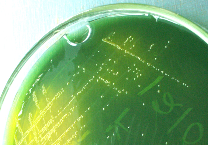
| Vibrio cholerae | TCBS (thiosulfate-citrate-bile salts-sucrose) agar | Sucrose-fermenting cholera vibrios give yellow colonies; enrich first in alkaline peptone broth |
| Neisseria gonorrhoeae | Thayer-Martin (modified) — also Martin-Lewis, NYC | Chocolate-agar base with vancomycin, colistin and nystatin; modified TM adds trimethoprim to stop Proteus swarming |
| Salmonella Typhi | Wilson and Blair (bismuth sulfite) agar; selenite/tetrathionate enrichment broth | Highly selective for typhoid bacilli from stool and sewage |
| Enteric gram-negatives | MacConkey (also EMB, Salmonella-Shigella, brilliant green) | Pink lactose fermenters vs pale non-fermenters |
How do stain and culture fit together for diphtheria, cholera and typhoid?
Diphtheria: sterile cotton swabs are taken from the tonsils or tonsillar beds (the membrane may need to be gently lifted). The smear is stained with Gram — C. diphtheriae can give a gram-variable result — and Albert or methylene blue for granules. Culture goes onto Loeffler medium and a tellurite medium; toxin production is then confirmed (tox gene PCR plus the Elek immunodiffusion test), because some strains carry the gene without making toxin.
Cholera: a stool or rectal swab is examined by dark-field microscopy, where the characteristic darting motility of vibrios is stopped by specific antisomatic antibody. Stool is plated on TCBS plus a non-selective medium, often after enrichment in alkaline peptone broth, and colonies are confirmed by slide agglutination.
Typhoid and other salmonellae: faeces, blood or other specimens are plated on several non-selective and selective agars — blood, MacConkey, eosin-methylene blue, bismuth sulfite, Salmonella-Shigella and brilliant green — and inoculated into an enrichment broth such as selenite or tetrathionate. Anything that grows in the broth is subcultured onto the agars.
What are the common traps with stains and media?
- Mordant vs decolouriser: iodine is the mordant; alcohol/acetone is the decolouriser. Mixing them up is the commonest wrong option.
- ZN counterstain is methylene blue; fluorochrome counterstain is potassium permanganate (preferred) or methylene blue.
- Acid-fast ≠ TB: Nocardia and coccidian parasites are acid-fast too.
- India ink is a negative stain; it is quick but less sensitive than cryptococcal antigen testing.
- Chocolate agar is non-selective; Thayer-Martin is the selective medium for gonococcus from contaminated sites.
- Enrichment broth ≠ enriched agar: selenite/tetrathionate and alkaline peptone are liquid enrichment media; blood/chocolate agar are enriched solid media.
- MacConkey colour: lactose fermenters pink; non-fermenters pale. Gram-positive organisms do not grow.