What is thalassaemia and which haemoglobins are involved?
Each adult haemoglobin molecule has two alpha-like and two non-alpha chains. In thalassaemia one type of chain is made in too small an amount. The unpaired excess chains precipitate in red cell precursors, causing ineffective erythropoiesis and haemolysis, and the cells that do mature are small and pale (microcytic, hypochromic).
| Haemoglobin | Chains | What happens in thalassaemia |
|---|---|---|
| HbA (main adult Hb) | α2β2 | Reduced or absent in beta-thalassaemia |
| HbA2 (minor adult Hb) | α2δ2 | Raised in beta-thalassaemia (> 3.5% in trait) |
| HbF (fetal Hb) | α2γ2 | Often raised in beta-thalassaemia |
| HbH (abnormal) | β4 | Forms when three alpha genes are lost |
| Hb Bart (abnormal) | γ4 | Forms when all four alpha genes are lost |
| Feature | Alpha-thalassaemia | Beta-thalassaemia |
|---|---|---|
| Gene and chromosome | HBA1 and HBA2 on chromosome 16 | HBB on chromosome 11 |
| Gene copies | Four alleles (two per chromosome) | Two alleles (one per chromosome) |
| Usual defect | Deletions | Point mutations (over 200 known); deletions are rare |
| Inheritance | Autosomal recessive | Autosomal recessive |
How do the alpha-thalassaemia syndromes differ?
Because there are four alpha alleles, severity rises step by step with each one lost. In fetal life the excess gamma chains form Hb Bart (γ4); after birth the excess beta chains form HbH (β4). Alpha-thalassaemia trait is thought to protect against malaria and is very common in Southeast Asia, the Mediterranean and the Middle East.
| Alleles lost | Genotype (example) | Name | Clinical picture |
|---|---|---|---|
| 1 | -α/αα | Silent carrier | Haematologically silent or very mild microcytosis |
| 2 | --/αα or -α/-α | Alpha-thalassaemia trait | Mild hypochromic microcytic anaemia with normal HbA2 and HbF |
| 3 | --/-α | HbH disease | Microcytic haemolytic anaemia, gallstones, haemolytic crises with infection or oxidant drugs; HbH inclusions on supravital stain |
| 4 | --/-- | Hb Bart hydrops fetalis | Prenatal generalised oedema, pleural and pericardial effusions from heart failure due to severe anaemia |
- HbH disease shows moderate reticulocytosis (3–6%), a fast-moving HbH band on electrophoresis and inclusion bodies of precipitated HbH after supravital staining.
- Even without regular transfusion, iron overload develops in HbH disease because anaemia increases gut iron absorption.
- HbA2 is normal in alpha-thalassaemia trait (and below 2% in HbH disease), so a normal HbA2 does not exclude alpha-thalassaemia — confirmation needs molecular testing.
How is beta-thalassaemia classified and how does it present?
Beta-thalassaemia is grouped by clinical severity, and the key dividing line between major and intermedia is dependence on regular transfusion.
| Form | Genetics | MCV | Clinical picture |
|---|---|---|---|
| Minor (trait, carrier) | One affected allele | 55–78 fL | Mild or no anaemia; often found on a routine blood count |
| Intermedia | Two affected alleles, milder | 50–70 fL | Anaemia not severe enough to need regular transfusion; marrow expansion compensates |
| Major (Cooley anaemia) | Two severely affected alleles | 50–70 fL | Presents at 6–24 months with pallor, poor weight gain, jaundice and hepatosplenomegaly; transfusion-dependent |

- Skeletal changes from marrow expansion: frontal bossing and maxillary protrusion (the 'thalassaemic facies'), long-bone deformities and osteoporosis.
- Extramedullary haematopoiesis causes hepatosplenomegaly and masses of erythroid marrow.
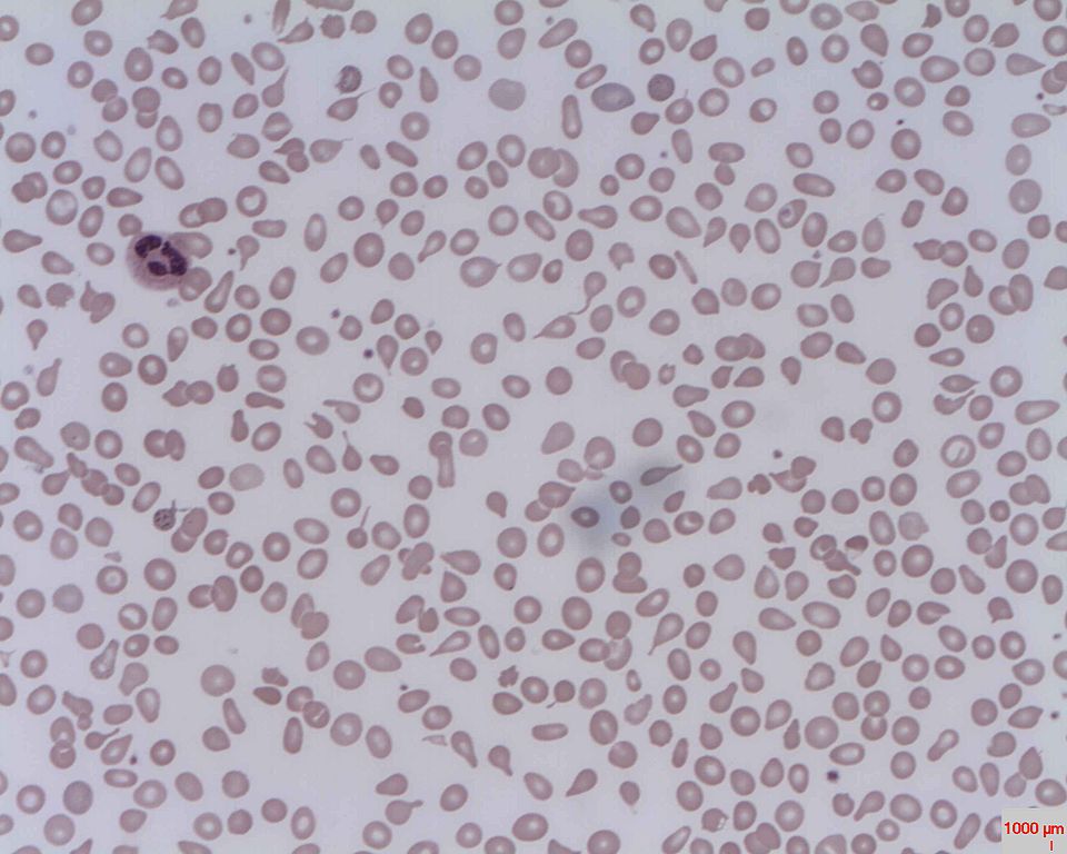
- Peripheral smear: microcytic hypochromic cells with target cells, teardrop cells and coarse basophilic stippling; nucleated red cells in major.
- Delta-beta thalassaemia behaves like beta-thalassaemia but HbA2 is not raised, because the delta chain is also affected.
Which tests confirm thalassaemia — what do HbA2 and HbF show?
Diagnosis of beta-thalassaemia needs haemoglobin electrophoresis or HPLC to show the abnormal proportions of HbA, HbA2 and HbF, together with normal iron studies so that iron deficiency is excluded.
| Haemoglobin | Normal | Beta-thalassaemia minor |
|---|---|---|
| HbA | 96–98% | 92–95% |
| HbF | < 1% | 0.5–4% |
| HbA2 | 2–3% | > 3.5% |

Sideroblastic anaemia is the other microcytic look-alike; thalassaemias lack ring sideroblasts in the marrow. For the full approach to a low MCV see anaemia classification.
What are the Mentzer index and the NESTROF test?
Mentzer index = MCV (fL) ÷ RBC count (millions/μL)
< 13 suggests beta-thalassaemia trait; > 13 suggests iron deficiency anaemia.
In thalassaemia trait the marrow makes many small cells, so the red cell count stays relatively high while the MCV is very low, giving a low ratio. In iron deficiency fewer cells are made, so the ratio is higher. The index is a screening aid only; confirmation needs HbA2 measurement and iron studies.
The NESTROF test (Naked Eye Single Tube Red cell Osmotic Fragility) is a cheap field screen used in antenatal clinics. Microcytic thalassaemic cells have decreased osmotic fragility. A drop of blood is mixed with 2 mL of 0.36% buffered saline; normal cells lyse and the tube becomes clear, so a line behind it is visible (negative). If the line is not visible, the cells resisted lysis and the test is positive. Positive mothers' husbands are then tested; if both are positive the pregnancy is 'at risk' of thalassaemia major.
How is thalassaemia major managed — transfusion and iron chelation?
Regular red cell transfusion, usually every 2–4 weeks, corrects anaemia, suppresses ineffective erythropoiesis and reduces excess gut iron absorption. GeneReviews gives a pretransfusion target of about 9.5–10.5 g/dL; StatPearls quotes 9–10 g/dL before and 13–14 g/dL after transfusion. Started by 24 months, this lets growth and development proceed normally into late childhood.
| Drug | Route and schedule | Key adverse effects / facts |
|---|---|---|
| Deferoxamine (desferrioxamine) | Slow subcutaneous infusion over about 12 hours by portable pump, 5–7 days a week | Ocular and auditory toxicity, growth restriction, rarely kidney damage; increases susceptibility to Yersinia infection |
| Deferiprone | Oral, 75–100 mg/kg/day | Neutropenia and agranulocytosis (needs blood count monitoring), arthropathy, GI upset; more cardioprotective than deferoxamine |
| Deferasirox | Oral, once daily | GI upset (pain, nausea, vomiting, diarrhoea) and a mild rise in serum creatinine; improves myocardial iron |
- Complications of iron overload: heart (arrhythmias, dilated cardiomyopathy), liver (fibrosis, cirrhosis), endocrine glands (diabetes, hypothyroidism, hypoparathyroidism, hypogonadism, pituitary and adrenal failure), and growth failure and delayed puberty in children.
- Deferoxamine and deferiprone can be combined for severe iron overload.
- Thalassaemia intermedia: folic acid, transfusion when needed, iron chelation and sometimes splenectomy.
Can thalassaemia be cured — HSCT, gene therapy and newer drugs?
| Option | Role |
|---|---|
| Haematopoietic stem cell transplant (HSCT) | Only established cure; with an HLA-identical sibling and no pre-transplant complications, disease-free survival exceeds 90% |
| Cord blood transplant from a related donor | Alternative graft source for HSCT |
| Gene therapy (autologous HSCT with gene addition or editing) | Emerging curative alternative to donor transplant |
| Luspatercept or mitapivat | Reduce transfusion requirement or improve anaemia, with variable effect |
| Hydroxyurea | Induces HbF; helps some patients |
What complications occur and how are patients monitored?
Complications come from three sources: the anaemia and marrow expansion itself, chronic haemolysis, and iron overload from transfusion and increased gut absorption. People with thalassaemia intermedia can develop jaundice and gallstones, hepatosplenomegaly, leg ulcers, pulmonary hypertension, extramedullary masses of erythroid marrow and an increased risk of thrombosis, even though they do not need regular transfusion.
| How often | What to check |
|---|---|
| Every month | Physical examination; response to transfusion and chelation side effects |
| Every 3 months | Serum ferritin and liver enzymes (ALT); growth and development in children |
| Every year | Eyes and hearing (chelator toxicity), heart, endocrine function (thyroid, parathyroid, pancreas, adrenal, pituitary) |
| Every year from about age 10 | Liver ultrasound and serum alpha-fetoprotein for early hepatocellular carcinoma; bone densitometry for osteoporosis |
In Hb Bart hydrops, the mother is also at risk: pre-eclampsia, polyhydramnios or oligohydramnios, antepartum haemorrhage and preterm delivery are common pregnancy complications, which is one more reason to screen at-risk couples before or early in pregnancy.
How do you tell thalassaemia trait from iron deficiency anaemia?
| Feature | Beta-thalassaemia trait | Iron deficiency anaemia |
|---|---|---|
| Serum ferritin | Normal | Low |
| Red cell count | Relatively high for the MCV | Low |
| Mentzer index | < 13 | > 13 |
| HbA2 | > 3.5% | Not raised |
| NESTROF | Positive (reduced osmotic fragility) | Used for carrier screening, not for iron deficiency |