What is Wilson's disease?
Wilson's disease (hepatolenticular degeneration) is a rare autosomal recessive disorder of copper metabolism. It was first described in 1912 by Kinnear Wilson as 'progressive lenticular degeneration'. Mutations in ATP7B, a copper-transporting ATPase in hepatocytes, stop the liver from excreting excess copper into bile. Copper first accumulates in the liver, then spills into the brain, cornea and other organs.
- Gene: ATP7B, chromosome 13 — disease needs two pathogenic variants (one on each allele). Hundreds of variants are known, which is why genetic testing is not always simple.
- Frequency: most often quoted as about 1 in 30,000; roughly 1 in 90 people may carry a disease-causing variant.
- Age: usual presentation 4–40 years, but cases have been confirmed from 3 to the early 70s. Males and females are affected equally, in all ethnic groups.
- Treatable: untreated it can be fatal — above all through acute liver failure — but lifelong copper-directed therapy works when started early.
Why does copper accumulate — and why is ceruloplasmin low?
About 95% of copper is excreted through the liver into bile and then stool; only a small amount leaves in urine. Normally ATP7B does two jobs in the hepatocyte: it pumps excess copper into bile and it loads copper onto newly made ceruloplasmin. Ceruloplasmin is a serum ferroxidase that carries around 90% of the copper leaving the liver.
- ATP7B fails → biliary copper excretion falls → copper is retained in hepatocytes.
- Copper is not incorporated into ceruloplasmin → the copper-free form (apoceruloplasmin) has a short half-life → low serum ceruloplasmin.
- Free copper generates oxidative damage to proteins, lipids, mitochondria and DNA methylation → hepatitis, steatosis, fibrosis, cirrhosis.
- Copper not bound to ceruloplasmin (non-ceruloplasmin copper) circulates, is filtered into urine (high urinary copper) and deposits in brain, cornea, kidney and red cells.
What are the hepatic, neurological and psychiatric features?
| System | Features | Exam pointer |
|---|---|---|
| Liver (often first) | Asymptomatic raised AST/ALT, acute hepatitis, steatosis, chronic hepatitis, cirrhosis with portal hypertension, acute liver failure | Liver symptoms usually appear in the first decade of life |
| Neurological | Tremor, dysarthria, hypophonia, dystonia, rigidity, parkinsonism, chorea (extrapyramidal) | Mean onset around 20 years or later; rare before 10 |
| Psychiatric | Mood or personality change, anxiety, auditory or visual hallucinations | Neuropsychiatric symptoms usually appear in the third or fourth decade |
| Eye | Kayser–Fleischer ring; sunflower cataract (rare) | Both regress with treatment |
| Blood | Coombs-negative haemolytic anaemia (copper damages RBC membranes); cytopenias from hypersplenism | Haemolysis + liver failure in a young patient = think Wilson |
| Kidney | Proximal tubular dysfunction (Fanconi syndrome), nephrolithiasis, hypouricaemia | Can cause rickets-like bone disease |
| Others | Cardiomyopathy and arrhythmias, arthritis, hypoparathyroidism, infertility, recurrent miscarriage | Recurrent miscarriage improves on treatment |
In the StatPearls summary, most patients present with liver-related symptoms in the first decade and neuropsychiatric symptoms in the third or fourth decade. Hepatic presentations are slightly more common in females and neurological ones in males, and female patients are more likely to present with acute liver failure.
What are the Kayser–Fleischer ring and the sunflower cataract?
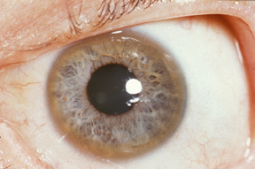
The Kayser–Fleischer (KF) ring is a golden-brown ring at the periphery of the cornea caused by copper deposited on the inner surface of the cornea in Descemet's membrane. Early rings need a slit-lamp examination by a skilled observer; only heavy deposits are visible to the naked eye. KF rings do not usually impair vision.
- Present in about 90% of patients with neurological disease, but absent in about 60% of patients with hepatic disease — and generally absent in children with mild liver disease.
- Not specific: also seen in primary biliary cholangitis and children with neonatal cholestasis.
- Fade with treatment and may reappear with non-adherence — a bedside marker of response.
- Sunflower cataract: a central opacity under the anterior lens capsule with ray-like spokes. Rare (about 1.2% of newly diagnosed patients in one series), does not reduce acuity and usually reverses with therapy.

Which tests diagnose Wilson's disease, and what are the cut-offs?
There is no single gold-standard test. AASLD recommends liver biochemistry, blood count and INR, serum ceruloplasmin, basal 24-hour urinary copper, slit-lamp (or optical tomography) examination for KF rings, neurological evaluation and ATP7B molecular testing, interpreted together.
| Test | Typical Wilson value | Pitfall |
|---|---|---|
| Serum ceruloplasmin | Usually < 14 mg/dL (normal > 20 mg/dL); very low (< 5 mg/dL) favours the diagnosis | Normal level does not exclude Wilson; it is an acute-phase protein that rises with inflammation, pregnancy and oestrogens; carriers, malabsorption and liver failure can lower it |
| Basal 24-h urinary copper | > 100 µg/24 h in symptomatic patients; > 40 µg/24 h warrants work-up in children and asymptomatic people | Raised in other causes of acute liver failure and cholestasis; reflects non-ceruloplasmin copper |
| Hepatic copper (dry weight) | > 250 µg/g in hepatitis, steatosis, cirrhosis or ALF; > 75 µg/g in asymptomatic disease | Needs liver biopsy; > 250 µg/g scores 2 Leipzig points |
| Slit lamp for KF ring | Present in most neurological cases | Often absent in hepatic and childhood disease |
| ATP7B genetic testing | Two pathogenic variants confirms | Not required for diagnosis; ideal for screening first-degree relatives of a proband |
How does the Leipzig score work?
The Leipzig score, developed at the 8th International Meeting on Wilson Disease in Leipzig, adds points for clinical, biochemical, histological and genetic findings. EASL and ESPGHAN favour it; AASLD uses a clinical–biochemical algorithm but also calls the score useful, and it has been validated in children and adults.
| Parameter | Finding | Points |
|---|---|---|
| KF ring | Present / absent | 2 / 0 |
| Neurological symptoms | Severe / mild / absent | 2 / 1 / 0 |
| Serum ceruloplasmin | < 0.1 g/L / 0.1–0.2 g/L / normal > 0.2 g/L | 2 / 1 / 0 |
| Coombs-negative haemolytic anaemia | Present / absent | 1 / 0 |
| Liver copper (no cholestasis) | > 250 µg/g dry weight / 50–249 µg/g / normal < 50 µg/g | 2 / 1 / −1 |
| Rhodanine-positive granules | Present | 1 |
| Urinary copper (no acute hepatitis) | > 2 × ULN, or normal but > 5 × ULN after D-penicillamine / 1–2 × ULN / normal | 2 / 1 / 0 |
| ATP7B mutation analysis | Two disease-causing variants / one / none | 4 / 1 / 0 |
Total ≥ 4 = diagnosis established · 3 = possible, more tests needed · ≤ 2 = very unlikely
Two deleterious ATP7B variants alone score 4, so they establish the diagnosis on their own.
How do you recognise Wilsonian acute liver failure?
About 3–5% of patients present with acute liver failure (ALF), often with silent underlying fibrosis or cirrhosis. The picture is distinctive: jaundice, Coombs-negative intravascular haemolysis with a sudden fall in haemoglobin, coagulopathy, ascites, progressing encephalopathy and rapidly worsening renal function.
- Alkaline phosphatase : total bilirubin < 4 (ALP is disproportionately low).
- AST : ALT > 2.2.
- Haemolysis markers: low haptoglobin, high reticulocytes, Coombs-negative.
- Ceruloplasmin may be low or even elevated; urinary copper > 100 µg/24 h.
How is Wilson's disease treated — penicillamine, trientine or zinc?
Every newly diagnosed patient needs lifelong copper-directed therapy; stopping treatment leads to new or recurrent hepatic and neuropsychiatric disease, liver failure or transplantation. Drugs either chelate copper and increase urinary excretion (D-penicillamine, trientine) or block intestinal absorption (zinc). A dietitian-planned copper intake below 0.9 mg/day complements, but never replaces, drugs.
| Drug | Mechanism | Adult dosing | Key cautions |
|---|---|---|---|
| D-penicillamine | Chelator → urinary copper excretion | Build up to ~1000–1500 mg/day (15–20 mg/kg/day, max 2000 mg) in 2–4 doses, empty stomach | Early sensitivity reactions in first 1–3 weeks (fever, rash, lymphadenopathy, cytopenias, proteinuria); paradoxical neurological worsening; inactivates pyridoxine — give vitamin B6 25–50 mg/day; stopped by almost one-third of patients |
| Trientine | Chelator, same mechanism | 20 mg/kg/day (max 2000 mg) in 2–3 doses | Better tolerated; neurological worsening less common; rare colitis or lupus-like reactions; take with plenty of fluid |
| Zinc salts | Induce enterocyte metallothionein, which traps dietary copper → lost in stool | 150 mg/day in 3 divided doses (adults and children > 50 kg), empty stomach | Gastric irritation; asymptomatic lipase/amylase rise; slow onset |
- Symptomatic patients: start with a chelator; trientine may be better tolerated than D-penicillamine.
- Asymptomatic (e.g. sibling found on screening): zinc is first-line, or a lower-dose chelator.
- Predominantly neurological disease: zinc can be used first-line because paradoxical neurological deterioration is uncommon with it.
- Maintenance (after > 1 year and a good response): lower-dose chelator or full-dose zinc.
- Monitoring: 24-hour urinary copper (on a chelator about 150–500 µg/24 h when stable; on zinc < 100 µg/24 h), liver tests, INR, blood count and urinalysis. Very low urinary copper with cytopenias or rising ferritin suggests overtreatment.
- Liver transplantation: for Wilsonian acute liver failure and for true treatment failure in advanced liver disease; data support its efficacy.
What are the common exam traps in Wilson's disease?
- Inheritance is autosomal recessive — not X-linked (that is Menkes) and not dominant.
- Ceruloplasmin is low, urinary copper is high — total serum copper is usually low, not high.
- A normal ceruloplasmin does not exclude Wilson's disease — it is an acute-phase reactant.
- Haemolysis is Coombs-negative (non-immune) — a positive DAT points elsewhere.
- KF ring lies in Descemet's membrane — not the stroma or epithelium — and is seen best on slit lamp.
- Brain MRI 'face of the giant panda' (increased T2 signal in the midbrain) is characteristic but seen only in advanced neurological disease.
- ALP : bilirubin < 4 and AST : ALT > 2.2 in a young patient with ALF and haemolysis = Wilson until proven otherwise.
- Asymptomatic sibling → zinc; ALF → transplant.
Wilson's disease turns up in Medicine, Pathology, Pediatrics and Ophthalmology papers. Work through the patterns on NEET PG PYQs, INI-CET PYQs and the most repeated topics list.