What hormones does each zone of the adrenal cortex make?
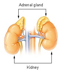
The adrenal gland has an outer cortex (from mesoderm) and an inner medulla (from neural crest). The cortex makes steroid hormones from cholesterol; the medulla makes the catecholamines epinephrine and norepinephrine. The cortex has three histological and functional zones, from outside in: zona glomerulosa, zona fasciculata and zona reticularis.
| Zone | Main hormone | Main regulator | Key enzyme point |
|---|---|---|---|
| Glomerulosa (outer) | Aldosterone (mineralocorticoid) | Renin-angiotensin system and serum potassium | Has aldosterone synthase; lacks 17α-hydroxylase |
| Fasciculata (middle) | Cortisol (glucocorticoid) | ACTH from the anterior pituitary | 17α-hydroxylase, 21-hydroxylase, 11β-hydroxylase lead to cortisol |
| Reticularis (inner) | DHEA and androstenedione (androgens) | ACTH | 17,20-lyase activity |
| Medulla | Epinephrine, norepinephrine | Sympathetic preganglionic fibres | Not part of the cortex |

How does the steroid pathway explain the adrenal enzyme defects?
All three zones start from cholesterol → pregnenolone. What each zone makes depends on which enzymes it has. The glomerulosa lacks 17α-hydroxylase, so its pregnenolone goes to progesterone → (21-hydroxylase) 11-deoxycorticosterone (DOC) → (11β-hydroxylase) corticosterone → (aldosterone synthase) aldosterone. Aldosterone synthase is present only in the glomerulosa and is driven by angiotensin II.
In the fasciculata, 17α-hydroxylase makes 17-hydroxyprogesterone (17-OHP); 21-hydroxylase converts 17-OHP to 11-deoxycortisol; 11β-hydroxylase converts 11-deoxycortisol to cortisol. In the reticularis, 17,20-lyase makes DHEA and androstenedione.
- Block an enzyme → the product falls and the substrate before the block piles up.
- Low cortisol removes negative feedback → ACTH rises → the glands enlarge (hyperplasia) and push extra precursor into any open pathway, often the androgen pathway.
- Whether blood pressure is high or low depends on whether a mineralocorticoid (DOC) accumulates before the block.
What causes Cushing syndrome and how does it present?
Cushing syndrome is the clinical picture of sustained excess glucocorticoid. The most common cause overall is iatrogenic — oral, inhaled, topical or injected glucocorticoids. Endogenous Cushing syndrome is split by the ACTH level into ACTH-dependent and ACTH-independent forms.
| Type | Cause | Share of endogenous cases | ACTH |
|---|---|---|---|
| ACTH-dependent | Cushing disease — pituitary ACTH-secreting adenoma (usually microadenoma) | 60% to 70% | Normal or high |
| ACTH-dependent | Ectopic ACTH from a non-pituitary tumour | 6% to 10% | High |
| ACTH-independent | Adrenal adenoma, carcinoma, nodular hyperplasia | 10% to 20% | Suppressed |
| Rare | Ectopic CRH | Under 1% | High |
- Moon face and dorsocervical fat pad (buffalo hump); supraclavicular fat pads are more specific than the buffalo hump.
- Thin skin with wide (over 1 cm) violaceous striae on the abdomen, thighs or breasts, and easy bruising.
- Proximal myopathy, osteoporosis, early hypertension and diabetes or hyperglycaemia.
- Hyperpigmentation only in ACTH-driven forms (pituitary or ectopic), not in adrenal tumours.
- Eye effects of steroid excess: cataract and open-angle glaucoma.
How is Cushing syndrome confirmed and localised?
First exclude exogenous steroids — including inhaled and topical ones. Then work up in three steps: confirm hypercortisolism, decide whether it is ACTH-dependent, and localise the source.
| Step | Test | How it is done / read |
|---|---|---|
| 1. Screen | Overnight 1 mg dexamethasone suppression test | 1 mg at 11 PM, serum cortisol at 8 AM; above the assay cut-off (commonly 1.8 µg/dL) is abnormal |
| 1. Screen | Late-night salivary cortisol | Loss of the normal midnight nadir |
| 1. Screen | 24-hour urinary free cortisol | Discard first morning void; a low eGFR can falsely lower it |
| 2. ACTH dependence | Plasma ACTH | Under 5 pg/mL → ACTH-independent (adrenal or exogenous); over 20 pg/mL → ACTH-dependent |
| 3. Pituitary vs ectopic | High-dose dexamethasone suppression (8 mg overnight or 2 mg 6-hourly for 2 days) | Cortisol falls by more than 50% → Cushing disease likely; ectopic ACTH does not suppress |
| 3. Pituitary vs ectopic | Inferior petrosal sinus sampling | The most validated way to localise ACTH when imaging and dynamic tests disagree |

How do Addison disease and adrenal crisis present and how are they treated?
Addison disease is primary adrenal insufficiency — destruction of both adrenal cortices, so cortisol and aldosterone are both low and ACTH is high. Autoimmune adrenalitis is the most common cause; 21-hydroxylase antibodies are its marker. Other causes are infections (tuberculosis, HIV, fungi), adrenal haemorrhage and infiltration. Adrenal crisis from bilateral adrenal haemorrhage in meningococcaemia is the Waterhouse-Friderichsen syndrome.
| Feature | Primary (Addison) | Secondary (pituitary) |
|---|---|---|
| ACTH | High | Low or inappropriately normal |
| Aldosterone | Low, with high renin | Normal |
| Hyperpigmentation | Present (palmar creases, gums, lips, pressure areas) | Absent — ACTH and MSH are low |
| Potassium | Hyperkalaemia | Usually normal |
- Labs: hyponatraemia, hyperkalaemia, hypoglycaemia.
- Diagnosis: low early-morning cortisol and a blunted ACTH (cosyntropin) stimulation test — a peak cortisol above 18 µg/dL is a normal response.
- Autoimmune polyglandular syndrome type 1: hypoparathyroidism, Addison disease and mucocutaneous candidiasis. Type 2: Addison disease with autoimmune thyroiditis (Schmidt syndrome).
Long-term: hydrocortisone in 2 or 3 divided doses plus fludrocortisone 0.05 to 0.2 mg daily, adjusted to keep renin in range. Sick-day rule: during fever or illness the hydrocortisone dose goes up about 2 to 3 times. Rifampicin speeds hydrocortisone clearance, so the dose needs to rise.
What are the types of congenital adrenal hyperplasia?
Congenital adrenal hyperplasia (CAH) is a group of inherited defects in cortisol synthesis. 21-hydroxylase deficiency (CYP21A2) accounts for about 95% of cases. About 75% of classic cases are salt-wasting and 25% simple virilising; non-classic forms are milder and present later.
| Enzyme | Blood pressure / salt | Androgens | Key lab |
|---|---|---|---|
| 21-hydroxylase | Salt-wasting: hypotension, hyponatraemia, hyperkalaemia | Excess — ambiguous genitalia in 46,XX girls | High 17-OHP |
| 11β-hydroxylase | Hypertension, hypokalaemia, low renin (DOC excess) | Excess — virilisation | High 11-deoxycortisol and DOC |
| 17α-hydroxylase | Hypertension, hypokalaemia, alkalosis | Deficient — female phenotype, absent puberty | High DOC and corticosterone |
- Newborn screening measures 17-hydroxyprogesterone. Classic 21-hydroxylase deficiency typically gives 17-OHP above 10,000 ng/dL; ambiguous values (200 to 1000 ng/dL) need a cosyntropin stimulation test.
- Affected boys are often not diagnosed until they present with a salt-wasting adrenal crisis.
- Treatment: glucocorticoid replacement (suppresses ACTH and the androgen drive), plus mineralocorticoid in salt-wasting forms.
What is Conn syndrome (primary hyperaldosteronism)?
Primary hyperaldosteronism is autonomous aldosterone production by the zona glomerulosa. Described by Conn in 1956, it is the most common cause of secondary hypertension, found in at least 10% of hypertensive patients and more than 20% of those with resistant hypertension.
- Causes: bilateral adrenal hyperplasia in about 60%; a unilateral aldosterone-producing adenoma in about 30%; carcinoma rarely.
- Aldosterone drives sodium reabsorption in the collecting duct → hypertension, with potassium and hydrogen loss → hypokalaemia and metabolic alkalosis. Hypokalaemia is not always present.
- Screening: aldosterone-to-renin ratio (high aldosterone with suppressed renin). Confirmation tests include saline loading and the captopril challenge.
- Lateralisation: CT, and adrenal vein sampling as the definitive test.
- Treatment: unilateral adenoma → laparoscopic adrenalectomy; bilateral disease → spironolactone first (eplerenone if gynaecomastia is a problem).
| Feature | Primary (Conn) | Secondary |
|---|---|---|
| Origin | Adrenal (autonomous) | Excess renin drive (renal artery stenosis, heart failure, cirrhosis) |
| Renin | Low | High |
| Aldosterone-to-renin ratio | High | Normal |
Which adrenal facts and traps are tested most often?
- Outermost zone = glomerulosa = aldosterone; innermost cortical zone = reticularis = androgens.
- Commonest cause of Cushing syndrome = exogenous steroids; commonest endogenous cause = pituitary adenoma (Cushing disease).
- Best screening tests = overnight 1 mg dexamethasone test, late-night salivary cortisol, 24-hour urinary free cortisol.
- High-dose dexamethasone suppression → pituitary; no suppression → ectopic ACTH or adrenal.
- Hyperpigmentation = high ACTH: Addison disease, Cushing disease, ectopic ACTH. Not seen in secondary adrenal insufficiency or adrenal adenoma.
- Drug of choice in adrenal crisis = hydrocortisone.
- Commonest CAH = 21-hydroxylase deficiency → raised 17-OHP; ambiguous genitalia in girls, salt-wasting in both sexes.
- Commonest cause of secondary hypertension = primary hyperaldosteronism; screen with aldosterone-to-renin ratio.
For the pharmacology of glucocorticoid replacement and steroid side-effects, see Corticosteroids; for mineralocorticoid antagonists in hypertension, see Antihypertensive Drugs.