What is renal tubular acidosis?
The kidneys keep acid-base balance in two ways: they reclaim filtered bicarbonate (mostly in the proximal tubule) and they excrete hydrogen ions with ammonia and phosphate buffers (in the distal tubule and collecting duct). Renal tubular acidosis (RTA) is the group of disorders in which one of these steps fails while the GFR is relatively preserved.
The result is always a non-anion-gap (hyperchloraemic) metabolic acidosis. Bicarbonate falls and chloride rises to keep the anion gap normal — so RTA belongs in the normal-gap column of every anion gap question, alongside diarrhoea and carbonic anhydrase inhibitors.
Two buffers carry acid out in the urine: ammonia, made by the proximal tubule and trapped as ammonium (NH4+) after H+ is secreted in the collecting duct, and phosphate (titratable acid). Ammonium excretion is the part the kidney can scale up in acidosis. Anything that stops H+ secretion, wastes bicarbonate or cuts ammonia production will therefore leave the patient acidotic even though filtration is fine. Each RTA type maps to one of these three steps, which is why learning the normal physiology makes the table easy to rebuild in the exam hall.
- Type 1 — distal RTA: the collecting duct cannot secrete H+.
- Type 2 — proximal RTA: the proximal tubule cannot reclaim bicarbonate.
- Type 3 — mixed: a vanishingly rare carbonic anhydrase II defect with features of both.
- Type 4 — hyperkalaemic RTA: aldosterone deficiency or resistance.
How do types 1, 2 and 4 RTA differ?
| Feature | Type 1 (distal) | Type 2 (proximal) | Type 4 (hyperkalaemic) |
|---|---|---|---|
| Basic defect | α-intercalated cells cannot secrete H+ (H+-ATPase, AE1 or back-leak) | Proximal HCO3- reabsorption fails — bicarbonate leak | Low aldosterone or aldosterone resistance → hyperkalaemia blocks ammoniagenesis |
| Plasma HCO3- | Can be very low (under 10 to 20 mEq/L) | 12 to 18 mEq/L (self-limiting) | Mild — above 17 mEq/L |
| Urine pH in acidosis | Always above 5.5 | Above 5.5 when HCO3- is above threshold; below 5.5 once it falls below | Below 5.5 |
| Plasma K+ | Low | Low | High |
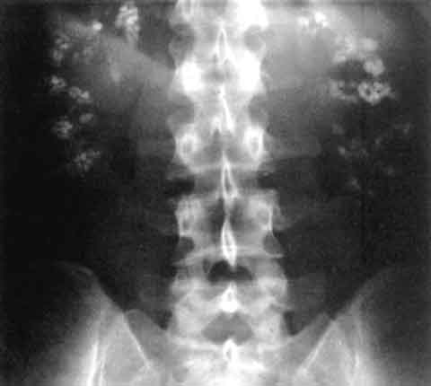
| Stones / nephrocalcinosis | Yes — calcium phosphate (hypercalciuria, hypocitraturia, alkaline urine) | No | No |
| Bone | Rickets / osteomalacia | Rickets / osteomalacia (phosphate loss) | Not typical |
| Associated | Sjogren, SLE, amphotericin B, lithium | Fanconi syndrome, myeloma, acetazolamide, tenofovir | Diabetic nephropathy, ACE inhibitors, spironolactone, NSAIDs |
| Alkali dose | Low (1 to 2 mEq/kg/day) | High (over 10 mmol/kg/day) | Treat potassium; fludrocortisone if aldosterone-deficient |
What goes wrong in type 1 (distal) RTA?
Normally the α-intercalated cells of the collecting duct secrete H+ into the urine through an H+-ATPase and H+/K+-ATPase, and return bicarbonate to blood through the basolateral chloride-bicarbonate exchanger (AE1, band 3). In distal RTA this machinery fails, so the kidney cannot lower urine pH even when the blood is acidic. Failure of the H+/K+-ATPase explains the hypokalaemia.
- Causes in adults: autoimmune disease is the commonest — Sjogren syndrome, SLE, rheumatoid arthritis, primary biliary cholangitis.
- Drugs and toxins: amphotericin B (H+ back-leak), lithium, toluene (glue sniffing).
- Inherited: AE1 (SLC4A1) mutations — autosomal dominant, mild; H+-ATPase subunit mutations — autosomal recessive, the ATP6V1B1 form comes with sensorineural deafness.
- Others: nephrocalcinosis states, medullary sponge kidney, obstructive uropathy, sickle cell disease, transplant rejection.
Clinical features: growth failure, rickets or osteomalacia, hypercalciuria with hypocitraturia (citrate is reabsorbed as a buffer), calcium phosphate stones, nephrocalcinosis and recurrent UTIs, and hypokalaemic muscle weakness or arrhythmia. Incomplete distal RTA should be suspected in any calcium-stone former whose urine pH stays at 5.5 or above without infection.

What goes wrong in type 2 (proximal) RTA?
The proximal tubule normally reclaims 85% to 90% of filtered bicarbonate. In proximal RTA the threshold for bicarbonate reabsorption is lowered. Bicarbonate spills into the urine until plasma bicarbonate falls to the new, lower threshold (typically 12 to 18 mEq/L); then the proximal tubule reclaims all of it again and the urine becomes acidic (pH under 5.5). That is why proximal RTA is self-limiting and why urine pH depends on whether the patient is on alkali.
- Commonest cause in adults: monoclonal gammopathy — light-chain disease, multiple myeloma, amyloidosis.
- Drugs: carbonic anhydrase inhibitors (acetazolamide, topiramate), outdated tetracyclines, aminoglycosides, valproate, tenofovir, ifosfamide, lead and other heavy metals.
- Often part of Fanconi syndrome — glycosuria with normal blood glucose, phosphaturia, aminoaciduria and uricosuria. Fanconi-associated diseases include Wilson disease, cystinosis, galactosaemia, hereditary fructose intolerance and von Gierke disease.
- Hypokalaemia comes from extra sodium bicarbonate delivered to the distal nephron, which increases potassium secretion.
Why does type 4 RTA cause hyperkalaemia?
Aldosterone acts on the collecting duct to reabsorb sodium and secrete potassium and hydrogen ions (see Corticosteroids for mineralocorticoid effects). When aldosterone is deficient or ineffective, potassium rises. Hyperkalaemia then suppresses ammonia production in the proximal tubule, so there is too little NH3 to trap secreted H+ as NH4+. The urine can still be acidified (pH under 5.5) — the problem is the amount of buffer, not the pump.
- Commonest cause in adults: hyporeninaemic hypoaldosteronism of diabetic nephropathy (damage to the juxtaglomerular apparatus), often with mild-to-moderate CKD.
- Drugs: potassium-sparing diuretics, ACE inhibitors, ARBs, renin inhibitors, beta-blockers, NSAIDs, calcineurin inhibitors (ciclosporin, tacrolimus), heparin, trimethoprim-sulfamethoxazole.
- Aldosterone resistance: tubulointerstitial disease, obstructive uropathy, pseudohypoaldosteronism.
- Type 4 is the most common RTA overall.
How is renal tubular acidosis diagnosed?
- Confirm metabolic acidosis on blood gas, with a normal anion gap and high chloride.
- Check that BUN and creatinine are near normal — acidosis from renal failure is not RTA.
- Measure plasma potassium — low points to type 1 or 2, high to type 4.
- Measure urine pH — inappropriately above 5.5 during acidosis suggests type 1.
- Calculate the urine anion gap = (Na+ + K+) − Cl−. A positive gap means little NH4Cl is being excreted → renal cause (RTA). Measure it only after the blood gas confirms a normal-gap acidosis.
- Rule out a urinary infection with urea-splitting organisms, which raises urine pH falsely, and severe volume depletion; urine sodium should be above 25 mEq/L.
| Test | Method | Result that confirms |
|---|---|---|
| Acid load (ammonium chloride) test | NH4Cl 100 mg/kg; urine pH hourly, plasma HCO3- 3-hourly | Urine pH stays above 5.5 despite plasma HCO3- below 21 mmol/L → type 1 |
| Bicarbonate infusion test | Infuse bicarbonate to raise plasma level above threshold | Fractional excretion of bicarbonate over 15% → type 2 |
| Urine sodium on salt restriction | Restrict dietary sodium | Persistently high urine Na → type 4 |
How is RTA treated and which traps appear in exams?
Treat the cause where possible (stop the drug, treat the autoimmune disease). Correct chronic acidosis with oral alkali — sodium bicarbonate or potassium citrate at 1 to 2 mEq/kg per day; potassium citrate is preferred when there is hypokalaemia, stones or nephrocalcinosis. Distal RTA is easy to correct with this dose because the healthy proximal tubule keeps the bicarbonate; proximal RTA needs far more.
- Normal-gap acidosis + urine pH above 5.5 + low K+ + stones → type 1.
- Normal-gap acidosis + low K+ + glycosuria with normal blood sugar, phosphaturia → type 2 with Fanconi syndrome.
- Normal-gap acidosis + high K+ in a diabetic on an ACE inhibitor → type 4.
- Acetazolamide produces a proximal RTA picture; amphotericin B a distal one.
- Sjogren syndrome is a classic cause of distal RTA and can present with hypokalaemic paralysis.
- Distal RTA with deafness → recessive H+-ATPase B1 subunit mutation.
- Type 3 (carbonic anhydrase II deficiency) = osteopetrosis, mixed RTA and cerebral calcification (marble brain disease).
- Before labelling RTA, exclude a urea-splitting urinary infection (falsely alkaline urine) and severe volume depletion (low distal sodium delivery reduces proton secretion).
Related reading: Anion Gap for the high-gap versus normal-gap split, Kussmaul Breathing for respiratory compensation, Rickets for the bone effects and Adrenal Cortex Hormones and Disorders for aldosterone.