What are the diagnostic criteria for diabetes mellitus?
Diabetes mellitus is chronic hyperglycaemia from defective insulin secretion, insulin action, or both. The American Diabetes Association (ADA) Standards of Care 2026 accept any one of four tests:
| Test | Diabetes | Prediabetes |
|---|---|---|
| HbA1c | 6.5% or more | 5.7–6.4% |
| Fasting plasma glucose (no calories for at least 8 h) | 126 mg/dL or more | 100–125 mg/dL (impaired fasting glucose) |
| 2-hour plasma glucose, 75 g OGTT | 200 mg/dL or more | 140–199 mg/dL (impaired glucose tolerance) |
| Random plasma glucose | 200 mg/dL or more with classic symptoms or a hyperglycaemic crisis | — |
Who should be screened, and how is gestational diabetes diagnosed?
ADA advises screening all adults from age 35, repeated at least every 3 years if normal. Testing should start earlier, at any age, in adults who are overweight — BMI of 25 or more, or 23 or more in Asian Americans — and have at least one additional risk factor (such as a first-degree relative with diabetes, hypertension, dyslipidaemia, previous gestational diabetes or physical inactivity).
| Time point | Plasma glucose |
|---|---|
| Fasting | 92 mg/dL |
| 1 hour | 180 mg/dL |
| 2 hours | 153 mg/dL |
How do type 1 and type 2 diabetes differ?
| Feature | Type 1 | Type 2 |
|---|---|---|
| Basic defect | Autoimmune beta-cell destruction → absolute insulin deficiency | Insulin resistance with progressive relative insulin deficiency |
| Features favouring it (ADA) | Age below 35, BMI below 25, glucose above 360 mg/dL at presentation | Older age, overweight, family history, features of metabolic syndrome |
| Autoantibodies | Present (islet autoantibodies) | Absent |
| Ketoacidosis | Common presentation | Less common; can occur under stress or with SGLT2 inhibitors |
| Treatment | Insulin from diagnosis | Lifestyle and oral or injectable agents; insulin later if needed |
ADA describes type 1 diabetes in three stages: stage 1 is two or more islet autoantibodies with normal glucose; stage 2 adds dysglycaemia without symptoms; stage 3 is clinical diabetes with symptoms. LADA (latent autoimmune diabetes in adults) is classified under type 1 — a slowly progressive autoimmune form that can initially look like type 2.
How do DKA and HHS differ, and how is DKA treated?
The 2024 international consensus report on hyperglycaemic crises updated the diagnostic criteria for diabetic ketoacidosis (DKA). Older textbook cut-offs (glucose above 250 mg/dL, bicarbonate below 15) still appear in some chapters; use the newer values.
| Feature | DKA (2024 consensus) | HHS (StatPearls) |
|---|---|---|
| Glucose | 200 mg/dL or more, or known diabetes | Above 600 mg/dL |
| Ketones | Beta-hydroxybutyrate 3.0 mmol/L or more | Minimal or absent |
| Acidosis | pH below 7.3 or bicarbonate below 18 mmol/L | Little or none |
| Effective serum osmolality | Variable | Above 320 mOsm/kg |
| Mortality | Below 1% | 5–10 times higher than DKA |
About 10% of DKA is euglycaemic — glucose is not markedly raised — classically in patients taking SGLT2 inhibitors. This is why the 2024 criteria allow 'known diabetes' in place of a glucose threshold.
- Fluids first to restore circulating volume.
- Check potassium before insulin: if K⁺ is below 3.5 mmol/L, hold insulin and replace potassium first — insulin drives potassium into cells and can cause fatal hypokalaemia.
- Fixed-rate IV insulin 0.1 U/kg/h.
- Add dextrose when glucose falls below 250 mg/dL, and keep the insulin running until ketoacidosis resolves.
- Look for and treat the trigger (infection, missed insulin, new diagnosis, SGLT2 inhibitor).
What are the features and treatment of diabetic retinopathy?
Microaneurysms are the earliest clinically visible lesion of diabetic retinopathy, followed by dot-blot haemorrhages, hard exudates and cotton-wool spots (non-proliferative). Retinal ischaemia then drives new vessel growth (proliferative retinopathy), which bleeds into the vitreous and can cause tractional detachment.

| Lesion | First-line treatment |
|---|---|
| Diabetic macular oedema (centre-involving) | Intravitreal anti-VEGF injections |
| Proliferative diabetic retinopathy | Panretinal photocoagulation (PRP); anti-VEGF is an alternative |
| Non-proliferative, no macular oedema | Glycaemic, BP and lipid control; regular review |

How is diabetic kidney disease detected and staged?
Diabetic kidney disease is screened with the urine albumin-to-creatinine ratio (UACR) and the eGFR. Because albumin excretion varies from day to day, an abnormal UACR is confirmed when two of three specimens are abnormal.
| UACR (mg/g creatinine) | Category | Old term |
|---|---|---|
| Below 30 | Normal to mildly increased | Normoalbuminuria |
| 30–299 | Moderately increased | Microalbuminuria |
| 300 or more | Severely increased | Macroalbuminuria |
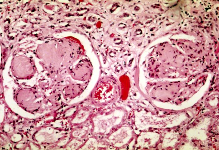
The pathological hallmark is glomerular basement membrane thickening and mesangial expansion, culminating in nodular glomerulosclerosis — the Kimmelstiel-Wilson nodules, which define class III of the pathological classification of diabetic nephropathy.
What are the neuropathic and macrovascular complications?
Distal symmetric polyneuropathy is the commonest diabetic neuropathy. It is length-dependent, so it starts in the toes and spreads upward in a stocking-glove pattern, with loss of protective sensation that leads to painless foot ulcers. Every patient needs at least an annual foot examination, including the 10-g monofilament test.
Atherosclerotic cardiovascular disease (ASCVD) — coronary disease, stroke and peripheral arterial disease — is the leading cause of death in people with diabetes. ADA sets a blood pressure target below 130/80 mmHg for most people with diabetes, alongside statin therapy and glycaemic control.
| Level | Definition |
|---|---|
| Level 1 | Glucose below 70 mg/dL and at or above 54 mg/dL |
| Level 2 | Glucose below 54 mg/dL — clinically significant |
| Level 3 | Severe event with altered mental or physical state needing another person's help, whatever the glucose |
Which diabetes numbers must you memorise?
Most diabetes questions turn on a single cut-off. This table pulls every number on the page into one place, grouped by the question it answers.
| Question | Number |
|---|---|
| Diagnosis — HbA1c / fasting / 2-h OGTT / random | 6.5% / 126 / 200 / 200 with symptoms (mg/dL) |
| Prediabetes — HbA1c / IFG / IGT | 5.7–6.4% / 100–125 / 140–199 (mg/dL) |
| Screening — start age / interval | 35 years / at least every 3 years |
| Earlier screening — BMI | 25 or more (23 or more in Asian Americans) plus a risk factor |
| Gestational diabetes — one-step 75 g OGTT | 92 / 180 / 153 mg/dL |
| DKA — glucose / beta-hydroxybutyrate / pH / bicarbonate | 200 or known diabetes / 3.0 mmol/L / below 7.3 / below 18 |
| DKA — insulin rate / add dextrose / hold insulin | 0.1 U/kg/h / glucose below 250 / K⁺ below 3.5 |
| HHS — glucose / effective osmolality | Above 600 mg/dL / above 320 mOsm/kg |
| Albuminuria — moderately / severely increased | UACR 30–299 / 300 or more mg/g |
| Hypoglycaemia — level 1 / level 2 | Below 70 / below 54 mg/dL |
| Blood pressure target | Below 130/80 mmHg |
| First eye exam — type 2 / type 1 | At diagnosis / within 5 years of onset |
What are the common exam traps on diabetes?
- HbA1c 5.7–6.4% is prediabetes, not diabetes; 6.5% is the diagnostic threshold.
- Fasting means at least 8 hours without calories.
- Random glucose of 200 needs symptoms (or a crisis) to diagnose diabetes on its own.
- DKA can be euglycaemic — think SGLT2 inhibitors.
- Never start insulin in DKA with potassium below 3.5.
- Earliest retinal lesion = microaneurysm; anti-VEGF for macular oedema, PRP for proliferative disease.
- UACR 30–299 = moderately increased albuminuria; confirm with 2 of 3 samples.
- Leading cause of death = cardiovascular disease, not kidney failure.