What are galactosaemia and glycogen storage diseases?
Both groups are inherited inborn errors of carbohydrate metabolism, and most are autosomal recessive. In galactosaemia the body cannot convert galactose, the sugar released from lactose in milk, into glucose. In glycogen storage diseases (GSDs) an enzyme or transporter for making or breaking down glycogen is defective, so glycogen either cannot be released as glucose or builds up abnormally. Manifestations range from neonatal life to adulthood.
The liver holds the highest percentage of glycogen by weight (about 10%), muscle about 2%, but because total muscle mass is larger, total muscle glycogen is roughly twice that of liver. That is why hepatic defects produce hypoglycaemia and hepatomegaly, while muscle defects give cramps, exercise intolerance and myoglobinuria.
| Feature | Galactosaemia | Glycogen storage diseases |
|---|---|---|
| Substrate | Galactose (from lactose) | Glycogen |
| Typical onset | Days after milk feeds begin | Neonate to adult depending on type |
| Hallmarks | Jaundice, hepatomegaly, bleeding, E. coli sepsis, cataract | Fasting hypoglycaemia, hepatomegaly, or exercise intolerance |
| Cornerstone of treatment | Lactose and galactose restriction | Frequent feeds / uncooked cornstarch for liver types; enzyme replacement in type II |
| Inheritance | Autosomal recessive | Mostly autosomal recessive; GSD IX (IXa) is X-linked |
Which enzyme is deficient in galactosaemia, and what builds up?
Classic galactosaemia is caused by deficiency of galactose-1-phosphate uridylyltransferase (GALT), encoded by the GALT gene. The normal pathway converts galactose to galactose-1-phosphate (galactokinase), then GALT converts galactose-1-phosphate and UDP-glucose into UDP-galactose and glucose-1-phosphate. With GALT absent, galactose-1-phosphate accumulates in red cells and tissues, and galactose is diverted to galactitol (a polyol made by aldose reductase).

Galactosaemia caused by GALT deficiency is divided into three phenotypes: classic galactosaemia, clinical variant galactosaemia (seen in African Americans and native Africans in South Africa, with residual GALT activity, sometimes missed by newborn screening that measures only blood galactose), and biochemical variant galactosaemia, of which Duarte variant is the example. Inheritance is autosomal recessive: each pregnancy of a couple with an affected child has a 25% chance of an affected baby.
How does galactosaemia present?
Within days of ingesting breast milk or lactose-containing formula, an infant with classic galactosaemia develops life-threatening complications: feeding problems, failure to thrive, hypoglycaemia, hepatocellular damage, bleeding diathesis and jaundice. Untreated infants are prone to E. coli sepsis. If a lactose-restricted diet is provided during the first ten days of life, the neonatal signs usually resolve quickly and liver failure, sepsis and neonatal death are prevented.
| Time | Problems |
|---|---|
| Neonatal (after milk) | Poor feeding, vomiting, jaundice, hepatomegaly, abnormal liver function, coagulopathy, hypoglycaemia, ascites, E. coli sepsis |
| Eyes | Cataracts (reported in 30% of 314 individuals in one series); nearly half were mild or transient and resolved with dietary treatment |
| Despite early diet | Speech problems, poor growth, poor intellectual function, extrapyramidal movement disorder with ataxia |
| Females | Hypergonadotropic hypogonadism / premature ovarian failure |
How is galactosaemia diagnosed and treated?
Diagnosis is established by elevated erythrocyte galactose-1-phosphate, reduced erythrocyte GALT activity and/or biallelic pathogenic variants in GALT. In classic galactosaemia erythrocyte galactose-1-phosphate is usually above 10 mg/dL (normal is below 1 mg/dL), plasma free galactose is usually above 10 mg/dL, and erythrocyte GALT activity is absent or barely detectable. In clinical variant galactosaemia GALT activity is close to or above 1% of control but probably never above 10-15%.
- Newborn screening (erythrocyte galactose-1-phosphate and blood galactose and/or GALT activity) detects virtually all affected infants if galactosaemia is in the panel.
- Screen-positive newborn: begin dietary intervention immediately while confirmatory testing is under way.
- Diet: restrict galactose and replace all milk products with lactose-free formulas (for example soy-based Isomil or Prosobee) containing non-galactose carbohydrates. Avoid breast milk, lactose-containing formula, cow's milk, dairy products and medicines with lactose.
- Surveillance: biochemical genetics review every three months in the first year, six-monthly in the second year, then yearly; monitor galactose-1-phosphate, urinary galactitol, cataracts, speech, development, movement disorder and, in females, ovarian function.
- Pregnancy: women with classic galactosaemia should keep a lactose-restricted diet.
How are glycogen storage diseases classified?
GSDs are numbered roughly in the order of recognition and grouped by the organ mainly affected. The key ideas: glycogen is a branched polymer with about 95% α-1,4 links and α-1,6 branch points. Synthase makes α-1,4 links, the branching enzyme (GBE1) makes branches, phosphorylase releases glucose-1-phosphate from α-1,4 links, and the debranching enzyme removes branches. Muscle lacks glucose-6-phosphatase, so it cannot release free glucose into the blood.
| Primarily affects | Types (enzyme) |
|---|---|
| Liver | 0a (glycogen synthase-2), Ia (glucose-6-phosphatase), Ib (glucose-6-phosphate transporter), III (debrancher), IV (branching enzyme), VI (liver phosphorylase), IXa (phosphorylase kinase), Fanconi-Bickel (GLUT2) |
| Skeletal muscle | V (muscle phosphorylase), VII (phosphofructokinase), X (phosphoglycerate mutase), XI (lactate dehydrogenase A), XII (aldolase A), XIII (β-enolase), XIV (phosphoglucomutase-1) |
| Skeletal and cardiac muscle | IIa (lysosomal acid maltase), IIb (LAMP-2), XV (glycogenin-1), 0b (muscle glycogen synthase) |
GSD type II is unique because it is also a lysosomal storage disease: the defective enzyme is lysosomal acid α-glucosidase (GAA), and glycogen accumulates inside lysosomes.
What are the enzyme, eponym and key feature of each GSD type?
| Type (eponym) | Defect | Key features |
|---|---|---|
| 0a | Liver glycogen synthase (GYS2) | Fasting ketotic hypoglycaemia; reduced liver glycogen (storage deficiency, not excess) |
| Ia (von Gierke) | Glucose-6-phosphatase (G6PC) | Hypoglycaemia, lactic acidosis, hypertriglyceridaemia, hyperuricaemia, hepatomegaly, renal dysfunction |
| Ib | Glucose-6-phosphate translocase (SLC37A4) | As Ia plus neutropenia, neutrophil dysfunction and inflammatory bowel disease |
| II (Pompe) | Lysosomal acid α-glucosidase (GAA) | Hypertrophic cardiomyopathy, hypotonia, motor delay; enzyme replacement therapy |
| III (Cori / Forbes) | Debranching enzyme (AGL) | Hypoglycaemia with ketosis, hyperlipidaemia, hepatomegaly, raised liver enzymes, myopathy; periportal fibrosis |
| IV (Andersen) | Branching enzyme (GBE1) | Abnormal polyglucosan bodies; hepatosplenomegaly, progressive cirrhosis, cardiomyopathy, hypotonia |
| V (McArdle) | Muscle phosphorylase (PYGM) | Exercise intolerance, cramps, rhabdomyolysis, myoglobinuria; second wind phenomenon |
| VI (Hers) | Liver phosphorylase (PYGL) | Hepatomegaly, hypoglycaemia with ketosis, raised transaminases, hyperlipidaemia, liver fibrosis |
| VII (Tarui) | Muscle phosphofructokinase (PFKM) | Haemolytic anaemia, muscle cramps with exertion, gout / hyperuricaemia |
| IXa (X-linked) | Liver phosphorylase kinase α2 (PHKA2) | Hepatomegaly, growth retardation, motor delay; type IX is the most common GSD subtype |
What is von Gierke disease (GSD type I)?
GSD I disrupts the final step of both glycogenolysis and gluconeogenesis. In hepatocytes, glucose-6-phosphate enters the endoplasmic reticulum through the translocase (G6PT), where glucose-6-phosphatase converts it to glucose. A defect in the enzyme (Ia) or the transporter (Ib) leaves glucose-6-phosphate in the cell, which is pushed into glycolysis, causing lactic acidosis, and into lipid and urate pathways, causing hyperlipidaemia and hyperuricaemia. Glycogen accumulates in liver, kidney and intestine.
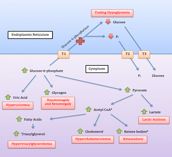
- Presentation: often at 3 to 6 months with hepatomegaly or fasting hypoglycaemia (seizures) when feeding intervals lengthen; recurrent hypoglycaemia can impair development.
- Laboratory: hypoglycaemia, lactic acidosis, hyperuricaemia, hypercholesterolaemia, hypertriglyceridaemia; neutropenia in Ib.
- Avoid the glucagon stimulation test: it can precipitate acidosis by raising blood lactate without raising glucose.
- Diagnosis: mutation analysis is first-line (G6PC sequencing if neutropenia is absent).
- Complications: short stature, osteopenia, nephropathy, gout, hepatocellular adenoma/carcinoma risk, and in Ib recurrent infections.
How do muscle GSDs and Pompe disease differ?
McArdle disease (GSD V) is deficiency of muscle glycogen phosphorylase (myophosphorylase), autosomal recessive, usually presenting in childhood or adolescence. Brief, moderate-to-intense exercise causes fatigue, cramps and weakness, with rhabdomyolysis and myoglobinuria (dark urine) in severe episodes; creatine kinase can exceed 1,000 U/L. A second wind phenomenon occurs, and it is not seen in other conditions with similar features. In the forearm exercise test, lactate and ammonia are compared before and after exercise; because glycogenolysis is blocked, no normal lactate rise occurs.
| Clue | Think of |
|---|---|
| Hypoglycaemia after a short fast | GSD I or III |
| Hypoglycaemia after an overnight fast | GSD 0, VI or IX |
| Ketosis (β-hydroxybutyrate above 2.5 mmol/L) with hepatomegaly, raised triglycerides | Hepatic GSD 0, III, VI, IX |
| Cramps after brief intense exercise, high CK, myoglobinuria | Muscle GSD V (fatty-acid oxidation defects typically appear after prolonged exercise) |
| Cardiomegaly / cardiomyopathy with hypotonia | GSD II (Pompe); also considered in III and IV |
How are GSDs diagnosed and managed?
Evaluation combines clinical features, biochemical tests (glucose, lactate, uric acid, lipids, liver enzymes, creatine kinase, urinary myoglobin) and genetic testing, which has reduced the need for liver or muscle biopsy. Biopsy patterns still appear in exams: liver biopsy in type I shows pale, swollen hepatocytes with steatosis and nuclear hyperglycogenation; fibrosis is prominent in types III, IV and VI; in McArdle disease muscle staining for phosphorylase activity is negative with subsarcolemmal PAS-positive glycogen deposits.
- No cure: treatment aims to prevent hypoglycaemia, hyperlactataemia, hyperuricaemia and hyperlipidaemia.
- Hypoglycaemia: frequent feeds of complex carbohydrate and uncooked cornstarch (a slow-releasing starch); in infants with type I, soy-based sugar-free formula every 2 to 3 hours with tube feeding overnight where needed.
- Acute hypoglycaemia: oral carbohydrate or intravenous glucose. Glucagon does not help hypoglycaemia caused by a GSD.
- Hyperuricaemia: allopurinol. Hyperlipidaemia: statins. Persistent lactic acidosis: citrate or bicarbonate.
- GSD Ib neutropenia: G-CSF, and empagliflozin is now supported for neutropenia and neutrophil dysfunction.
- GSD II: enzyme replacement therapy. Liver transplant is considered for hepatic failure or malignancy, but it does not correct cardiomyopathy.
What are the common traps and quick comparisons?
- Substrate confusion: galactosaemia = GALT (not galactokinase) in the classic form; the toxic metabolite is galactose-1-phosphate.
- Reducing sugar vs enzyme: diagnosis rests on erythrocyte galactose-1-phosphate and GALT activity, not on dietary history alone.
- Deficiency vs accumulation: GSD 0 has too little glycogen; most other types have too much, and type IV has abnormal glycogen (polyglucosan).
- Lysosomal GSD: type II is the only one that is also a lysosomal storage disease.
- Muscle cannot release glucose: muscle has no glucose-6-phosphatase, so muscle GSDs do not cause hypoglycaemia.
- X-linked GSD: type IX (IXa) is X-linked; the rest of this page's types are autosomal recessive.
Related metabolism pages: glycolysis steps and regulation, lysosomal storage disorders and amino acid disorders.