What is neonatal jaundice and why are newborns prone to it?
Neonatal jaundice is yellow discoloration of the skin, sclerae and mucous membranes caused by raised total serum bilirubin (TSB) — neonatal hyperbilirubinaemia. It is the most frequent medical condition of the first two weeks of life: about 60% of term and 80% of preterm newborns become clinically jaundiced in the first week, but only about 10% need phototherapy.
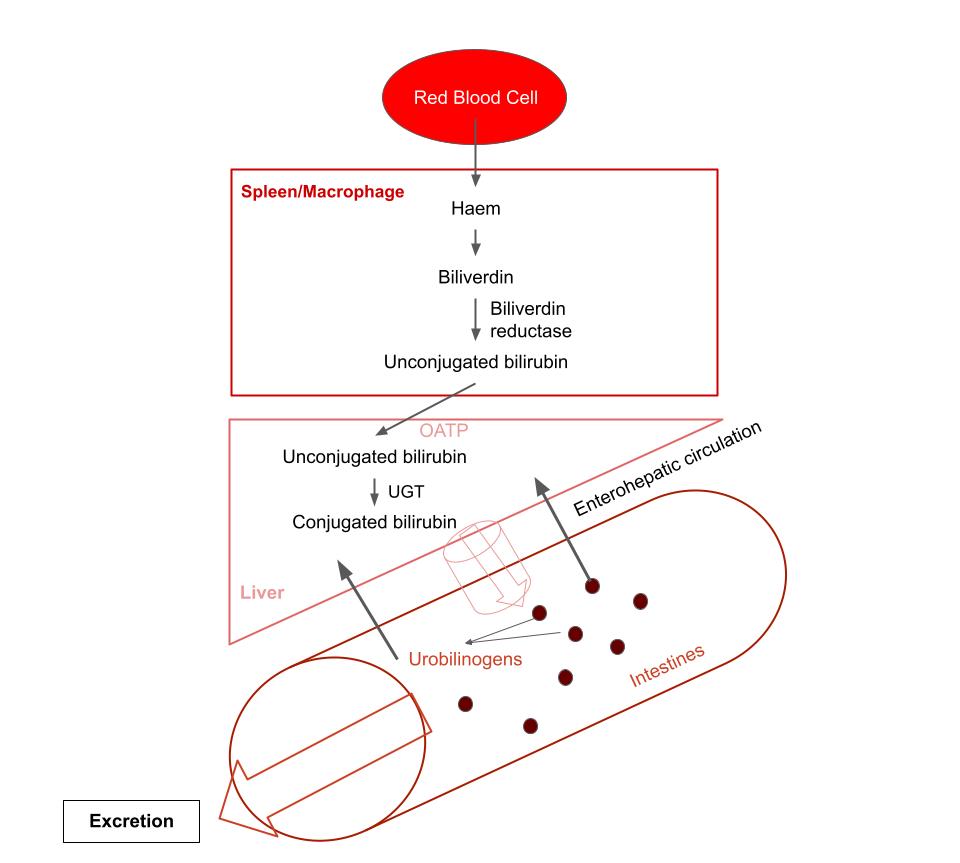
- More bilirubin is made: higher red-cell mass and haemoglobin at birth, and a shorter red-cell life span.
- Less is conjugated: newborn UDP-glucuronosyltransferase (UGT1A1) activity is about 1% of adult activity.
- More is reabsorbed: increased enterohepatic circulation — gut beta-glucuronidase deconjugates bilirubin, which is reabsorbed.
How do you tell physiological from pathological jaundice?
| Feature | Physiological | Pathological |
|---|---|---|
| Onset | After 24 hours of life | Within the first 24 hours |
| Peak | At about 48–96 hours; peak TSB in healthy term babies about 5–6 mg/dL | TSB above the 95th percentile on the hour-specific nomogram |
| Rate of rise | Slow | ≥ 5 mg/dL per day or > 0.2 mg/dL per hour |
| Type of bilirubin | Unconjugated | Unconjugated, or conjugated > 1.0 mg/dL |
| Duration | Resolves by 2–3 weeks | Prolonged: > 14 days at ≥ 37 weeks, or > 21 days below 37 weeks (NICE) |
| Baby | Well, feeding, gaining weight | Pallor, bruising/cephalohaematoma, hepatosplenomegaly, poor feeding, lethargy, pale stools or dark urine |
Physiological jaundice is a diagnosis of exclusion and accounts for about 75% of neonatal hyperbilirubinaemia. Remember that it is defined by pattern (timing, rate of rise, level for age in hours, well baby) rather than by any single number.
What causes neonatal jaundice, by time of onset?
| Onset | Causes to think of | Clue |
|---|---|---|
| First 24 hours | Haemolysis — Rh or ABO incompatibility, G6PD deficiency, hereditary spherocytosis | Always pathological; Coombs (DAT) test, blood groups, smear, reticulocytes |
| After 24 hours, first week | Physiological jaundice; breastfeeding (suboptimal intake) jaundice; cephalohaematoma or bruising; polycythaemia (IUGR, infant of diabetic mother, delayed cord clamping); Crigler-Najjar type I (severe, first few days) | Check feeding, weight loss, hydration |
| Late first week to week 2 | Breast milk jaundice | Thriving, well-fed baby on breast milk |
| Prolonged (> 14 days term, > 21 days preterm) | Breast milk jaundice; congenital hypothyroidism; biliary atresia (presents at 2–4 weeks with pale stools); neonatal hepatitis, galactosaemia, alpha-1 antitrypsin deficiency | Always measure conjugated bilirubin; look at stool and urine colour |
| Any time | Sepsis, urinary tract infection, drugs that displace bilirubin from albumin, intestinal obstruction, pyloric stenosis | Unwell baby |
- Exaggerated haemolysis (immune or non-immune) is the most common cause of pathological hyperbilirubinaemia.
- Among babies with severe hyperbilirubinaemia (TSB > 25 mg/dL, about 1 in 2,500 births), ABO incompatibility followed by G6PD deficiency are the most frequently identified causes.
- ABO incompatibility exists in about 15% of pregnancies, but haemolytic disease occurs in only about 4% of those babies. Rh disease is usually more severe and can cause hydrops fetalis.
- G6PD deficiency is X-linked recessive and the most common red-cell enzyme defect; hereditary spherocytosis is the commonest membrane defect (mostly autosomal dominant).
How is jaundice assessed — Kramer zones, TcB and TSB?
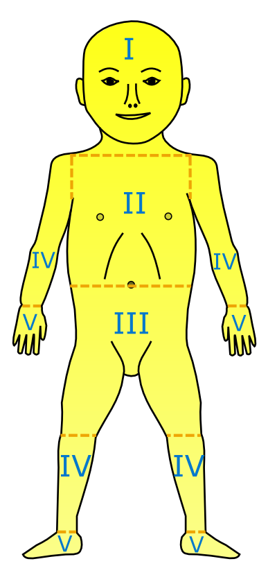
Jaundice spreads cephalocaudally — face first, then trunk, then limbs. Kramer (1969) described five dermal zones; the further down the body the yellow colour extends, the higher the bilirubin tends to be.
| Zone | Area jaundiced |
|---|---|
| 1 | Head and neck |
| 2 | Upper trunk (above the umbilicus) |
| 3 | Lower trunk and thighs (below the umbilicus) |
| 4 | Arms and lower legs |
| 5 | Palms and soles |
- Look properly: examine the naked baby in bright, preferably natural light; check the sclerae and gums and blanch the skin. Jaundice is harder to see in darker skin (NICE).
- Do not rely on visual inspection alone to estimate the level (NICE). The AAP recommends visual screening every 12 hours until discharge and a bilirubin measurement between 24 and 48 hours of age, or before discharge if earlier.
- Transcutaneous bilirubin (TcB): a screening tool for babies ≥ 35 weeks and > 24 hours old; less reliable in dark skin and after phototherapy. Check serum bilirubin if TcB exceeds the 95th percentile or 75% of the phototherapy threshold (StatPearls), or > 250 µmol/L (NICE).
- Serum bilirubin (TSB) is required in the first 24 hours, below 35 weeks, and at or above treatment thresholds.
What changed with the AAP 2022 guideline?
The American Academy of Pediatrics 2022 clinical practice guideline covers infants 35 weeks or more of gestation. Treatment thresholds are read from curves using three inputs: gestational age, age in hours and the presence of neurotoxicity risk factors. Compared with the previous guideline, it raised both the phototherapy and the exchange-transfusion thresholds, and the higher thresholds have not been linked to more bilirubin neurotoxicity.
| Risk factors for developing significant hyperbilirubinaemia | Neurotoxicity risk factors (lower the treatment threshold) |
|---|---|
| Gestational age < 38 weeks; jaundice in the first 24 hours | Gestational age < 38 weeks |
| Pre-discharge TSB or TcB close to the phototherapy threshold; phototherapy before discharge | Serum albumin < 3.0 g/dL |
| Haemolysis or a rapidly rising TSB/TcB; family history of red-cell disorders such as G6PD deficiency | Isoimmune haemolytic disease (positive DAT), G6PD deficiency or another haemolytic condition |
| Parent or sibling who needed phototherapy or exchange transfusion | Sepsis |
| Exclusive breastfeeding with suboptimal intake; scalp haematoma or significant bruising; Down syndrome; macrosomic infant of a diabetic mother | Significant clinical instability in the previous 24 hours |
How does phototherapy work and how is it given?
Phototherapy is first-line treatment for pathological unconjugated hyperbilirubinaemia. Bilirubin absorbs blue-green light of 460–490 nm; the light converts it by photoisomerisation into lumirubin, a water-soluble structural isomer excreted in bile and urine without hepatic conjugation. Efficacy depends on the light dose (irradiance), wavelength and the exposed surface area.
- Expose the maximum skin surface; shield the eyes; keep interruptions to a minimum.
- Maintain hydration — most lumirubin leaves in the urine. Support breastfeeding; supplement with expressed breast milk or formula if intake is poor, but not water or dextrose water.
- Intensify (more lights, higher irradiance) if bilirubin is rising rapidly or approaching the exchange threshold.
- Do not use sunlight as treatment (NICE).
- After stopping, a small rebound rise is expected; it rarely needs phototherapy to restart.
| Effect | Note |
|---|---|
| Dehydration, rash | Monitor fluids and weight |
| Hypocalcaemia, retinal damage | Eye shields prevent retinal injury |
| Delayed PDA closure in preterms; oxidative haemolysis; allergic reactions | — |
| Bronze baby syndrome | Bronze-grey skin, mucosa and urine, a rare effect when phototherapy is given to a baby with conjugated hyperbilirubinaemia; resolves after stopping |
When are exchange transfusion and IVIG used?
Exchange transfusion is the second-line treatment. It is indicated when TSB reaches the exchange threshold for gestation, age in hours and risk factors, when phototherapy fails, and in any baby with clinical signs of acute bilirubin encephalopathy (NICE). It removes both bilirubin and the haemolysing antibodies.
- Double-volume exchange — about 160–180 mL/kg, i.e. twice the baby's blood volume — using cross-matched blood, usually through umbilical catheters. NICE advises against a single-volume exchange and against albumin priming, and says phototherapy must continue during and after the exchange.
- TSB falls to about 60% of the pre-exchange level immediately, then rebounds to 70–80% as extravascular bilirubin re-equilibrates.
- Complications: hypocalcaemia, hyperkalaemia, arrhythmias, thrombocytopenia, blood-borne infection, portal vein thrombosis, graft-versus-host disease and necrotising enterocolitis.
- IVIG in isoimmune (Rh or ABO) haemolysis: the AAP suggests it when TSB stays within 2–3 mg/dL of the exchange level despite intensive phototherapy; NICE gives 500 mg/kg over 4 hours when bilirubin keeps rising by more than 8.5 µmol/L per hour.
What is the difference between breastfeeding jaundice and breast milk jaundice?
| Feature | Breastfeeding (suboptimal intake) jaundice | Breast milk jaundice |
|---|---|---|
| Timing | First week of life | Starts late in the first week, peaks in the second week; may last up to 8–12 weeks (about 3 months) |
| Mechanism | Inadequate milk intake → dehydration, slower gut motility and less bilirubin lost in the stool | Factors in the milk: pregnanediol inhibits UGT; milk beta-glucuronidase deconjugates bilirubin in the gut |
| Baby | Weight loss, dehydration, sometimes hypernatraemia | Thriving — adequate intake and good weight gain |
| Management | Improve feeding frequency and technique; supplement with expressed milk or formula if needed | Continue breastfeeding; it resolves on its own; exclude pathological causes first |
Why must conjugated jaundice be investigated urgently?
Conjugated hyperbilirubinaemia (neonatal cholestasis) — direct bilirubin > 1.0 mg/dL — is almost always pathological. Biliary atresia is the most common cause (about 25–40% of cases), followed by infections and parenteral-nutrition cholestasis. It classically presents at 2–4 weeks with jaundice, pale (acholic) stools and dark urine.
- Ultrasound: absent or small gallbladder and the triangular cord sign (high sensitivity, almost 100% specificity for biliary atresia).
- Hepatobiliary scintigraphy: no tracer excretion at 24 hours suggests obstruction; phenobarbital priming improves sensitivity.
- Liver biopsy is the gold standard for neonatal cholestasis (bile ductular proliferation, bile plugs, portal fibrosis in biliary atresia).
- Kasai portoenterostomy gives the best results within 60 days: more than 70% establish bile flow, versus fewer than 25% if surgery is after 90 days. Biliary atresia remains the commonest indication for paediatric liver transplant.
- Other causes: Alagille syndrome (paucity of bile ducts, butterfly vertebrae, posterior embryotoxon), alpha-1 antitrypsin deficiency (PAS-positive granules), galactosaemia (cataract, E. coli sepsis), TORCH infections, choledochal cyst.