What is a corneal ulcer and what are the causes?
A corneal ulcer (microbial keratitis) is an infection of the corneal tissue in which the epithelium is breached and the stroma becomes infiltrated and necrotic. Bacteria, fungi, protozoa and viruses can all invade, but bacteria are the most feared because they cause rapidly progressive, vision-threatening keratitis. The eye is normally protected by anatomical barriers (bony orbital rim, lids, intact epithelium), mechanical barriers (tear film, lacrimal drainage) and antimicrobial tear components (IgA, lysozyme, lactoferrin, complement). When these fail, infection follows.
- Contact lens wear — a major cause: overnight wear, poor hygiene, tap-water rinse, sharing, swimming with lenses.
- Trauma and foreign body, chemical injury, previous ocular surgery, and drugs such as topical corticosteroids.
- Local disease: dry eye, trichiasis, blepharitis, chronic dacryocystitis, lagophthalmos, neurotrophic keratopathy, bullous keratopathy, recurrent erosions, prior viral keratitis.
- Systemic: diabetes, malnutrition, xerophthalmia, immunosuppression, Stevens–Johnson syndrome, facial and trigeminal nerve palsy, alcoholism.
Where the cornea sits. The wall of the eyeball has three coats: the fibrous tunic (outermost), the vascular tunic (uvea) and the retina. The fibrous tunic is the transparent cornea in front and the opaque sclera behind. The corneal surface is non-keratinised stratified squamous epithelium, the middle coat is collagen with fibroblasts, and the inner surface is a simple (squamous) endothelial layer — which is why an ulcer that breaks the epithelium exposes the stroma.
What are the features of a bacterial corneal ulcer?
The most common species are Staphylococcus aureus, S. epidermidis, Streptococcus pneumoniae, Pseudomonas aeruginosa and Enterobacteriaceae. Symptoms are pain, redness, watering, purulent discharge, photophobia and reduced vision. The ulcer passes through four stages: progressive infiltration, active ulceration, regression and cicatrisation.
| Organism | Characteristic feature |
|---|---|
| *Pseudomonas aeruginosa* | Greenish-yellow discharge, rapid stromal melt, ring infiltrate, hypopyon, ground-glass cornea; early descemetocele or perforation |
| *Streptococcus pneumoniae* | Central deep oval ulcer with a progressive (undermined) edge; hypopyon ulcer = ulcus serpens; source is often chronic dacryocystitis |
| Staphylococcus aureus | Oval yellowish-white stromal abscess, in compromised corneas (bullous keratopathy, dry eye, rosacea); biofilm on contact lenses |
| Moraxella | Indolent paracentral oval ulcer with undermined necrotic edge in debilitated, alcoholic or diabetic patients |
| Nocardia | Wreath-like (necklace) pinhead infiltrates after soil trauma |
| Non-tuberculous mycobacteria | Slowly progressive, crack-windshield radiating infiltrate, after LASIK or contaminated instruments |
| N. gonorrhoeae | Hyperpurulent conjunctivitis with rapid stromal infiltration |

How does fungal keratitis differ, and what is the treatment?
Fungal keratitis accounts for 40–50% of microbial keratitis in some series and is typically linked to trauma with vegetative matter (farmers, field work), contact lenses, ocular surface disease and topical steroids. Of the 100+ implicated species, Fusarium, Aspergillus and Candida are the commonest; in India Aspergillus was the most commonly isolated species in several studies (one showed more than 55%), followed by Fusarium. Fusarium is more linked to trauma and contact lenses; Candida to ocular surface disease and steroids.
| Feature | Bacterial | Fungal |
|---|---|---|
| History | Contact lens, trauma | Vegetative or soil trauma, farming |
| Ulcer | Suppurative, yellow-white, sloughing edge | Raised firm slough, feathery margins, satellite lesions, hyphae beyond the ulcer |
| Endothelium | Plaque in severe cases | Endothelial plaque; fungus can breach Descemet membrane |
| Quick stain | Gram stain | 10% KOH (sensitivity over 90%), calcofluor white |
| Drug | Fortified cephalosporin + aminoglycoside, or fluoroquinolone | Natamycin 5% for filamentous fungi; amphotericin B for yeasts |
Topical antifungals: natamycin 5% is the first treatment of choice for filamentous fungi; amphotericin B 0.15–0.3% is preferred for yeasts (Candida); voriconazole 1% penetrates better and can be given intrastromally or intracamerally; others are econazole, itraconazole and miconazole. All antifungals are largely fungistatic, so treatment is prolonged and dosing is hourly in the first 48 hours before tapering. Because drugs penetrate poorly, regular scraping of necrotic slough improves penetration. KOH smear is fast and highly sensitive; Gram and Giemsa are about 50% sensitive; cultures (Sabouraud agar) may take days to weeks.
What are Acanthamoeba keratitis and herpetic keratitis?
Acanthamoeba keratitis is a protozoal infection seen in contact-lens wearers (tap-water exposure, swimming, poor lens hygiene). It is intensely painful because the organism damages corneal nerves, producing radial keratoneuritis; late disease shows a ring-like stromal infiltrate (about 50% of advanced cases). It is frequently misdiagnosed — 75–90% of cases, mostly as herpetic keratitis (about 48%) or fungal keratitis (about 25%). Diagnosis uses scraping: calcofluor white (cysts as light-green double-walled structures) or Gram stain, culture on non-nutrient agar overlaid with *E. coli*, PCR and confocal microscopy. The cyst form is resistant to most drugs; first-line treatment is a biguanide (PHMB 0.02% or chlorhexidine 0.02%, escalated if unresponsive) with or without a diamidine.
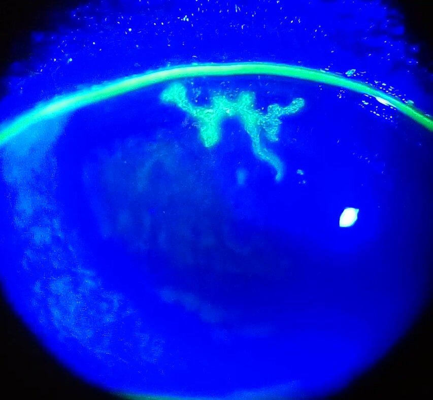
Herpes simplex keratitis follows reactivation of latent HSV. Presentations are dendritic ulcers, geographic ulcers, stromal keratitis, disciform keratitis and neurotrophic keratopathy; stromal keratitis is the most common and is largely immune-mediated. HSV epithelial keratitis starts as small dot-like lesions that merge into branched dendrites, which may enlarge into geographic ulcers. Reduced corneal sensation, scarring and blindness are the complications. For epithelial keratitis use acyclovir 3% ointment or ganciclovir 0.15% gel five times daily; trifluridine can also be used. About 99% of ulcers heal within 2 weeks.
How is a corneal ulcer investigated?
Corneal scraping from the margin and base of the ulcer is the key investigation. Indications are a large central infiltrate with stromal involvement or melt, a chronic ulcer unresponsive to broad-spectrum antibiotics, previous corneal surgery, atypical features and multiple infiltrates. It is done under topical anaesthesia — preservative-free proparacaine is preferred, as preservatives reduce bacterial viability — using a Bard-Parker no. 11 or 15 blade, a 26 G needle or a sterile spatula. Smears go for Gram stain (and KOH/calcofluor/acid-fast as needed); scrapings are inoculated onto culture media. Smear results are available within about an hour; culture in 48–72 hours.
| Organism | Stain / medium |
|---|---|
| Bacteria | Gram stain; blood agar (aerobes), thioglycolate broth |
| Neisseria, Moraxella, Haemophilus | Chocolate agar; Thayer–Martin agar for Neisseria |
| Mycobacterium | Acid-fast stain (pink); Löwenstein–Jensen medium |
| Fungi | 10% KOH, calcofluor white, Giemsa, PAS, GMS; Sabouraud agar |
| Acanthamoeba | Calcofluor white; non-nutrient agar with E. coli |
Other tests: contact lens and solution cultures (if not cleaned), conjunctival swab on calcium alginate, corneal biopsy when two smears and cultures are negative and the ulcer progresses, anterior chamber paracentesis when cultures are negative, B-scan ultrasound when corneal haze or exudate prevents a fundus view (for example after therapeutic keratoplasty), and PCR/confocal microscopy in selected cases. Revise stains and culture media for the microbiology link.
How is a bacterial corneal ulcer treated and what are the complications?
Start broad-spectrum antibiotics covering Gram-positive and Gram-negative bacteria once the smear is taken, then switch to targeted therapy when culture is back. Peripheral ulcers below 3 mm outside the visual axis may be treated with monotherapy; larger, deep stromal ulcers need two drugs. Drugs: fortified cefazolin 5% (Gram-positives), fortified tobramycin 0.3% or gentamicin (Gram-negatives), fortified vancomycin 5% (MRSA), and fluoroquinolones (ciprofloxacin, ofloxacin, moxifloxacin, gatifloxacin) as monotherapy — moxifloxacin and gatifloxacin are now favoured because resistance to ciprofloxacin and ofloxacin is growing.
- Cycloplegics reduce ciliary spasm, pain and synechiae; timolol 0.5% is the safest antiglaucoma drug if pressure is raised. Miotics and prostaglandin analogues should be avoided because they aggravate inflammation.
- Systemic antibiotics have a limited role — scleritis, endophthalmitis or non-resolving ulcers.
- Supportive: lubricants, analgesia, dark goggles, avoid rubbing and water splash.
| Complication | What happens | Treatment |
|---|---|---|
| Descemetocele | Ulcer reaches Descemet membrane, which resists and bulges out | Avoid coughing and straining, which can perforate it; glue plus bandage lens for impending perforation |
| Perforation | Aqueous gushes out, pain relieved; small perforation plugged by iris → adherent leucoma | Cyanoacrylate glue + bandage contact lens, amniotic membrane graft; larger than 2 mm needs keratoplasty; Gunderson conjunctival flap if no donor cornea |
| Anterior staphyloma | Whole cornea sloughs; a thin pseudocornea cannot withstand pressure and bulges with plastered iris | Partial or total ectatic scar; late complication |
| Non-healing ulcer (2 weeks) | Persistent infection | Therapeutic penetrating keratoplasty (TPK) |
| Scarring | Nebular, macular, leucomatous opacity by depth | Optical keratoplasty after the eye has been quiet for several months |
What are the types of keratoplasty and which is used when?
Keratoplasty replaces diseased cornea with donor tissue. Penetrating keratoplasty (PKP) is full thickness (all five corneal layers). Lamellar techniques replace only the diseased layer: DALK (deep anterior lamellar keratoplasty) replaces epithelium, Bowman layer and stroma while keeping the host endothelium; endothelial keratoplasty (DSEK/DSAEK, DMEK) replaces only endothelium with Descemet membrane (plus a thin stromal layer in DSAEK). Lamellar surgery is now preferred over PKP for many indications because it gives better outcomes and fewer complications.
| Procedure | Layers replaced | Typical indication |
|---|---|---|
| PKP (penetrating) | Full thickness | Scars, advanced keratoconus, dystrophies, therapeutic use in infection |
| DALK | Epithelium, Bowman layer, stroma; host Descemet and endothelium kept | Keratoconus and anterior stromal disease |
| DSEK / DSAEK | Endothelium + Descemet + thin posterior stroma (automated with microtome in DSAEK) | Fuchs dystrophy, pseudophakic bullous keratopathy |
| DMEK | Endothelium + Descemet membrane only | Endothelial failure; rejection about 1.9% on average in an AAO review |
| Tectonic / therapeutic / optical / cosmetic | PKP aim: restore globe integrity / remove infection / restore vision / improve appearance | Perforation / non-healing ulcer / scar / opacity |

Prognosis by indication (PKP): excellent (over 90% success) for keratoconus, lattice and granular dystrophy and early Fuchs; very good (80–90%) for pseudophakic and aphakic bullous keratopathy and herpetic keratitis; fair (50–80%) for active keratitis and perforation; poor (below 50%) for ocular pemphigoid, Stevens–Johnson syndrome, neurotrophic keratitis and multiple graft failures. Therapeutic PKP for an infected ulcer uses a graft about 0.5 mm larger than the host bed; optical PKP is done after the infection has been quiet for 6–8 months. Contraindications include severe dry eye, SJS/TEN, advanced ocular surface disease and vascularisation in more than two quadrants.
What do you need to know about donor cornea and graft rejection?
The cornea is immune privileged: it has no blood vessels, no lymphatics, and anterior chamber–associated immune deviation (ACAID), which explains the high success of grafts. Donor corneas are enucleated within about 6 hours of death; a donor endothelial cell count of at least 2000 cells/mm² is required, and storage media range from a moist chamber (48 hours) and M-K medium (up to 4 days) to organ culture (35 days) and cryopreservation (up to a year).
| Term | Meaning |
|---|---|
| Primary graft failure | Corneal oedema from the first postoperative day (poor tissue, surgical trauma) |
| Graft rejection | Immune response to donor tissue, diagnosed only when the graft has been clear for at least 2 weeks; about 18–21% of graft recipients have a rejection episode |
| Types | Epithelial, subepithelial, stromal and endothelial (the commonest); the Khodadoust line is the endothelial rejection line; keratic precipitates, oedema and neovascularisation also occur |
| High-risk host | Two or more quadrants of deep vascularisation, previous rejected graft, inflamed eye, glaucoma or ocular surface disease |
| Treatment | Intensive topical corticosteroid (e.g. dexamethasone 0.1%) reverses most endothelial rejection; systemic steroid in selected cases |