What is xerophthalmia?
Xerophthalmia ('dry eye') covers all the eye signs of vitamin A deficiency (VAD): night blindness, conjunctival and corneal xerosis, Bitot's spots, corneal ulceration, keratomalacia and retinopathy (StatPearls). WHO calls VAD the world's leading preventable cause of childhood blindness.
The eye signs are only the tip of the iceberg: many children in the same community are vitamin A deficient with normal-looking eyes. VAD also weakens defences against infection, so children with any eye sign are at high risk of dying — in an Indonesian study, children with night blindness were almost three times as likely to die, and those with night blindness plus Bitot's spots almost nine times (Gilbert, Community Eye Health 2013).
Why does vitamin A deficiency damage the eye?
Vitamin A does two different jobs in the eye, and its deficiency produces two families of signs:
- Retina — vision in dim light. Retinal is part of the visual pigment rhodopsin in rods. StatPearls notes that the rod system is much more sensitive to deficiency than the cone pigment iodopsin, so night blindness comes early.
- Surface epithelium. Without vitamin A the conjunctiva undergoes keratinising squamous metaplasia — it becomes more like skin than a mucous membrane. Loss of goblet cells and mucin, and failing tears (StatPearls), give xerosis, Bitot's spots and, when acute and severe, corneal melting.
| Cause | How it leads to deficiency |
|---|---|
| Poor dietary intake | The main cause in developing countries; can reach epidemic levels in young children (StatPearls) |
| Measles | Rapidly depletes vitamin A stores — a classic trigger of keratomalacia |
| Diarrhoea and other infections | Increase use and loss of vitamin A; can tip a deficient child into acute corneal disease |
| Severe acute malnutrition | Children with keratomalacia are often malnourished (Gilbert 2013) |
| Defects of absorption, metabolism or storage | Sporadic cases: chronic liver disease, alcoholism, upper GI surgery, inflammatory bowel disease, pancreatitis (StatPearls) |
What is the WHO classification of xerophthalmia?
| Grade | Sign | Peak age (years) | Type of deficiency | Mortality risk |
|---|---|---|---|---|
| XN | Night blindness | 2–6; adult women | Longstanding; not blinding | + |
| X1A | Conjunctival xerosis | 3–6 | Longstanding; not blinding | + |
| X1B | Bitot's spots | 3–6 | Longstanding; not blinding | + |
| X2 | Corneal xerosis | 1–4 | Acute; can be blinding | ++ |
| X3A | Corneal ulcer / keratomalacia < 1/3 of cornea | 1–4 | Severe acute; blinding | +++ |
| X3B | Corneal ulcer / keratomalacia ≥ 1/3 of cornea | 1–4 | Severe acute; blinding | ++++ |
| XS | Corneal scar (from X3) | > 2 | Consequence of ulceration | +/– |
| XF | Xerophthalmic fundus | Adults | Longstanding; rare; not blinding | – |
What do Bitot's spots, corneal xerosis and keratomalacia look like?
- Night blindness (XN): children cannot complain, so mothers notice the child becomes less active and fearful after sunset. It also affects pregnant and lactating women. Local names for it usually exist where VAD is common.
- Conjunctival xerosis (X1A): dry, lustreless, slightly wrinkled bulbar conjunctiva — hard to detect and not a very reliable sign.
- Bitot's spots (X1B): slightly elevated, white foamy (cheesy) patches of keratin on the bulbar conjunctiva near the limbus at the 3 or 9 o'clock positions, more often temporal. They can be partly wiped away but do not disappear completely even after treatment, so they do not prove current deficiency.
- Corneal xerosis (X2): dull, dry cornea from failure of tears and mucus — a sign of acute deficiency, with high infection risk.
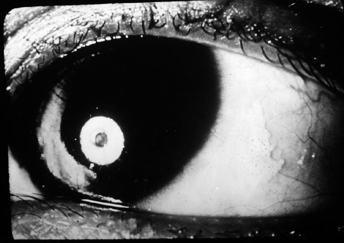
- Corneal ulcer (X3A) and keratomalacia (X3B): a punched-out or fluffy ulcer; in keratomalacia more than a third of the cornea becomes oedematous and melts through necrosis, sometimes within days. The eye can look surprisingly white unless secondarily infected.
- End results (XS): corneal scarring, staphyloma or phthisis bulbi. Signs are usually bilateral, so blindness follows.

How is xerophthalmia treated?
Any child with eye signs of VAD (or recent measles in a malnourished child) gets high-dose oral vitamin A on three occasions. The WHO Pocket Book of Hospital Care for Children gives the age-based doses:
| Age | Dose on day 1 | Day 2 | Day 14 |
|---|---|---|---|
| < 6 months | 50 000 IU | 50 000 IU | 50 000 IU |
| 6–12 months | 100 000 IU | 100 000 IU | 100 000 IU |
| > 12 months | 200 000 IU | 200 000 IU | 200 000 IU |
- If the first dose was given at the referring centre, treat on days 1 and 14 only (WHO Pocket Book).
- For corneal clouding or ulceration, add: chloramphenicol or tetracycline eye drops four times daily for 7–10 days, atropine eye drops one drop three times daily for 3–5 days, saline-soaked eye pads and a bandage — to prevent corneal rupture and extrusion of the lens.
- Treat the cause too: feeding, measles care, diarrhoea management and treatment of severe acute malnutrition.
- StatPearls summarises the WHO scheme as 200 000 IU orally, repeated the next day and again several weeks later.
What is India's vitamin A prophylaxis schedule?
Prevention in India is built into the Universal Immunization Programme. The MoHFW Immunization Handbook for Health Workers lists nine oral doses of vitamin A between 9 months and 5 years:
| Dose | When | Amount | Route |
|---|---|---|---|
| 1st | At 9 months (with measles-rubella 1st dose) | 1 ml = 1 lakh IU | Oral |
| 2nd | At 16 months (with DPT booster / MR 2nd dose) | 2 ml = 2 lakh IU | Oral |
| 3rd to 9th | One dose every 6 months up to 5 years | 2 ml = 2 lakh IU each | Oral |
- The 2nd to 9th doses can be given to children aged 1–5 years in biannual rounds in collaboration with ICDS (Handbook footnote).
- An opened vitamin A bottle should be used within 8 weeks; write the date of opening on the label.
- Total in the programme: 1 lakh IU once + 2 lakh IU × 8 = 17 lakh IU over the first five years.
When is vitamin A deficiency a public health problem?
WHO uses population prevalence of clinical and biochemical indicators to decide whether VAD is a public health problem:
| Indicator | Population | Cut-off |
|---|---|---|
| Bitot's spots (X1B) | Children under 6 years | ≥ 0.5% = major public health problem (StatPearls) |
| Night blindness (XN) | Pregnant women | ≥ 5% = moderate public health problem (WHO) |
| Serum retinol < 0.70 µmol/L | Preschool-age children | < 2% none · 2–9% mild · 10–19% moderate · ≥ 20% severe (WHO) |
WHO estimates that 250 000–500 000 vitamin A-deficient children go blind every year and that half of them die within 12 months of losing their sight. Because deficiency also raises deaths from diarrhoea and measles, prophylaxis is as much a child-survival measure as an eye programme.
How is xerophthalmia asked in NEET PG and INI-CET?
- Image of a foamy temporal conjunctival patch → Bitot's spot → X1B.
- Grade a description: corneal ulcer under a third of the cornea → X3A; a third or more / melting → X3B; corneal scar → XS.
- Early symptom → night blindness (XN), which WHO calls one of the first signs of deficiency.
- Treatment schedule: days 1, 2 and 14; 2 lakh IU (200 000 IU) for a child older than 1 year.
- Prophylaxis (India): 1 lakh IU at 9 months, then 2 lakh IU every 6 months to 5 years — 9 doses.
- Public-health criterion: Bitot's spots ≥ 0.5% in under-6s.
- Trigger: measles or diarrhoea in a malnourished toddler → acute keratomalacia.