What are the parts, wall layers and position of the uterus?
The uterus is a hollow, pear-shaped muscular organ that lies posterior to the bladder and anterior to the rectum. From above downward it has four parts: the fundus (the broad curved top where the uterine tubes join), the corpus (body), the isthmus (the narrowed lower segment) and the cervix, which projects into the vagina. The cavity is an inverted triangle on coronal section. An adult uterus measures about 8 cm in length, 5 cm in width and 4 cm in thickness.
| Layer | Description | Clinical note |
|---|---|---|
| Endometrium | Inner lining: superficial functional layer and deeper basal layer | Functional layer is shed in menstruation; damage to the basal layer causes Asherman syndrome (intrauterine adhesions) |
| Myometrium | Middle layer of smooth muscle | Produces contractions in labour |
| Perimetrium (serosa) | Outer thin layer | Peritoneal covering |

Position is a classic exam point. The long axis of the cervix does not line up with the body. Anteversion is the forward tilt of the whole uterus over the vagina (seen in about 80% of women); anteflexion is the forward bend of the body on the cervix. The opposite terms are retroversion (about 20%) and retroflexion. A retroverted or retroflexed uterus may contribute to pelvic pain, dyspareunia and infertility, and in pregnancy it can lead to uterine incarceration.
How do the cervix-to-body ratio and uterine size change with age?
| Stage | Cervix : body ratio | Notes |
|---|---|---|
| Prepubertal | 2 : 1 (cervix longer than the body) | Small uterus |
| Reproductive years | 1 : 2 (body twice the cervix) | Mature size; a multiparous uterus is larger than a nulliparous one |
| After menopause | 2 : 1 again | Uterus atrophies; body smaller than the cervix |
Embryology explains many anomalies. Around the 8th week the paramesonephric (Müllerian) ducts and the mesonephric (Wolffian) ducts are both present. In females, without a Y chromosome or testicular testosterone, the Müllerian ducts persist: the first two segments of each form the fallopian tube, and the fused third segments canalise to form the uterus, cervix and upper one-fifth of the vagina. Failure of fusion or canalisation produces anomalies such as a septate or didelphys uterus. See Müllerian anomalies.
What supports the uterus in the pelvis?
Uterine support has three tiers. Primary support is the condensation of the pelvic fascia and parametrium around the uterus into true ligaments: the pubocervical ligament anteriorly, the cardinal ligament laterally and the uterosacral ligament posteriorly. The cardinal and uterosacral ligaments provide apical support to the uterus and upper vagina. Secondary support comes from peritoneal folds, which are less important. Inferior support comes from the pelvic diaphragm, urogenital diaphragm and perineal body.
| Group | Structures | Direction |
|---|---|---|
| Primary (true) ligaments | Pubocervical; cardinal (Mackenrodt); uterosacral | Anterior; lateral; posterior |
| Secondary (peritoneal) supports | Uterovesical fold; rectouterine fold; broad ligament | Anterior; posterior; lateral |
| Inferior support | Pelvic diaphragm, urogenital diaphragm, perineal body | Below |
The cardinal ligament is a paired, fan-shaped condensation of the parametrium and endopelvic fascia at the base of the broad ligament. It passes from the lateral wall of the cervix and vaginal fornix to the pelvic sidewall near the origin of the internal iliac vessels. Its alternative names are the Mackenrodt, transverse cervical, lateral cervical and paracervical ligament. It gives hammock-like support that holds the cervix in position and prevents downward displacement through the vagina, and it contains blood vessels (mainly veins), autonomic nerves from the inferior hypogastric plexus, and lymphatics.
What are the ovarian ligaments, relations and structure?
The ovary lies in the ovarian fossa, bounded by the bifurcation of the external and internal iliac arteries. The medial umbilical ligament is in front of it, and the ureter and internal iliac artery lie behind it. The normal ovary measures about 2 cm wide, 3.5 cm long and 1 cm thick, with a volume that peaks near 7.7 mL at age 20 and falls to about 2.8 mL after menopause.
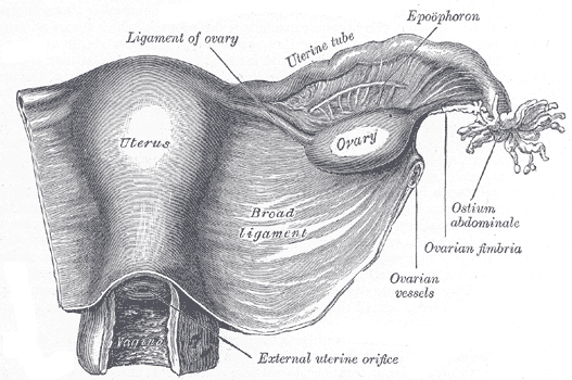
| Ligament | Connects | Contents / origin |
|---|---|---|
| Suspensory ligament (infundibulopelvic) | Ovary and tube to the pelvic wall; posterior extension of the broad ligament | Carries the ovarian artery, ovarian vein and the sympathetic and parasympathetic plexus |
| Proper ligament of the ovary | Ovary to the uterus | Remnant of the gubernaculum; contains no vessels |
Microscopically the ovary has an outer germinal (simple cuboidal) epithelium, beneath it the tunica albuginea (collagen), then the cortex with follicles at different stages, and the central medulla with loose connective tissue and the large vessels (the hilum). The ovary arises from the intermediate mesoderm; unlike the testis its descent stops in the pelvis, because the gubernaculum attaches to the uterus, and the remnant becomes the proper ligament of the ovary.
What is the blood supply of the uterus and ovary?
The uterine arteries, branches of the internal iliac artery, are the main supply of the uterus. Inside the myometrium they give arcuate arteries, which give radial arteries; at the endometrium the radial arteries divide into basal and spiral arteries, which sustain the endometrium in menstruation and the placental bed in pregnancy.
| Organ | Arterial supply | Venous drainage |
|---|---|---|
| Uterus | Uterine artery (main); ovarian artery anastomoses with it in the broad ligament | Uterine venous plexus |
| Ovary | Dual: ovarian artery (from the abdominal aorta just below the renal artery, about L2) and uterine artery | Right ovarian vein → inferior vena cava; left ovarian vein → left renal vein |
The ovarian artery runs in the suspensory ligament, enters the mesovarium and may anastomose with the uterine artery within the broad ligament. The ovarian vein also drains the parametrium, cervix, mesosalpinx and pampiniform plexus, and has anastomoses with the uterine, vesical, rectal and vulvar venous plexuses.
What is the lymphatic drainage of the uterus, cervix and ovary?
| Organ | Main drainage | Other routes |
|---|---|---|
| Ovary | Para-aortic nodes (around L2), along the ovarian vessels | Lateral vessels to hypogastric nodes; along the round ligament to external iliac and inguinal nodes |
| Fundus of uterus | Para-aortic nodes (same as ovary and tube) | Along the round ligament to the superficial inguinal nodes |
| Lower uterine segment | External and internal iliac nodes, along the uterine vessels | — |
| Cervix | External iliac and obturator nodes are the most common sentinel sites in mapping studies | Internal iliac, common iliac, presacral, parametrial and para-aortic nodes less often |
The ovarian pathways matter clinically because they are routes of spread of ovarian cancer: it can reach para-aortic nodes high in the abdomen and, via the round ligament, the groin. Lymphatic vessels and nodes also lie inside the cardinal ligament and can be involved in cervical cancer, even out to the pelvic sidewall, so radical surgery removes the cardinal ligament widely.
What is the nerve supply and referred pain of the uterus and cervix?
The internal pelvic organs receive both sympathetic and parasympathetic fibres. Sympathetic supply to the uterus is from T11 and T12 through the hypogastric plexus; parasympathetic fibres come from S2 to S4. Sensory signals from the uterus travel along the hypogastric nerves to T10 to L1, so uterine pain is referred to the lower back and lower abdomen. Sensation from the vagina reaches S2 to S4 through the pelvic nerves, giving gluteal and posterior leg pain. The cervix sends pain signals through both the hypogastric and pelvic nerves, and cervical dilation is a major source of labour pain.
| Organ | Sympathetic | Parasympathetic | Pain afferents |
|---|---|---|---|
| Uterus | T11–T12 (hypogastric plexus) | S2–S4 | T10–L1 via hypogastric nerves |
| Cervix | Hypogastric plexus | S2–S4 (pelvic nerves) | Hypogastric and pelvic nerves |
| Vagina | — | S2–S4 | S2–S4 via pelvic nerves |
What are the common exam traps?
- Cardinal ligament = Mackenrodt = transverse cervical ligament. It is the main lateral support and contains the uterine vessels.
- The ureter passes under the uterine artery near the cervix; the artery crosses the ureter superiorly.
- The proper ligament of the ovary has no vessels; the suspensory ligament carries the ovarian vessels.
- Ovarian artery comes from the aorta (about L2), not the internal iliac; the uterine artery is the internal iliac branch.
- Right ovarian vein → IVC; left ovarian vein → left renal vein.
- Ovary and fundus → para-aortic nodes; cervix and lower uterus → iliac/obturator nodes; round ligament → inguinal nodes.
- Cervix-to-body ratio is 2 : 1 before puberty, 1 : 2 in reproductive life and 2 : 1 after menopause.
Related pages: practise previous papers at NEET PG Anatomy PYQs and NEET PG OBG PYQs, and see the most repeated topics. Also revise the pouch of Douglas and genital prolapse (POP-Q).