How are ovarian tumours classified?
The WHO classification sorts ovarian neoplasms by the tissue they come from. The ovary has a surface (Müllerian-type) epithelium, germ cells, and the sex cords and stroma that surround each follicle; a fourth group is metastatic disease from other organs. Each group has a typical age, a typical hormone or marker, and a few eponymous histological features that examiners love.
| Group | Main members | Typical age | Marker / hormone clue |
|---|---|---|---|
| Surface epithelial | High-grade serous (commonest carcinoma), low-grade serous, mucinous, endometrioid, clear cell; benign, borderline and malignant forms | Older women; high-grade serous peaks at 60–65 years | CA-125, HE4 |
| Germ cell | Mature cystic teratoma (dermoid), immature teratoma, dysgerminoma, yolk sac tumour, embryonal carcinoma, choriocarcinoma | Children, adolescents and young women | AFP, hCG, LDH |
| Sex cord-stromal | Adult and juvenile granulosa cell tumour, Sertoli-Leydig cell tumour, fibroma, thecoma | Mostly first three decades; adult granulosa peaks at 50–55 | Inhibin; oestrogen or androgen excess |
| Metastatic | Krukenberg tumour (signet-ring adenocarcinoma, usually from the stomach) | Depends on primary | Often bilateral |
Which tumour marker goes with which ovarian tumour?
| Marker | Tumour(s) | Remember |
|---|---|---|
| CA-125 | Malignant surface epithelial tumours (also often raised in yolk sac tumour) | Made by Müllerian epithelium; raised in only about 50% of early-stage cancers |
| HE4 | Epithelial ovarian cancer | Specificity about 96%; more sensitive than CA-125 in early stage |
| AFP | Yolk sac tumour, immature teratoma, embryonal carcinoma; rarely Sertoli-Leydig | Think yolk sac = fetal protein |
| β-hCG | Non-gestational choriocarcinoma, embryonal carcinoma; rarely dysgerminoma | May cause abnormal uterine bleeding or precocious puberty |
| LDH | Dysgerminoma | Commonest malignant germ cell tumour |
| Inhibin | Granulosa cell tumour (juvenile and adult) | Also stains tumour cells on immunohistochemistry |
What are the features of surface epithelial ovarian tumours?
Epithelial ovarian cancer is the leading cause of death among gynaecological cancers in the United States. The five main histotypes are high-grade serous, low-grade serous, clear cell, endometrioid and mucinous carcinoma; transitional-cell carcinoma is now filed under high-grade serous. High-grade serous carcinoma is the commonest; it carries TP53 mutations, shows psammoma bodies, and much of it is now thought to start in the fallopian tube (serous tubal intraepithelial carcinoma).
- Risk factors — older age, early menarche, late menopause, nulliparity, family history, obesity, endometriosis, smoking, HRT, perineal talc.
- Hereditary syndromes — BRCA1/BRCA2, Lynch syndrome (mismatch-repair genes MLH1, MSH2, MSH6, PMS2), Li-Fraumeni (TP53), Peutz-Jeghers (STK11).
- Protective factors — anything that reduces ovulation: oral contraceptives, multiparity, breastfeeding, and bilateral tubal ligation or salpingectomy.
- Spread — mainly across the peritoneum (omentum, visceral surfaces); lymphatic spread goes first to para-aortic and paracaval nodes.
- Prognosis — about 90% of early-stage disease is cured, but most patients present at an advanced stage; stage I recurs in under 10%, stage IV in about 90%.
What are the high-yield facts about ovarian germ cell tumours?
Germ cell tumours mostly affect young women and girls. Non-epithelial tumours make up about 10% of ovarian cancers, and malignant germ cell tumours about 5%. Because the patients are young, fertility-sparing surgery is the cornerstone for stage I disease, and these tumours are highly sensitive to platinum chemotherapy — the standard regimen is BEP (bleomycin, etoposide, cisplatin).
| Tumour | Key features | Marker |
|---|---|---|
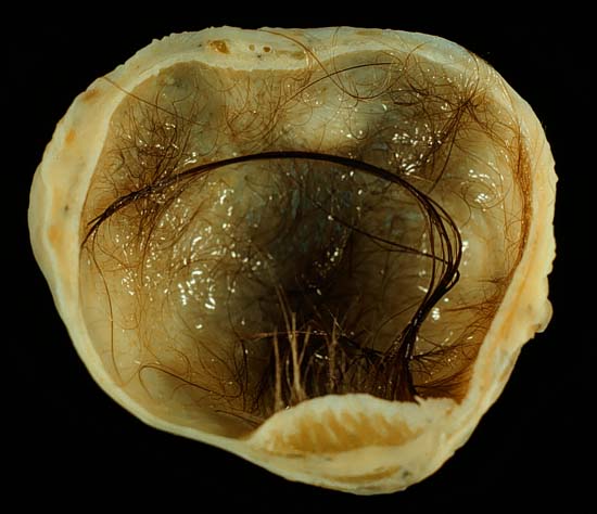
| Mature cystic teratoma (dermoid) | Commonest ovarian neoplasm; women aged 20–40; hair, sebum, teeth; Rokitansky nodule on ultrasound; torsion is a common complication; malignant change in 1–2%, most often squamous cell carcinoma | None |
| Monodermal teratoma | Struma ovarii (thyroid tissue), carcinoid, neural tumours | — |
| Immature teratoma | Contains immature (neural) tissue; malignant | AFP may rise |
| Dysgerminoma | Commonest malignant germ cell tumour; 10–15% bilateral; linked with gonadal dysgenesis | LDH |
| Yolk sac (endodermal sinus) tumour | Schiller-Duval bodies (glomerulus-like structure with a central vessel); worst prognosis of the pure types | AFP (CA-125 often raised) |
| Embryonal carcinoma | Often hormonally active | hCG, AFP |
| Choriocarcinoma (non-gestational) | Trophoblastic differentiation | hCG |

Which sex cord-stromal tumours produce hormones?
Sex cord-stromal tumours differ from the other groups because they often announce themselves with hormones — precocious puberty, menstrual change, hirsutism or virilisation. Most present in the first three decades, except the adult granulosa cell tumour, which peaks at 50–55 years.
| Tumour | Hormone effect | Hallmarks |
|---|---|---|
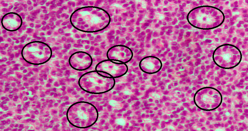
| Adult granulosa cell tumour | Oestrogen — postmenopausal bleeding; endometrial hyperplasia in 25–50% | Call-Exner bodies, grooved 'coffee-bean' nuclei, inhibin positive; FOXL2 mutation in over 90% |
| Juvenile granulosa cell tumour | Oestrogen — precocious puberty in girls | About two-thirds of childhood sex cord-stromal tumours; usually confined to an intact capsule |
| Sertoli-Leydig cell tumour | Androgen — hirsutism, virilisation | DICER1 mutations; about 20% of childhood sex cord-stromal tumours |
| Fibroma | Usually none | Benign; fibroma + ascites + pleural effusion = Meigs syndrome |
What is a Krukenberg tumour?
A Krukenberg tumour is a metastatic signet-ring cell adenocarcinoma of the ovary. The primary is in the stomach in about 70% of cases (most often a pyloric adenocarcinoma); gastric and colorectal cancers together account for almost 90%. Breast, appendix, biliary tract and pancreas are other sources. Bilateral involvement is common, and immunohistochemistry helps separate it from a primary ovarian cancer.

How do you assess whether an adnexal mass is malignant?
| Feature | Suggests malignant | Suggests benign |
|---|---|---|
| Consistency | Solid or with solid components | Purely cystic |
| Size | Large (≥ 8–10 cm) | Smaller |
| Composition | Heterogeneous | Homogeneous |
| Papillary projections | More likely | Less likely |
| Torsion | Less likely | More likely |
RMI = U × M × serum CA-125 (U/mL)
U = ultrasound score from features such as multilocularity, solid areas, bilaterality, ascites and metastases. M = menopausal score: 3 if postmenopausal (more than a year of amenorrhoea, or over 50 after hysterectomy), 1 if premenopausal.
The Risk of Malignancy Index (RMI) combines the three most useful pieces of information — imaging, menopausal status and CA-125 — into one number used to decide which women need referral to a gynaecological oncologist. A higher RMI means a higher chance of cancer; the exact cut-off varies between versions and studies.
How are ovarian tumours managed in outline?
- Epithelial ovarian cancer is staged surgically using the FIGO and TNM systems, and primary debulking (cytoreductive) surgery aims to remove all visible disease.
- When advanced disease is unlikely to be completely resected, neoadjuvant chemotherapy is given first and surgical cytoreduction follows.
- Young women with stage I, low-grade epithelial cancer who want children may have fertility-sparing surgery with surgical staging and preservation of the uterus.
- Malignant germ cell tumours: fertility-sparing surgery for stage I, often followed by surveillance; BEP chemotherapy for higher stages and most non-dysgerminomatous types. Survival is excellent — about 98% at 5 years for dysgerminoma in one Italian series, but lower for yolk sac tumour (about 70%).
- Mature cystic teratoma is removed surgically because of the risk of torsion and the small risk of malignant change.
Which ovarian tumour facts are asked most in NEET PG and INI-CET?
- Commonest ovarian neoplasm → mature cystic teratoma; commonest malignant germ cell tumour → dysgerminoma; commonest carcinoma → high-grade serous.
- Schiller-Duval bodies → yolk sac tumour (AFP). Call-Exner bodies and coffee-bean nuclei → granulosa cell tumour (inhibin). Psammoma bodies → serous carcinoma. Signet-ring cells → Krukenberg.
- Postmenopausal bleeding with an ovarian mass and endometrial hyperplasia → oestrogen-secreting granulosa cell tumour.
- Virilisation with an ovarian mass → Sertoli-Leydig cell tumour.
- Ovarian mass + ascites + pleural effusion that clear after surgery → Meigs syndrome (fibroma).
- Thyroid tissue in a teratoma → struma ovarii; malignant change in a dermoid → usually squamous cell carcinoma.
- Malignant germ cell tumour chemotherapy → BEP; fertility-sparing surgery for stage I.