What is the third stage of labour?
Labour has three stages. The third stage begins immediately after the baby is born and ends with the expulsion of the placenta and membranes (NICE NG235). Its goals are a safe, complete placental delivery, prevention of postpartum haemorrhage and sensible management of the cord.
StatPearls notes that the placenta is usually expelled spontaneously within 5 to 30 minutes. A third stage longer than 30 minutes raises the risk of postpartum haemorrhage and may call for manual removal or other interventions.
How does the placenta separate from the uterine wall?
After the baby is delivered the uterus shrinks sharply. The placental site does not shrink with it, so the placenta is sheared off the shrinking wall through the decidua basalis, and a retroplacental haematoma collects behind it. The membranes, thrown into folds as the cavity shrinks, are peeled off afterwards (Williams Obstetrics).
Bleeding from the placental bed is controlled mainly by myometrial contraction. The muscle compresses the avulsed spiral arteries at the implantation site, and clotting then obliterates the vessel lumens. Williams Obstetrics stresses that fatal haemorrhage from the implantation site is unlikely if the myometrium contracts vigorously — which is why a firm, contracted uterus is the centre of third-stage care and atony is the leading cause of PPH.
What is the difference between Schultze and Duncan mechanisms?
| Feature | Schultze (central) | Matthews Duncan (marginal) |
|---|---|---|
| Where separation starts | At the centre of the placenta | At the periphery (margin) |
| Retroplacental blood | Collects in the inverted membrane sac behind the placenta | Collects between the membranes and uterine wall and escapes from the vagina |
| Surface seen first at the introitus | Fetal (shiny) surface, like an inverted umbrella | Maternal (rough) surface, placenta comes edgeways |
| Visible bleeding | Concealed until the placenta is out | Revealed throughout separation |
| How usual | The usual pattern | Less common |
The NCBI MedGen definitions match this: the Schultze mechanism is separation that begins at the placental centre; the Duncan mechanism is expulsion with the maternal rough side first 'rather than the usual fetal side'.

What are the signs of placental separation?
StatPearls lists three cardinal signs of separation from the uterine wall. Williams Obstetrics adds a fourth — the uterus rises — because the separated placenta passes into the lower segment and vagina and its bulk pushes the uterine body up.
- A sudden gush of blood from the vagina.
- Lengthening of the umbilical cord outside the vulva as the placenta descends (a clamp on the cord moves away from the introitus).
- The fundus becomes globular and firmer on palpation (it was discoid before separation).
- The uterus rises in the abdomen.
What does active management of the third stage include?
Active management of the third stage of labour (AMTSL) reduces PPH. The components and their current status under the WHO 2012 PPH recommendations, with the WHO 2014 cord-clamping guideline and NICE NG235:
| Component | Current recommendation | Key details |
|---|---|---|
| Uterotonic for every birth | Core component (strong) | Oxytocin 10 IU IM or IV is the drug of choice (WHO). NICE: 10 units IM after vaginal birth |
| Alternatives if no oxytocin | Recommended where oxytocin is unavailable | Ergometrine / methylergometrine, fixed oxytocin–ergometrine, or misoprostol |
| Delayed cord clamping | Recommended | Not earlier than 1 minute (WHO 2014); WHO 2012 said about 1–3 minutes. Early clamping generally contraindicated |
| Controlled cord traction | Optional with skilled attendants | Contraindicated where unskilled attendants conduct births; after signs of separation |
| Uterine massage | Not routine after oxytocin | Continuous massage not recommended to prevent PPH if oxytocin given; check tone instead |
StatPearls adds a practical timing rule: oxytocin can be given from delivery of the anterior shoulder onwards, but not before, as it could worsen an unrecognised shoulder dystocia. NICE gives it immediately after the birth and before the cord is clamped and cut.
Physiological (expectant) management — the alternative NICE describes — uses no routine uterotonic, does not clamp the cord until pulsation stops, and lets the placenta deliver by maternal effort. NICE advises women that active management carries a lower risk of PPH and blood transfusion.
How is controlled cord traction (Brandt–Andrews) done?
Controlled cord traction (CCT) is often called the Brandt–Andrews manoeuvre. It is done only after the uterotonic has been given and signs of separation are present, and NICE limits it to active management.
- Give the uterotonic first and confirm the signs of separation — traction comes only after both (NICE).
- Make sure the uterus is firmly contracted; the other hand on the lower abdomen steadies the uterus so it is not dragged down.
- Apply gentle, sustained traction on the cord — never a jerk; stop if the placenta does not descend.
- Deliver the placenta carefully so the membranes are not torn off and left behind, then check uterine tone and inspect the placenta for completeness.

When is the placenta called retained and how is it managed?
NICE diagnoses a prolonged third stage if it is not completed within 30 minutes of the birth with active management, or within 60 minutes with physiological management. WHO 2012 likewise acts after 30 minutes in an actively managed third stage.
| Step | WHO 2012 | NICE NG235 |
|---|---|---|
| First step | Additional oxytocin 10 IU IM/IV with controlled cord traction | Secure IV access; explain why |
| Umbilical vein injection | Insufficient evidence to recommend | Do not use umbilical vein agents |
| IV oxytocics | — | Not routinely; give if the woman is bleeding excessively |
| Bleeding with retained placenta | Expedite manual removal | Arrange transfer to an obstetric unit |
| Antibiotics | Single prophylactic dose for manual removal | — |
Williams Obstetrics groups causes into three: placenta adherens (contractions too weak to detach it), a detached but trapped placenta behind a constricted lower segment, and a morbidly adherent placenta. Recurring risks include stillbirth, previous caesarean, previous retention and preterm delivery.
What is the placenta accreta spectrum?
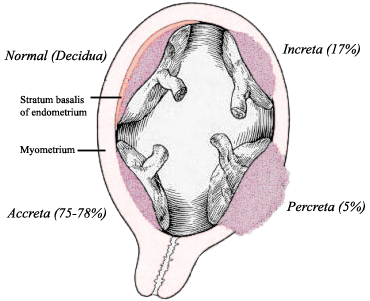
In placenta accreta the placenta is abnormally adherent to the myometrium because the decidua basalis is partly or completely absent and the fibrinoid (Nitabuch) layer is incompletely developed (StatPearls). The plane of separation simply does not exist, so the placenta cannot shear off.
| Type | Depth of villous invasion | Share of cases (StatPearls) |
|---|---|---|
| Accreta (vera) | Villi attach to the myometrium without invading it | Most cases |
| Increta | Villi penetrate into the myometrium, not the serosa | About 15–17% |
| Percreta | Villi penetrate through to the serosa and may invade bladder or rectum | About 5–7% |
- Biggest risk: a previous caesarean section plus a current placenta praevia. Risk climbs with each caesarean — StatPearls quotes about 3% with one, 11% with two and 40% with three previous caesareans when praevia is present.
- Ultrasound clues: loss of the retroplacental clear zone, vascular lacunae, myometrial thinning and interruption of the bladder line.
- Management: planned delivery; hysterectomy has been the traditional treatment, though uterus-conserving approaches are now used. Attempting to pull the placenta off causes torrential bleeding.
How is placental separation asked in NEET PG and INI-CET?
- Mechanism identification: 'maternal surface delivered first, blood trickles out' → Matthews Duncan; 'shiny fetal surface first, bleeding concealed' → Schultze.
- Signs of separation: gush of blood, cord lengthening, globular firm fundus, uterus rising. An option such as 'uterus becomes flabby' is the trap.
- Components of AMTSL: uterotonic, delayed cord clamping, controlled cord traction — and which is the most important (uterotonic).
- Drug and dose: oxytocin 10 IU IM (or IV) — the uterotonic of choice.
- Time limits: retained placenta after 30 minutes (active) or 60 minutes (physiological, NICE).
- Complications: uterine inversion from traction before separation; accreta spectrum with previous caesarean plus praevia.