What is the anatomy of the prostate?
The prostate is a dense fibromuscular gland shaped like an inverted cone: the base lies against the neck of the urinary bladder and the apex rests on the external urethral sphincter. It sits in the lesser pelvis, wrapping around the proximal urethra, and is enclosed in a fibrous capsule with its nerves and vascular plexus, which is in turn surrounded by a visceral layer of pelvic fascia.
| Direction | Relation |
|---|---|
| Anterior | Pubic symphysis, separated by retropubic fat and the prostatic venous plexus |
| Posterior | Rectum, separated by Denonvilliers' fascia |
| Inferior | External urethral sphincter |
| Lateral | Levator ani, covered by endopelvic fascia |
The gland is also traversed by the proximal urethra and the two ejaculatory ducts, which enter as they emerge from the seminal vesicles and converge on the urethra at the seminal colliculus (verumontanum). Embryologically the prostate develops as epithelial buds growing from the wall of the urogenital sinus; the buds branch, canalise and form ducts and acini, while the surrounding mesenchyme forms the stroma and smooth muscle. See development of the genitourinary system.
What are the zones and lobes of the prostate?
Many anatomy textbooks describe five lobes — anterior, posterior, two lateral and a median lobe — while clinicians speak of two lateral lobes and a median lobe. The histologically based zonal description is the one that matters for pathology.
| Zone | Position and size | Clinical importance |
|---|---|---|
| Peripheral zone | The largest, about 70% of the gland; surrounds most of the central zone and part of the distal prostatic urethra | Site of most carcinomas (about 75%); the part palpable on DRE |
| Central zone | Forms the base of the gland, around the ejaculatory ducts | — |
| Transition zone | Small glandular zone around the urethra between the bladder and the verumontanum | Site of BPH |
| Anterior fibromuscular stroma | Non-glandular muscle and fibrous tissue around the apex | Contains no glands |

What is the blood supply, drainage and nerve supply of the prostate?
- Arteries: the inferior vesical artery is the major supply, with branches from the middle rectal and internal pudendal arteries.
- Veins: the prostatic venous plexus drains to the internal iliac veins.
- Lymphatics: internal iliac and sacral nodes.
- Nerves: autonomic fibres from the inferior hypogastric plexus — sympathetic input via the hypogastric nerve and parasympathetic input via the pelvic nerve; both plexuses carry sensory fibres.
What is benign prostatic hyperplasia and how does it arise?
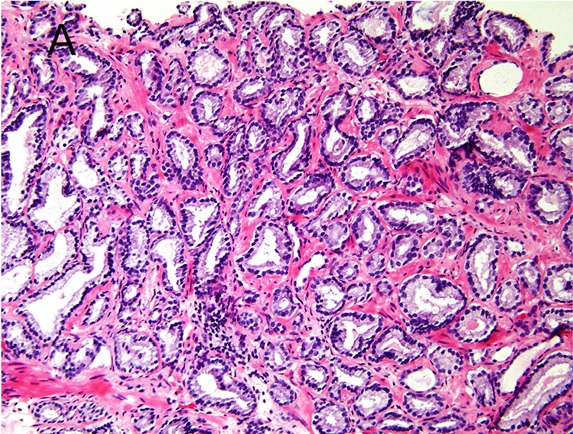
BPH is non-malignant proliferation of prostatic tissue and a common cause of lower urinary tract symptoms (LUTS) in older men. Histologically it is a hyperplastic process with increased numbers of both glandular and stromal cells in the periurethral and transition zones: the periurethral zone shows hyperplastic stromal nodules and the transition zone shows glandular nodular proliferation. The enlargement is nodular, not diffuse.

- Mechanism: a loss of balance between cell proliferation and apoptosis favours proliferation.
- Androgens: testicular androgens are required. DHT drives growth, and testosterone is converted to DHT by 5-alpha-reductase type 2 in stromal cells, which accounts for about 90% of total intraprostatic androgens.
- Static component: increased gland volume. Dynamic component: prostatic smooth muscle tone — the reason alpha-blockers relax the gland and 5-alpha-reductase inhibitors shrink it.
- Risk factors: age (about half of men over 50 show evidence of BPH), metabolic syndrome and obesity (higher prostate volumes), diabetes and insulin use, and chronic inflammation.
- BPH is not a risk factor for prostate cancer, although it can raise PSA.
How does BPH present and how is it evaluated?
LUTS divide into storage symptoms (frequency, nocturia, urgency) and voiding symptoms (weak or intermittent stream, hesitancy, straining, prolonged micturition, incomplete emptying). BPH accounts for more than two-thirds of cases of acute urinary retention. Diuretics for hypertension or heart failure can worsen frequency and nocturia.
| Test | Purpose |
|---|---|
| IPSS / AUA symptom score | Grades LUTS: 0 to 9 mild, 10 to 19 moderate, 20 to 35 severe |
| Digital rectal examination | Size and consistency; a hard nodule or asymmetry raises concern for cancer |
| Urinalysis | Infection, haematuria, glycosuria |
| Post-void residual and flow rate | Bladder emptying and obstruction |
| PSA | Used where cancer is suspected; roughly tracks volume (a benign 35 cc prostate gives about 1.5 ng/mL) |
| Voiding diary | Separates nocturnal polyuria from small frequent voids |
How is BPH treated — alpha-blockers, 5-ARIs and surgery?
| Drug class | Examples | Mechanism and key points |
|---|---|---|
| Alpha-1 blockers | Tamsulosin, alfuzosin | Relax stromal and bladder-neck smooth muscle (the dynamic component) and improve flow and symptoms. Side effects: ejaculatory problems, intraoperative floppy iris syndrome — warn the ophthalmologist before cataract or glaucoma surgery |
| 5-alpha-reductase inhibitors | Finasteride, dutasteride | Block conversion of testosterone to DHT; reduce volume by up to 25%, with maximal effect at about 6 months; benefit mainly in prostates larger than about 30 g; lower PSA by about 50% |
| PDE-5 inhibitor | Tadalafil | Roughly equivalent to tamsulosin 0.4 mg for symptoms; useful when erectile dysfunction coexists; no floppy iris risk |
Severe BPH that has failed medical therapy, or recurrent retention, is treated surgically, classically by transurethral resection of the prostate (TURP): an instrument is passed up the urethra and trims tissue to enlarge the channel. Starting a 5-ARI 2 to 4 weeks before surgery reduces bleeding. See adrenergic drugs for the receptor pharmacology.
What is prostate carcinoma and how does it differ from BPH?
Prostate cancer is usually an adenocarcinoma, arising in glandular tissue, most often in the peripheral zone. Most tumours grow slowly, are low-grade and cause no early symptoms; late features include bone pain, anaemia, spinal cord compression and renal failure from bilateral ureteric obstruction. Metastatic disease often causes severe bone pain in the vertebrae, pelvis, hips and ribs. Risk factors include family history, higher BMI, smoking, BRCA2 mutation (BRCA1 to a lesser degree) and over 100 other variants.
| Feature | BPH | Carcinoma |
|---|---|---|
| Zone | Transition and periurethral | Peripheral (about 75%) |
| Pattern | Nodular hyperplasia of glands and stroma | Adenocarcinoma, graded by Gleason |
| Presentation | LUTS, retention | Usually silent; bone pain, cord compression if advanced |
| DRE | Smooth, enlarged | Hard nodule, asymmetry |
| PSA | May rise modestly | Higher; above 20 ng/mL has a positive predictive value of 65% for metastatic and skeletal disease |
| Malignant potential | None; not a cancer risk factor | Malignant |
Up to 14% of patients have metastatic disease at diagnosis. Prostatic MRI with PI-RADS scoring and MRI-guided biopsy are now standard, and PSMA PET is emerging as the leading imaging modality for staging because PSMA is over-expressed in 90% to 100% of prostate cancer cells. F-18 sodium fluoride PET is a sensitive tracer for skeletal metastases. See renal tumours and neoplasia nomenclature for related pathology.
What are the Gleason score and grade groups?
The Gleason system grades prostate cancer by the architecture of the glands, not by individual cell features. Patterns are graded 1 to 5. The score is the sum of the most common (primary) pattern and the second most common pattern, so a tumour is reported as, for example, 3+4 = 7. If only one pattern is seen it is doubled (3+3 = 6, the commonest score). The primary pattern comes first: 3+4 and 4+3 are different.
| Grade group | Gleason score | Histology | Category |
|---|---|---|---|
| 1 | 6 or less (3+3) | Only discrete, well-formed glands | Low grade |
| 2 | 3+4 = 7 | Mostly well-formed glands with fewer poorly formed, fused or cribriform glands | Intermediate |
| 3 | 4+3 = 7 | Mostly poorly formed, fused or cribriform glands with fewer well-formed glands | High |
| 4 | 8 | Only poorly formed, fused or cribriform glands (or other combinations lacking glands) | High |
| 5 | 9 or 10 | Lack of gland formation, or necrosis | High |
What is PSA and how is it interpreted?
Prostate-specific antigen is a serine protease produced by the columnar epithelium of prostatic tissue. Its physiological job is to break down semenogelin and fibronectin, which reduces seminal viscosity (liquefies the ejaculate) and so assists sperm migration. PSA is organ-specific, not cancer-specific: BPH, prostatitis and other causes also raise it.
| Measure | Interpretation |
|---|---|
| Total PSA | A value of 4.0 ng/mL or higher is the consensus standard for further evaluation; at that cut-off sensitivity for prostate cancer is about 91%. No value guarantees cancer is absent |
| Age-specific range | PSA rises with age (about 3.2% a year in healthy 60-year-olds), so a single cut-off is imperfect |
| Free PSA % | Used when total PSA is 4 to 10 ng/mL; a low ratio raises cancer risk (below 10% carries about 50% risk; above 25% below 10%) |
| PSA density | Total PSA divided by prostate volume; 0.15 or higher is a higher-risk marker on active surveillance |
| PSA velocity | A rise of no more than about 0.75 ng/mL a year is acceptable; faster is suspicious |
| After radical prostatectomy | All prostatic tissue should be gone, so any detectable PSA suggests residual (presumed malignant) tissue |
Screening is controversial because of overdiagnosis and overtreatment, but most professional bodies endorse informed PSA screening; see screening tests for how sensitivity and specificity apply.