What is rhinitis and why are allergic and atrophic types exam favourites?
Rhinitis is inflammation of the nasal mucosa. In ENT papers two opposite pictures are paired again and again: allergic rhinitis, in which an over-reactive, swollen, watery mucosa blocks the nose, and atrophic rhinitis, in which a wasted, dry, crusted mucosa leaves the nose too wide. Examiners love the contrast because the same symptom word, 'nasal obstruction', has opposite explanations in the two diseases.
| Feature | Allergic rhinitis | Atrophic rhinitis (ozaena) |
|---|---|---|
| Basic process | IgE-mediated type 1 hypersensitivity, Th2 inflammation | Chronic progressive atrophy of mucosa and underlying turbinate bone |
| Nasal cavity | Narrowed by swollen, pale-bluish turbinates | Roomy, wide cavity with thin shrunken turbinates |
| Secretion | Thin, clear, watery | Thick, dry, greenish crusts with foul smell (fetor) |
| Smell sense | Usually preserved | Patient is often anosmic and so unaware of own smell |
| Typical patient | Atopic, family history, asthma or eczema | Young adult, female preponderance, poor hygiene and low socio-economic status |
| First-line treatment | Allergen avoidance + intranasal corticosteroid | Crust removal, moisturising, treat infection; surgery (Young's operation) in refractory cases |
How does allergic rhinitis develop? (early and late phase)
StatPearls defines allergic rhinitis (AR) as an atopic disease with nasal congestion, clear rhinorrhoea, sneezing, postnasal drip and nasal itching. It affects roughly one in six people, and under the 'unified airway' concept it is part of a systemic allergic response that also includes asthma and atopic dermatitis.
- Sensitisation — inhaled allergen (pollen, dust mite, animal dander, mould) drives a Th2 response and allergen-specific IgE that coats mast cells and basophils.
- Early phase (5–15 minutes) — re-exposure cross-links IgE and the mast cell degranulates: histamine (the key mediator), leukotrienes and prostaglandins are released. Histamine causes sneezing (via the trigeminal nerve) and rhinorrhoea (stimulating mucous glands); leukotrienes and prostaglandins act on vessels to cause congestion.
- Late phase (4–6 hours) — mast-cell cytokines IL-4 and IL-13 recruit eosinophils, T lymphocytes and basophils to the mucosa, causing oedema and sustained blockage.
- Priming and hyper-reactivity — with repeated inflammation the mucosa responds to non-specific triggers such as tobacco smoke and cold air.

Allergic rhinitis is classically seasonal (intermittent) or perennial (persistent); StatPearls quotes about 20 per cent seasonal, 40 per cent perennial and 40 per cent mixed. Seasonal disease is commoner in children and perennial in adults. Risk factors include a family history of atopy, male sex and raised allergen-specific IgE; twin concordance is about 45–60 per cent in monozygotic and about 25 per cent in dizygotic twins.
What are the clinical signs and how is allergic rhinitis diagnosed?
The history carries most of the diagnosis: paroxysmal sneezing, watery discharge, itching of nose, eyes or palate, blocked nose and postnasal drip, often with a family history of atopy or personal asthma. Intermittent disease gives sneezing, rhinorrhoea and watery eyes; chronic disease gives mainly blockage and postnasal drip.
- Allergic shiners — dark circles under the eyes from venous congestion.
- Transverse supratip nasal crease ('allergic salute' line), more often seen in children, from repeated upward rubbing of the nose.
- Mouth breathing, frequent sniffing and throat clearing.
- On anterior rhinoscopy — pale or bluish, boggy inferior turbinates, thin clear secretions and sometimes cobblestoning of the mucosa.
- Eustachian tube dysfunction on pneumatic otoscopy, and signs of coexisting asthma or dermatitis.
Diagnosis is clinical. A good response to a trial of nasal steroid supports it. A formal diagnosis uses allergen-specific serum IgE or skin-prick testing; guidelines reserve testing for patients who do not respond to empirical treatment or in whom a specific allergen must be identified. Skin testing is slightly more sensitive than serum testing, but is contraindicated in uncontrolled severe asthma, unstable cardiovascular disease, pregnancy and concurrent beta-blocker therapy, and antihistamines, H2 blockers, tricyclics and omalizumab interfere with it. Imaging is not routine and is used mainly to exclude sinusitis.
What is the treatment ladder for allergic rhinitis?
- Allergen avoidance — dust-mite covers, hot-water washing of bedding, HEPA vacuum cleaners; pet isolation helps but dander can persist for weeks after the animal is removed.
- Intranasal corticosteroids (beclometasone, budesonide, fluticasone, mometasone, triamcinolone) — first-line, superior to antihistamines for reducing inflammation. Peak effect takes days, so use is regular; aim the spray laterally, away from the septum, to limit irritation and epistaxis.
- Second-generation oral antihistamines (loratadine, desloratadine, cetirizine, fexofenadine) — effective for sneezing, itching and rhinorrhoea with less sedation than first-generation drugs (chlorpheniramine, diphenhydramine, hydroxyzine). Intranasal antihistamines can be combined with a nasal steroid.
- Leukotriene receptor antagonists (montelukast, zafirlukast) — less effective than nasal steroids; used as add-on, especially with coexisting asthma.
- Sodium cromoglycate — mast-cell stabiliser, reasonable but weaker option.
- Allergen immunotherapy — for patients not controlled by avoidance and drugs; maintenance for about 3–5 years gives a prolonged protective effect.
- Surgery — for nasal polyps, inferior turbinate hypertrophy with intractable blockage, or deviated septum; not for the allergy itself.
What is atrophic rhinitis (ozaena) and what causes it?
Primary atrophic rhinitis is a chronic nasal disease with progressive atrophy of the mucosa and the underlying bone of the turbinates, producing a wide, roomy nasal cavity filled with thick greenish crusts and a characteristic foul odour (ozaena). It is described as common in tropical countries, in low socio-economic groups with poor hygiene, and as having a female preponderance; one Indian study of young and middle-aged adults reports about 1 per cent prevalence.
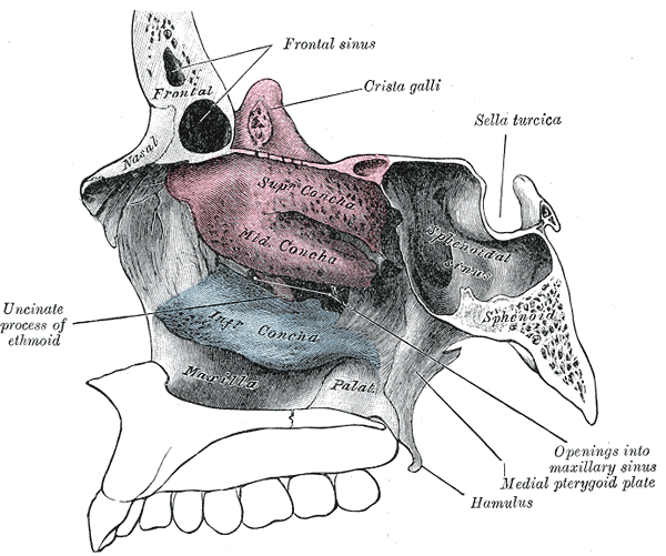
The cause is still debated. Published reviews list heredity (polygenic inheritance in 15–30 per cent), chronic infection, nutritional deficiency (iron, fat-soluble vitamins, protein), autonomic dysfunction, oestrogen imbalance (disease peaks near puberty and menopause), immune dysfunction, biofilms and a developmental tendency to poorly pneumatised maxillary sinuses and congenitally spacious nasal cavities. None has been proven to be a direct cause.
Histologically the disease shows loss of the mucus blanket and cilia, squamous metaplasia of the respiratory epithelium and, in the commoner type (about 50–80 per cent), endarteritis obliterans with periarteritis and fibrosis; a second type (about 20–50 per cent) shows capillary vasodilatation. Secondary atrophic rhinitis follows surgery (for example excessive turbinectomy), trauma, infection, radiation or immune disease, and is diagnosed by excluding those causes.
What are the clinical features and triad of atrophic rhinitis?
Atrophic rhinitis is diagnosed on the triad of fetor, greenish crusts and a roomy nasal cavity. Patients may complain of paradoxical nasal obstruction (they feel blocked despite a wide cavity, because the airflow sensation is lost and crusts fill the space), crusting, epistaxis when crusts are removed, headache and anosmia.
- Fetor — foul smell noticed by others; the patient is often anosmic ('merciful anosmia') and unaware of it, though social ostracisation is common.
- Greenish crusts adherent to atrophic mucosa; removal causes bleeding.
- Roomy nasal cavity on rhinoscopy or endoscopy: shrunken turbinates, visible posterior pharyngeal wall and sometimes an anterior septal perforation.
- Bone remodelling on sinus CT: widened cavities, thin turbinates and sinuses that are often small or poorly pneumatised.
- Nasal swab for culture (often Klebsiella ozaenae) and a mucosal biopsy in selected cases.
| Point | Primary | Secondary |
|---|---|---|
| Cause | Idiopathic or constitutional, chronic infection, nutritional and hormonal factors | Surgery (over-resection of turbinates), trauma, infection, radiation, immune disease |
| Course | Slow, progressive atrophy | Follows a defined insult; empty-nose syndrome is a related picture after turbinectomy |
| Diagnosis | Clinical triad after exclusion of secondary causes | History of the precipitating event |
How is atrophic rhinitis treated, and what is Young's operation?
No single regimen has been proven ideal, because the cause is multifactorial. Published reviews describe a combination approach: control dryness and crusting with saline irrigation and lubricants (reducing evaporation from the mucosa), culture-directed antibiotics (with limited success), correction of nutritional deficiency (multivitamin, iron) and treatment of any underlying infection. Newer experimental approaches include nasal sprays of manuka honey to restore the mucosal microbiome, with endoscopic and symptom improvement in a small AIIMS Bhubaneswar trial.
- Medical (first): regular nasal douching to clear crusts, moisturising agents, targeted antibiotics, vitamin and iron correction.
- Surgical — narrowing the nasal cavity: the aim is to reduce airflow, drying and crust formation.
- Young's operation — complete closure of the nostril(s) (plastic closure of the nasal vestibule) for several months, after which it is reopened; the idea is that the mucosa recovers once it is no longer exposed to airflow. Used in severe, refractory disease.
- Modified Young's operation — closure leaving a 1–2 mm residual aperture, so some odorants still reach the olfactory region; the Indian series that used it re-opened the nostril after about 8 months.
- Submucosal implants to narrow the cavity — for example bioactive glass inserted through a sublabial approach, reported to reduce crusting and heal the mucosa in early case reports.
Which other rhinitis types can be confused with these two?
| Type | Key point |
|---|---|
| Rhinitis medicamentosa | Rebound congestion after more than about a week of topical decongestants; stop the drug and use nasal steroid |
| Vasomotor (non-allergic) rhinitis | Autonomic imbalance; nasal blockage and watery discharge triggered by temperature, humidity or odours with negative allergy tests |
| Infective (viral) rhinitis | Common cold; self-limiting, purulent phase suggests bacterial superinfection |
| Rhinitis sicca | Dry crusts at the anterior septum in dry climates, without bone atrophy or ozaena |
| Specific granulomatous rhinitis | Lupus, leprosy, syphilis, rhinosporidiosis and granulomatosis with polyangiitis — crusting, destruction or septal perforation with known aetiologies |
If a patient has both a runny nose and sinus symptoms, remember that allergic rhinitis predisposes to sinusitis and nasal polyps. Review sinusitis and nasal polyps for the follow-on questions, and epistaxis and Little's area for bleeding after crust removal.
What should you memorise for the exam?
- Allergic rhinitis: type 1 hypersensitivity, IgE, mast cells, histamine; early phase 5–15 minutes, late phase 4–6 hours with IL-4 and IL-13 and eosinophils.
- Signs: allergic shiners, transverse nasal crease, pale bluish boggy turbinates, thin watery discharge; associated with asthma and atopic dermatitis.
- First-line drug: intranasal corticosteroid; decongestants not beyond one week; immunotherapy lasts 3–5 years.
- Atrophic rhinitis: triad of fetor, greenish crusts, roomy nose; female preponderance; low socio-economic status; Klebsiella ozaenae; anosmia.
- Treatment: irrigation, lubricants, antibiotics, vitamins; Young's operation (closure) or modified Young's (1–2 mm aperture) when refractory.