Where do the paranasal sinuses drain and what is the osteomeatal complex?
Adults have four paired paranasal sinuses — maxillary, ethmoid, frontal and sphenoid — lined by pseudostratified columnar epithelium. Only the ethmoid and maxillary sinuses are present at birth; the frontal sinuses appear at about 5–6 years and reach full size after puberty, and the sphenoid begins to pneumatise around 5 years.
| Site | What drains there |
|---|---|
| Superior meatus / sphenoethmoidal recess | Sphenoid sinus and posterior ethmoid cells |
| Middle meatus | Frontal, anterior ethmoid and maxillary sinuses |
| Inferior meatus | Nasolacrimal duct |
The osteomeatal complex (OMC) is not a single structure but the region of the lateral nasal wall, lateral to the middle turbinate, where the anterior ethmoid, maxillary and frontal sinuses drain. It includes the anterior ethmoid cells, maxillary ostium, ethmoid infundibulum, frontal recess, middle meatus, hiatus semilunaris, bulla ethmoidalis and uncinate process. Obstruction here is the common pathway for most sinusitis — the target of functional endoscopic sinus surgery.

How is sinusitis classified by duration?
| Type | Duration |
|---|---|
| Acute | Less than 4 weeks |
| Subacute | 4–12 weeks |
| Chronic | More than 12 weeks |
| Recurrent acute | Four episodes, each under 4 weeks, with complete resolution between them |
Because the nasal and sinus linings are continuous, the term rhinosinusitis is often preferred. Sinusitis is thought to start with obstruction of drainage, mucosal swelling and impaired mucociliary clearance: rhinitis, a deviated septum, polyps, enlarged turbinates, foreign bodies or tumours can all trigger it.
When is acute sinusitis bacterial?
Viruses cause most acute rhinosinusitis (rhinovirus, influenza, parainfluenza, adenovirus, coronavirus, RSV). The three cardinal symptoms are purulent nasal discharge together with nasal obstruction or facial pain, pressure or fullness. An isolated headache is not typical, except in sphenoid sinusitis.
- Suspect acute bacterial rhinosinusitis when key symptoms last more than 10 days, or worsen after initial improvement — 'double worsening'.
- Organisms: Streptococcus pneumoniae, Haemophilus influenzae, Moraxella catarrhalis; less often S. pyogenes, S. aureus and anaerobes (dental source). Nosocomial cases involve Pseudomonas and other Gram-negatives.
- Examination: mucopurulent discharge from the middle meatus/OMC on anterior rhinoscopy or endoscopy; endoscopic culture from the middle meatus correlates with maxillary puncture cultures.
- Imaging is not routine; plain films (Waters, Caldwell, lateral) may show opacification or air-fluid levels but are of limited value. CT is reserved for complications, immunocompromise or failed treatment.
- Treatment: mostly medical. Trials comparing amoxicillin or amoxicillin–clavulanate with cephalosporins or macrolides showed no difference in clinical resolution. Surgery only for failure, rapid worsening, abscess or orbital/intracranial complications.
How is chronic rhinosinusitis diagnosed and treated?
Chronic rhinosinusitis (CRS) needs at least 12 weeks of symptoms with objective evidence of mucosal disease (endoscopy or CT). Fever and pain are often mild or absent, so it is frequently missed.
- Two or more of: thick or discoloured drainage (anterior or posterior), nasal congestion, facial pain/pressure/fullness, reduced sense of smell.
- Predisposing factors: allergic rhinitis, asthma, nasal polyps, impaired mucociliary clearance, immunodeficiency, dental disease, cystic fibrosis, aspirin-exacerbated respiratory disease (Samter triad), granulomatosis with polyangiitis, EGPA, sarcoidosis.
- Investigations: thin-cut coronal CT of the sinuses and nasal endoscopy (with culture or biopsy); MRI for intracranial, orbital or fungal complications.
- Medical treatment: saline irrigation, topical intranasal corticosteroids, targeted antibiotics; dupilumab for severe or refractory disease with polyps.
- Surgery: functional endoscopic sinus surgery (FESS), balloon sinuplasty in selected cases, or open approaches.
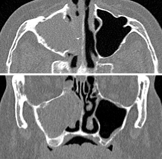
What are the types of fungal sinusitis?
Fungi are found in the noses of almost everyone; disease depends on the host response. Fungal rhinosinusitis is split into non-invasive and invasive forms, and allergic fungal rhinosinusitis (AFRS) is the commonest type.
| Type | Host | Key features |
|---|---|---|
| Fungal ball (non-invasive) | Immunocompetent, typically women | Mostly the maxillary sinus; often asymptomatic; may follow dental work or sinus surgery |
| Allergic fungal rhinosinusitis (non-invasive) | Immunocompetent young adults, usually 20–30 years | Type 1 hypersensitivity, nasal polyps, eosinophilic (allergic) mucin with Charcot–Leyden crystals, IgE often above 1000 U/mL; CT 'double density' sign; ethmoids most often involved |
| Acute invasive | Diabetes, chemotherapy, steroids, immunosuppression | Course under 30 days; vascular invasion, tissue necrosis; Mucor/Rhizopus or Aspergillus |
| Chronic invasive | Invasive forms mostly affect the immunocompromised | Slowly progressive course |
| Granulomatous invasive | Most often immunocompetent | Defined by submucosal granulomatous inflammation |
AFRS is diagnosed with the Bent and Kuhn criteria. All five major criteria are needed: type 1 hypersensitivity, nasal polyposis, characteristic CT findings, eosinophilic mucin without tissue invasion and positive fungal stain of sinus contents. Minor criteria (asthma, unilateral predominance, bone erosion, positive culture, Charcot–Leyden crystals, eosinophilia) are supportive. Aspergillus is the classic cause; more recent series recover dematiaceous fungi such as Bipolaris and Curvularia most often.
Treatment follows the type. Saprophytic crusting can be managed with saline douching; fungal ball and AFRS need surgery plus medical care; dupilumab has recently been approved in the US for AFRS. For invasive disease, systemic antifungals are adjunctive to surgery, amphotericin B is the usual first-line drug because it covers both Mucor and Aspergillus, topical antifungals are not recommended, and the underlying immunodeficiency must be corrected.
Why is rhino-orbito-cerebral mucormycosis an emergency?
Mucormycosis (Mucorales — Mucor, Rhizopus) is an aggressive invasive infection of immunocompromised hosts. Poorly controlled diabetes, especially diabetic ketoacidosis, is the most important risk factor — over 50% of cases in Indian series. Others are haematological malignancy, transplantation, prolonged neutropenia, glucocorticoids, iron overload (deferoxamine therapy), burns and severe COVID-19.
During the pandemic, COVID-19-associated mucormycosis surged, and India reported 57% of published cases in a meta-analysis — the 'black fungus' outbreak. Hyperglycaemia, steroid use and severe COVID combined to create the ideal host.
- Clinical: facial pain and swelling, nasal blockage, then darkening of skin; black eschar on the nasal turbinates or palate; necrosis of the turbinates; proptosis (the commonest orbital sign), ophthalmoplegia and visual loss; cranial nerve palsies with cerebral spread.
- Histopathology: broad, irregular, aseptate (pauciseptate) hyphae branching at about 90°, with necrosis and haemorrhage.
- Treatment: correct the predisposition (control glucose and ketoacidosis, reduce steroids), urgent surgical debridement, and liposomal amphotericin B (preferred over conventional amphotericin because it is far less nephrotoxic).
| Feature | Mucorales | Aspergillus |
|---|---|---|
| Width | Broad (5–10 µm) | Narrow (3–5 µm) |
| Septation | Non-septate or pauciseptate | Regularly septate |
| Branching | Right angle (90°) | Acute angle (45°) |
| Appearance | Ribbon-like, irregular | Uniform, tubular |
What are the complications of sinusitis and the Chandler classification?
The orbit is the commonest site of complication because it is separated from the ethmoid sinus only by the thin lamina papyracea. Infection spreads directly through this bone or its dehiscences, or along valveless ethmoidal and ophthalmic veins. The Chandler classification grades orbital complications from least to most severe.
| Stage | Name | Key findings |
|---|---|---|
| I | Preseptal (periorbital) cellulitis | Eyelid and periocular tissue anterior to the orbital septum |
| II | Orbital cellulitis | Orbital fat and extraocular muscles behind the septum: pain on eye movement, ophthalmoplegia, proptosis, impaired vision |
| III | Subperiosteal abscess | Collection beneath the periosteum of the orbital wall |
| IV | Orbital abscess | Collection within the orbital soft tissue |
| V | Cavernous sinus thrombosis | Spread through valveless veins to the cavernous sinus |
- Intracranial (rare): meningitis, epidural or subdural abscess/empyema (subdural empyema has a high mortality).
- Pott's puffy tumour: subperiosteal abscess of the frontal bone with osteomyelitis, from frontal sinusitis via valveless diploic veins.
- Mucocele and osteomyelitis are other local complications.