NEET-PG ENT Previous Year Questions (2017–2026)

44 NEET-PG ENT PYQs from 2017 to 2026, tagged by topic, with an explanation for every question. ENT had 7 of the 180 questions in NEET-PG 2026.

Last verified: 24 September 2026 against Kinase question bank and NEET-PG 2026 recall paper. natboard.edu.in · nmc.org.in

Quick Answer

NEET-PG ENT had 7 of 180 questions (3.9%) in the NEET-PG 2026 paper — up to 28 marks. Kinase has 44 NEET-PG ENT PYQs from 2017 to 2026; the most-asked topic is Larynx and Applied ENT (12 questions).

ENT in NEET-PG at a glance

Questions in NEET-PG 20267 of 180 (3.9%)
Maximum marks at stake28 of 720
NEET-PG ENT PYQs on Kinase44 (2.7% of all NEET-PG PYQs)
Years covered2017–2026
INICET ENT PYQs on Kinase91

Most-asked ENT topics in NEET-PG PYQs

TopicNEET-PG PYQsShare of subject
Larynx and Applied ENT1227%
Ear1125%
Nose1125%
Pharynx49%
Instruments25%

Across NEET PG and INI-CET papers: Most repeated ENT topics →

Sample NEET-PG ENT PYQs with answers

Question 1 · NEET PG Aug 2026

An adolescent boy is investigated for recurrent unilateral epistaxis and nasal obstruction, and a vascular nasopharyngeal mass is found. Biopsy is strongly discouraged because it can provoke torrential haemorrhage. The best explanation for this bleeding tendency is:

  1. A.Erodes internal carotid artery
  2. B.Vessels lack a muscle coat
  3. C.Releases a fibrinolytic factor
  4. D.Tumour lacks a capsule
Show answer and explanation

Answer: B. Vessels lack a muscle coat

Correct answer: B. Vessels lack a muscle coat.

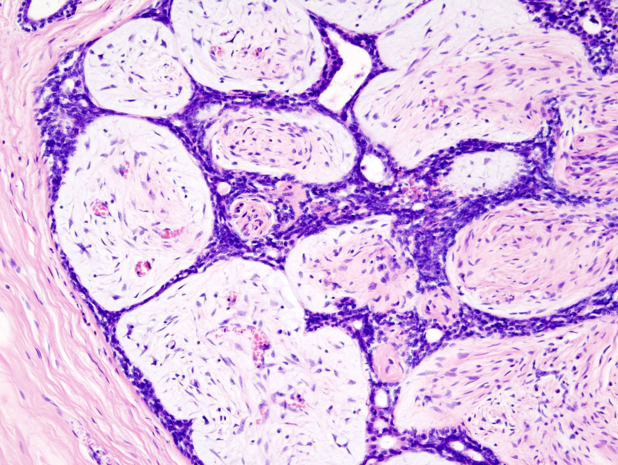
Juvenile nasopharyngeal angiofibroma is a fibrovascular tumour in which the vessels are irregular, thin-walled channels lined only by endothelium and lacking a complete smooth-muscle media. Because these vessels cannot contract when cut, biopsy produces torrential bleeding, so diagnosis relies on contrast imaging and angiography rather than tissue sampling.
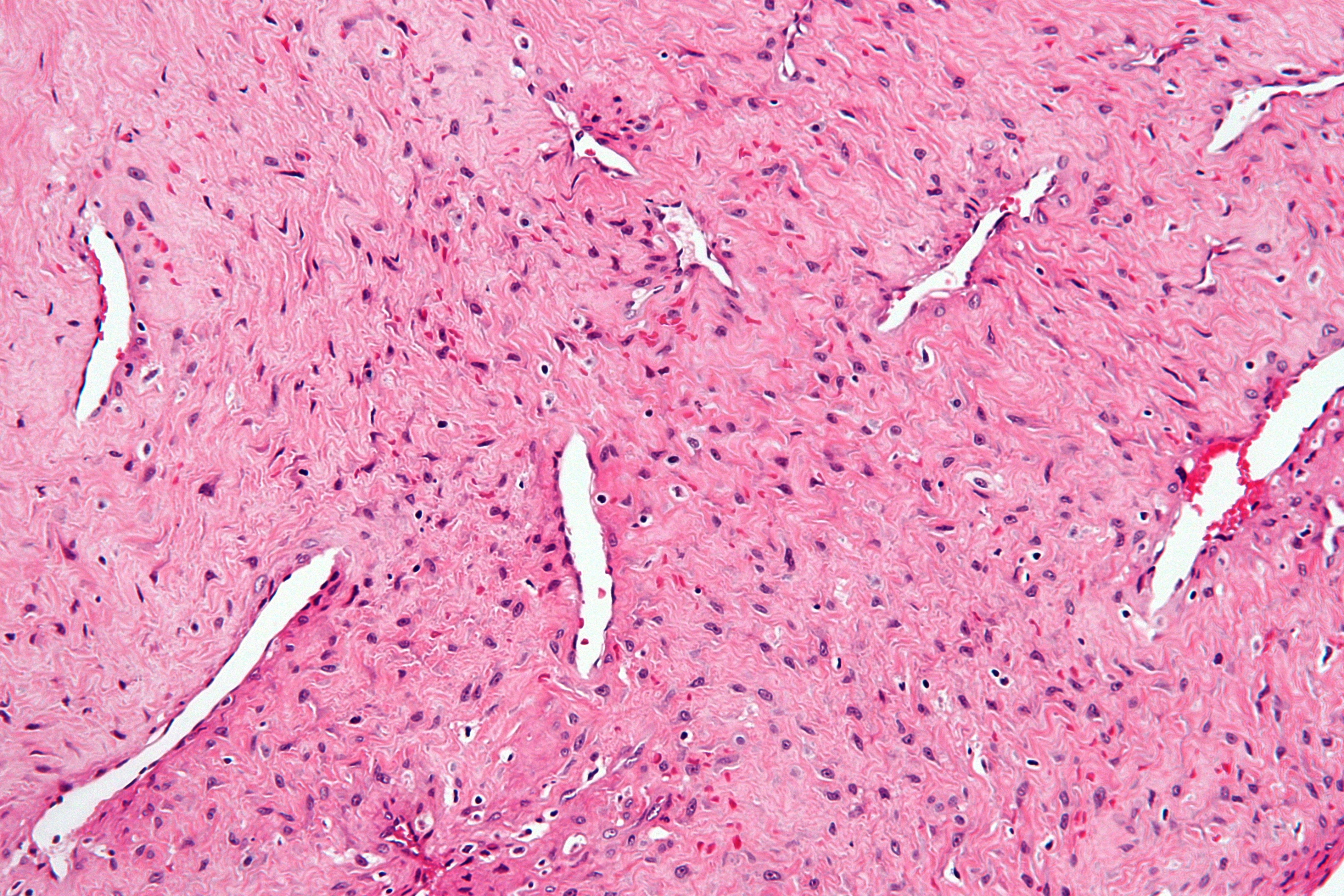
JNA histology thin-walled vesselsFibrous stroma with thin-walled, muscle-poor vessels of angiofibroma.

Incorrect Options:

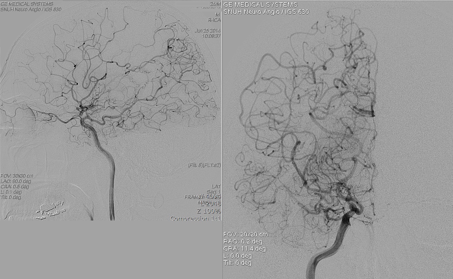
  • A. Erodes internal carotid artery: advanced tumour may reach the skull base, but routine biopsy bleeding is from its own defective vessels, not carotid erosion. Internal carotid angiographyAngiography of the internal carotid artery.
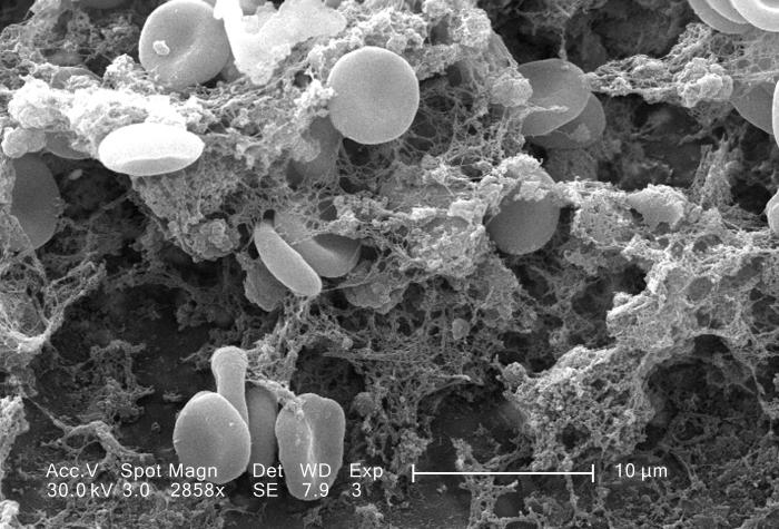
  • C. Releases a fibrinolytic factor: the tumour does not secrete a fibrinolysin; bleeding is structural, not a clotting defect. Fibrin clot meshFibrin strands enmeshing red cells (a fibrinolysin would dissolve these).
  • D. Tumour lacks a capsule: the angiofibroma is indeed unencapsulated, but absence of a capsule does not by itself cause bleeding. Encapsulated tumour for contrastA well-circumscribed encapsulated tumour, shown for contrast.
High-Yield Facts:

  • Classic patient: adolescent male with recurrent epistaxis and nasal obstruction.
  • Holman-Miller sign (anterior bowing of the posterior maxillary wall) on imaging.
  • Diagnosis by contrast imaging and angiography; biopsy is contraindicated.
  • Treatment: pre-operative embolisation then surgical excision.
Question 2 · NEET PG Aug 2026

A woman applying for a disability benefit reports profound hearing loss. On pure-tone audiometry her thresholds are grossly inconsistent, improving by more than 30 dB on repeat testing, and there is no shadow curve. Otoacoustic emissions and acoustic reflexes are normal and the Stenger test is positive. What is the diagnosis?

  1. A.Presbycusis
  2. B.Non-organic hearing loss
  3. C.Noise-induced hearing loss
  4. D.Otosclerosis
Show answer and explanation

Answer: B. Non-organic hearing loss

Correct answer: B. Non-organic (functional/malingering) hearing loss.

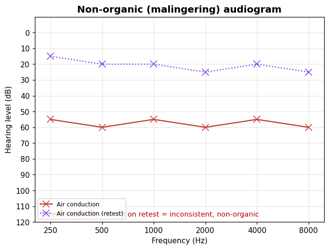
Hearing thresholds that are inconsistent and non-reproducible (large shifts on retest, no shadow curve) in the presence of normal objective tests (otoacoustic emissions, acoustic reflexes) and a positive Stenger test indicate a non-organic loss. The objective measures confirm the cochlea and auditory pathway are working, so the claimed loss is functional or feigned.

Audiogram showing inconsistent thresholds on retest
Large threshold shifts on retest point to a non-organic loss.

Incorrect Options:

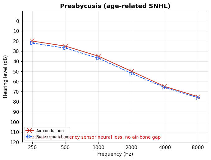
  • A. Presbycusis: a real, reproducible bilateral high-frequency sensorineural loss of ageing. Down-sloping audiogram of presbycusis
    Presbycusis: consistent high-frequency SNHL.
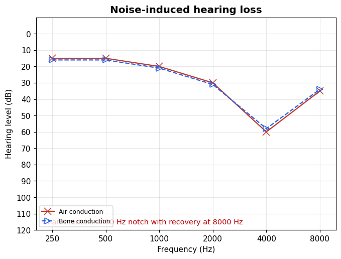
  • C. Noise-induced hearing loss: shows a reproducible 4000 Hz notch, not fluctuating thresholds. Audiogram with a 4000 Hz notch
    NIHL: characteristic 4 kHz notch.
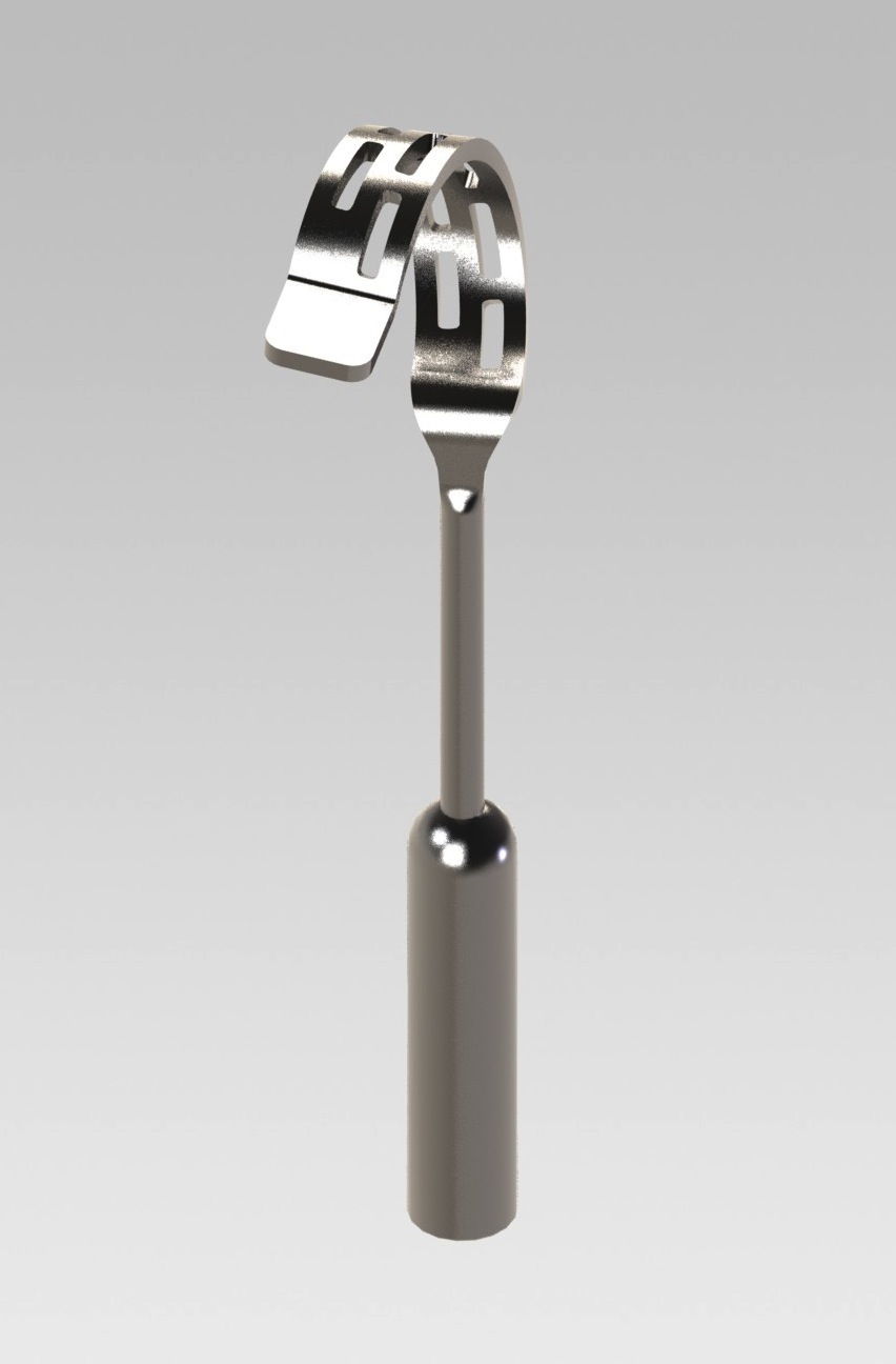
  • D. Otosclerosis: a conductive loss with an air-bone gap and Carhart notch, with consistent, reproducible thresholds. Stapes prosthesis used in otosclerosis surgery
    Otosclerosis: reproducible conductive loss, treated by stapedectomy.

High-Yield Facts:

  • Suspect non-organic loss when thresholds are inconsistent or contradicted by objective tests.
  • Objective tests (OAE, acoustic reflexes, brainstem-evoked response) bypass voluntary responses.
  • A positive Stenger test unmasks a feigned unilateral or asymmetric loss.
  • Carhart notch (2 kHz bone-conduction dip) is a feature of otosclerosis, which disappears after stapedectomy.
Question 3 · NEET PG Aug 2026

A 45-year-old man reports six months of ringing and steadily worsening hearing confined to his right ear. Examination reveals reduced sensation over the posterior wall of the right external auditory canal (a positive Hitzelberger sign), and pure-tone audiometry confirms a unilateral sensorineural loss. Which investigation will most reliably confirm the suspected lesion?

  1. A.Brainstem evoked audiometry
  2. B.Contrast MRI of the CP angle
  3. C.HRCT of the temporal bone
  4. D.Tympanometry
Show answer and explanation

Answer: B. Contrast MRI of the CP angle

Correct answer: B. Gadolinium-enhanced MRI of the cerebellopontine (CP) angle.

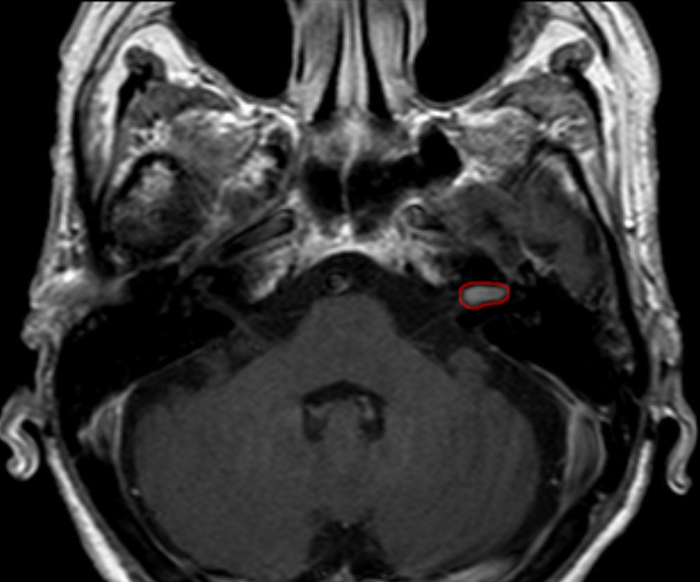
Unilateral tinnitus with progressive sensorineural hearing loss and a positive Hitzelberger sign (numbness of the posterior external auditory canal wall from involvement of the sensory fibres of the facial nerve) is the classic picture of a vestibular schwannoma (acoustic neuroma) at the CP angle. Gadolinium-enhanced MRI is the investigation of choice: it resolves even intracanalicular tumours a few millimetres across and shows the enhancing lesion filling the internal auditory canal and extending into the cistern (the classic ice-cream cone appearance).

Gadolinium-enhanced axial MRI showing an enhancing intracanalicular vestibular schwannoma at the cerebellopontine angle

Contrast MRI: a small enhancing schwannoma (outlined) at the right internal auditory canal / CP angle.

Incorrect Options:

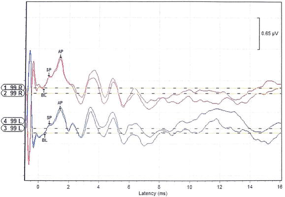
  • Brainstem evoked audiometry (BERA): only a screening test - a normal trace does not exclude a small tumour, so it cannot confirm the diagnosis. Auditory evoked-response waveform with latency in milliseconds Auditory evoked-potential waveform (SP/AP peaks against latency) - a screening electrophysiology test.
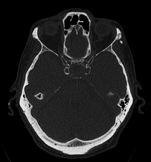
  • HRCT of the temporal bone: excellent for bony detail and cholesteatoma but poor for a small soft-tissue tumour in the canal. Axial CT of the temporal bone in bone window Axial HRCT temporal bone (bone window) - shows osseous anatomy, not the soft-tissue tumour.
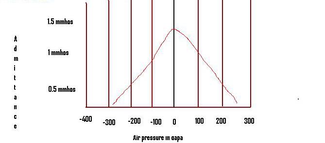
  • Tympanometry: assesses middle-ear compliance; normal here because the lesion is retrocochlear. Type A tympanogram curve of admittance against ear-canal pressure Tympanogram (admittance vs pressure) - a middle-ear test, unaffected by a retrocochlear tumour.

High-Yield Facts:

  • Vestibular schwannoma arises from Schwann cells of the vestibular division of cranial nerve VIII, usually within the internal auditory canal.
  • Gadolinium is essential - non-contrast MRI can miss tiny intracanalicular tumours.
  • Bilateral vestibular schwannomas are pathognomonic of neurofibromatosis type 2.
  • Early facial-canal sensory loss (Hitzelberger sign) precedes overt facial weakness.

ENT concept guides

Browse the full concept library and clinical image atlas.

Practise the rest: open the Kinase question bank, choose ENT, set the exam to NEET-PG and switch on PYQs only. Questions you get wrong go into Fix My Weakness for targeted revision.

Frequently Asked Questions

How many ENT questions were in NEET-PG 2026?▼
7 of the 180 questions in the NEET-PG 2026 paper (30 August 2026) were ENT — 3.9% of the paper, worth up to 28 marks at +4 per correct answer. This is from the recall paper in the Kinase question bank.
How many NEET-PG ENT PYQs does Kinase have?▼
Kinase has 44 NEET-PG ENT previous year questions from 2017 to 2026, each with an explanation, plus 91 INICET ENT PYQs for extra practice.
What are the most-asked ENT topics in NEET-PG?▼
In Kinase's NEET-PG ENT PYQs, the most-asked topics are Larynx and Applied ENT, Ear, Nose. Larynx and Applied ENT alone accounts for 12 of the 44 questions.
Are NEET-PG ENT PYQs the same as INICET PYQs?▼
Both exams test the MBBS syllabus, so most ENT concepts overlap. The marking differs: NEET-PG is +4 / −1 and INICET is +1 / −1/3. Kinase lets you practise either exam's PYQs, or both together.
How should I practise ENT PYQs for NEET-PG?▼
Start with the most-asked topics above, do a daily set of ENT PYQs, and read every explanation — including the ones you got right. Kinase's Fix My Weakness then builds practice sets from the topics you miss most.
44 ENT PYQs
2017–2026 papers
Topic + year tagged

Ready to Start Practicing?

ENT was 7 questions (up to 28 marks) in NEET-PG 2026.

Start Free Practice

No credit card required