What kind of organism is Staphylococcus aureus?
Staphylococcus aureus is a gram-positive coccus that typically appears in grape-like clusters, although single cells, pairs and short chains are also seen. It is nonmotile, non-spore-forming and a facultative anaerobe. Clinically it ranges from trivial skin infection and self-limited food poisoning to bacteraemia, endocarditis, osteomyelitis, necrotising pneumonia and toxic shock. Worldwide it is the leading cause of death from bacterial bloodstream infection.
Think of the organism in three layers for exams: how the laboratory identifies it (catalase, coagulase, clumping factor, DNase, mannitol), how it causes disease (surface proteins, enzymes and toxins) and how resistance changes treatment (penicillinase, then MRSA through mecA and PBP2a). Most single-best-answer questions test a one-line link from one of these layers.
Which laboratory tests identify S. aureus?
The first bench step after a Gram stain is the catalase test: staphylococci are catalase positive, streptococci catalase negative. Among staphylococci, coagulase separates S. aureus (coagulase positive) from the coagulase-negative staphylococci such as S. epidermidis, which comprise over 30 other species and are usually less virulent.

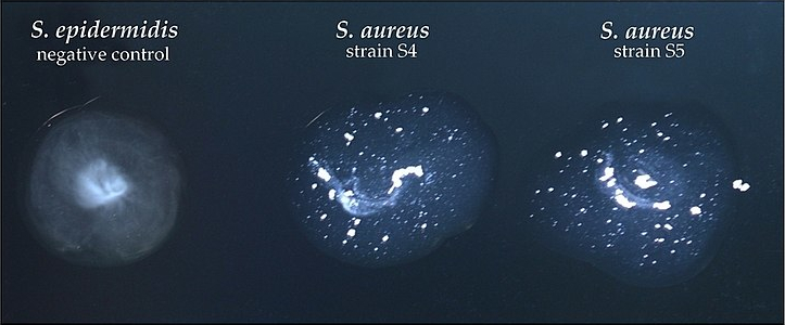
Two different molecules hide behind the word coagulase. Free coagulase is secreted and is detected by the tube coagulase test, in which a clot forms in plasma after incubation. Clumping factor is a fibrinogen-binding protein on the cell surface, loosely called bound coagulase, and is detected by the slide test or latex kits. Genetic studies show the two are distinct, and some clinical isolates lack one or both, which can make identification difficult.
| Test | Result in S. aureus | What it detects / why it matters |
|---|---|---|
| Gram stain | Gram-positive cocci in clusters | First clue; streptococci form chains |
| Catalase | Positive | Separates staphylococci from streptococci |
| Tube coagulase | Positive (clot in plasma) | Free (secreted) coagulase |
| Slide coagulase / latex | Positive (clumping) | Clumping factor and protein A on the surface |
| Thermostable DNase | Positive | Heat-stable nuclease; confirms S. aureus |
| Mannitol salt agar | Grows; mannitol fermented (yellow) | 7.5% sodium chloride selects salt-tolerant staphylococci |
For contaminated specimens such as nasal swabs, mannitol salt agar contains 7.5% sodium chloride, which lets salt-tolerant staphylococci grow while suppressing many other organisms. S. aureus ferments mannitol and produces acid that turns the phenol red indicator yellow. Latex agglutination kits coated with IgG and fibrinogen detect protein A and clumping factor together for rapid confirmation.

Which surface proteins and enzymes make S. aureus virulent?
Virulence comes from a long list of surface and secreted molecules. The high-yield ones each have a single job that examiners like to match:
| Factor | Location | Action |
|---|---|---|
| Protein A | Cell wall | Binds the Fc portion of IgG, so antibody cannot opsonise; protects from phagocytosis |
| Clumping factor | Cell surface | Binds fibrinogen; attachment to clots and damaged tissue |
| Coagulase | Secreted | Converts fibrinogen to fibrin; promotes thrombus and walls off infection |
| Catalase | Enzyme | Breaks down hydrogen peroxide; blunts the neutrophil oxidative burst |
| Capsule | Surface | Interferes with phagocytosis |
| Biofilm | Surface matrix | Shields bacteria from immunity and antibiotics on valves, bone and implants |
| Haemolysins (alpha, beta, gamma, delta) | Secreted | Alpha toxin is a major virulence factor; systemic release causes septic shock |
Biofilm formation explains why S. aureus is the commonest cause of chronic infections of heart valves, bone, implants and prosthetic devices. Bacteria inside a biofilm are protected from both immune attack and antibiotics, so removal of infected hardware is often needed.
How do enterotoxins and TSST-1 cause disease?
The enterotoxins and toxic shock syndrome toxin-1 (TSST-1) are superantigens: proteins that activate a very large pool of T lymphocytes at once, releasing a cytokine storm (TNF-alpha, IL-1, IL-2) that leads to shock and multiorgan failure. A conventional antigen activates only a tiny fraction of T cells.
Staphylococcal food poisoning is an intoxication, not an infection. A food handler carrying S. aureus contaminates food (sliced meats, puddings, pastries, sandwiches); the organism multiplies at room temperature and forms preformed, heat-stable enterotoxin. Cooking kills the bacteria but does not destroy the toxin. Symptoms — sudden nausea, vomiting, cramps and diarrhoea — start within 30 minutes to 8 hours and usually last 24 hours or less. Antibiotics have no role because they do not affect the toxin.
Toxic shock syndrome follows systemic absorption of TSST-1 or enterotoxins. Menstrual TSS rose to prominence with super-absorbent tampons; this is not a true infection but toxin absorption from organisms growing in the tampon. Enterotoxins B and C cause about half of non-menstrual TSS. TSST-1 itself has no emetic activity.
| Toxin | Type | Classic disease | Exam hook |
|---|---|---|---|
| Enterotoxins (A–E, G) | Superantigen; heat stable | Food poisoning; non-menstrual TSS | Short incubation, vomiting, no fever focus |
| TSST-1 | Superantigen | Toxic shock syndrome | Tampons; shock with rash and multiorgan failure |
| Exfoliative toxins A and B | Serine proteases | SSSS; bullous impetigo | Cleave desmoglein-1 |
| Panton–Valentine leukocidin | Pore-forming leukocidin | Necrotising pneumonia, skin abscesses | Kills leukocytes; leukopenia in severe pneumonia |
| Alpha toxin | Haemolysin | Septic shock, tissue damage | Major virulence factor |
Why does staphylococcal scalded skin syndrome split the skin?
Exfoliative (epidermolytic) toxins A and B are serine proteases released at a local focus of infection. In staphylococcal scalded skin syndrome (SSSS) the toxin spreads through the blood and cleaves desmoglein-1, so keratinocytes separate in the stratum granulosum — a superficial, intraepidermal split. In bullous impetigo the same toxin acts only at the infection site.
SSSS mostly affects children younger than 5 years, particularly before 3 years, and adults with immunosuppression or severe kidney impairment, who clear the toxin less efficiently. Exfoliation begins in the flexures, with periorificial crusting and radial fissuring around the mouth and eyes, and a positive Nikolsky sign. Healing is usually without scarring because the split is superficial. Neonates in the first days of life are relatively protected because desmoglein-3 dominates their epidermis and is not targeted.
Blood and blister cultures are usually negative in children because the toxin, not the organism, travels in the blood; the organism may be recovered from the nasopharynx, conjunctiva, perianal area or a skin focus. Treatment is prompt anti-staphylococcal therapy (for example cefazolin, nafcillin or oxacillin) plus fluid, temperature and wound care. Clindamycin can reduce toxin production, but up to half of SSSS strains are resistant, so it is not used alone.
| Feature | SSSS | Toxic epidermal necrolysis |
|---|---|---|
| Cause | Staphylococcal exfoliative toxin | Usually drugs |
| Level of split | Stratum granulosum (superficial) | Full-thickness epidermal necrosis |
| Mucosa | Typically spared | Usually involved |
| Usual age | Young children | Adults |
What does Panton–Valentine leukocidin do?
Panton–Valentine leukocidin (PVL) is cytotoxic to leukocytes and alveolar epithelium. PVL-producing strains are prone to recurrent skin abscesses and furunculosis, necrotising pneumonia, osteomyelitis and bacteraemia, and community-associated MRSA is a recognised cause of life-threatening necrotising pneumonia in otherwise healthy people.
Necrotising pneumonia presents with severe respiratory symptoms, high fever, haemoptysis and hypotension, progressing quickly to septic shock with leukopenia and a raised C-reactive protein. Leukopenia with rapidly progressive necrotising pneumonia should make you think of a PVL-producing strain.
- PVL: kills white cells → abscesses, necrotising pneumonia.
- Exfoliative toxin: cleaves desmoglein-1 → SSSS.
- TSST-1: superantigen → toxic shock.
- Enterotoxin: superantigen, heat stable → food poisoning.
- Alpha toxin: haemolysin → tissue damage and septic shock.
How does MRSA resist beta-lactams?
Plasmid-mediated penicillinase is present in over 80% of isolates, so plain penicillin rarely works. Penicillinase-resistant beta-lactams (methicillin, oxacillin, nafcillin) and cephalosporins overcame this, until MRSA emerged.
MRSA carries the mecA (or mecC) gene on a mobile element, the staphylococcal cassette chromosome mec (SCCmec). mecA encodes an altered penicillin-binding protein, PBP2a, which binds beta-lactams with low affinity, so cell-wall synthesis continues despite the drug. Resistance is therefore target modification, not drug destruction — which is why adding a beta-lactamase inhibitor does not help.
In the laboratory MRSA is confirmed by detecting mecA (PCR) or PBP2a (latex agglutination), or by phenotypic testing. Cefoxitin disc or broth testing is preferred to oxacillin because cefoxitin is a better inducer of mecA and gives clearer end points; incubation is at 35 °C, since higher temperatures may miss resistance. A nasal MRSA PCR is a useful screening test: a negative result has a high value for ruling out MRSA.
How are MSSA and MRSA infections treated?
For MSSA, cefazolin or an anti-staphylococcal penicillin (nafcillin, oxacillin, cloxacillin) remains preferred, with vancomycin or daptomycin as alternatives in beta-lactam allergy.
For MRSA, the glycopeptide vancomycin and the lipopeptide daptomycin remain active against most strains; the anti-MRSA cephalosporins ceftaroline and ceftobiprole are also recommended options. Linezolid (an oxazolidinone acting on the 50S ribosome) also retains activity. For uncomplicated skin and soft-tissue infection, oral co-trimoxazole, doxycycline/minocycline or clindamycin are used empirically. Clindamycin and linezolid are not used for endocarditis because outcomes are poorer than with cell-wall-active drugs.
| Situation | Preferred agents |
|---|---|
| MSSA bacteraemia or deep infection | Cefazolin or anti-staphylococcal penicillin |
| MRSA bacteraemia or endocarditis | Vancomycin or daptomycin |
| MRSA uncomplicated skin infection | Co-trimoxazole, doxycycline or clindamycin (oral) |
| Toxin-mediated disease | Add a protein-synthesis inhibitor such as clindamycin to suppress toxin (with resistance caveat) |
| Food poisoning | Supportive; no antibiotics |
Vancomycin-intermediate S. aureus (VISA) has a thickened cell wall that traps the drug and may look susceptible on routine testing. It is one reason vancomycin retains activity against most, but not all, MRSA strains.
Where does S. aureus colonise and how is carriage controlled?
About 20–30% of people are persistently colonised and roughly another 30% intermittently. The anterior nares and oropharynx are the main niches, with skin, axilla, groin and perineum also involved. Carriage is a risk factor for later skin colonisation and infection, including surgical-site infection.
Prevention rests on hand hygiene, screening for colonisation and targeted decolonisation. Topical mupirocin in the nose combined with chlorhexidine body washes or dilute bleach baths is frequently effective, though often only temporarily. Decolonising both the index case and household members reduces recurrent skin infections in children.