What are SJS and TEN and how are they classified?
Stevens–Johnson syndrome (SJS) and toxic epidermal necrolysis (TEN) are rare, life-threatening severe cutaneous adverse reactions (SCARs). They are the same disease at different extents: keratinocytes die across the full thickness of the epidermis, which separates from the dermis, and mucous membranes are almost always involved. They are classified by the percentage of body surface area (BSA) that is detached.
| Category | Detached BSA | Approximate mortality |
|---|---|---|
| SJS | < 10% | Up to 10% |
| SJS/TEN overlap | 10–30% | Intermediate |
| TEN | > 30% | At least 30% |
SJS/TEN is rare — about 1–5 cases per million people per year — and commoner in adults than in children, probably because adults take more of the trigger drugs. Rates vary between countries with the use of drugs such as antitubercular and anti-HIV medicines and with genetic background. Newer data show a clear mortality jump above 10% BSA, so some authors have proposed redefining TEN as 10% or more; the classic exam cut-offs remain 10% and 30%.
Which drugs cause SJS/TEN, and what is the latent period?
More than 80% of cases are caused by drugs; infections, tumours and vaccination are less common triggers, and some cases are idiopathic. Symptoms usually begin 4–28 days after starting the drug (almost always within 8 weeks). On re-exposure, immune memory can bring the reaction back within hours.
| Group | Drugs |
|---|---|
| Antiepileptics | Lamotrigine, carbamazepine, phenytoin, valproic acid, phenobarbital |
| Anti-gout | Allopurinol |
| Antibiotics | Trimethoprim–sulfamethoxazole, aminopenicillins, cephalosporins, tetracyclines |
| Antiretroviral | Nevirapine (NNRTI) |
| Analgesics | NSAIDs |
Which HLA alleles predispose to SJS/TEN?
SJS/TEN is a type IV (T-cell-mediated) hypersensitivity reaction (see hypersensitivity reactions). The drug is presented on particular HLA class I molecules to CD8+ cytotoxic T cells, which kill keratinocytes through the Fas–Fas ligand pathway, perforin/granzyme and granulysin. Granulysin levels in blister fluid and serum track severity, though they are not specific.
| Allele | Drug | Exam point |
|---|---|---|
| HLA-B*15:02 | Carbamazepine, oxcarbazepine | Highest in East Asian (6.9%) and South/Central Asian (4.6%) populations; first described in Han Chinese |
| HLA-A*31:01 | Carbamazepine | SJS/TEN, DRESS and maculopapular exanthema |
| HLA-B*58:01 | Allopurinol | Present in all 51 Han Chinese patients with allopurinol SCAR vs 15% of tolerant patients |
| HLA-B*57:01 | Abacavir | Abacavir hypersensitivity reaction |

What are the clinical features of SJS/TEN?
- Prodrome: an influenza-like illness — fever, malaise, myalgia, upper respiratory symptoms — 1–21 days before the rash.
- Mucosa first: mucosal erythema and erosions occur in about 90% and usually precede the skin rash by 1–3 days; two or more mucosal sites are involved in about 90%. The oropharynx, eyes and genitalia are most often affected.
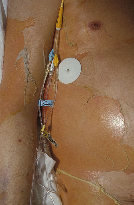
- Skin: painful erythematous, atypical targetoid or purpuric macules, mainly on the face and trunk, that merge, blister into flaccid bullae and slough in sheets.
- Nikolsky sign positive: the epidermis slides off with gentle lateral pressure.
- Eyes: conjunctivitis and lid oedema, up to corneal ulceration — needs daily ophthalmology review.
- Systemic: fluid and protein loss, sepsis, multiorgan failure and gastrointestinal bleeding.
Late sequelae (DermNet): patchy hyper- and hypopigmentation, scarring, nail loss, scarred genitalia (phimosis, vaginal adhesions), joint contractures and chronic lung disease such as bronchiolitis.
How is SJS/TEN confirmed and what are the differentials?
- Skin biopsy (urgent frozen section): full-thickness epidermal necrosis, subepidermal bullae and individual keratinocyte necrosis; in TEN the necrosis is extensive and the epidermis detaches.
- Direct immunofluorescence on a punch biopsy: negative.
- Bloods: anaemia, lymphopenia, neutropenia, raised transaminases, hypoalbuminaemia, renal function, glucose and bicarbonate (for SCORTEN).
- Patch testing after recovery may identify the culprit in about half of patients.
| Condition | How it differs |
|---|---|
| Erythema multiforme major | Usually < 10% BSA; symmetric acral target lesions, with or without blisters |
| Staphylococcal scalded skin syndrome | Generalised blistering mimic — skin biopsy is used to exclude it |
| DRESS | Later onset (2–8 weeks), facial swelling, eosinophilia, organ involvement, little epidermal loss |
| AGEP | Rapid sheets of sterile pinpoint pustules, starting in the flexures |
What is SCORTEN and how is it scored?
SCORTEN (Severity-of-Illness Score for Toxic Epidermal Necrolysis) uses 7 independent variables, each scoring 1 point, assessed within the first 24 hours of admission.
- Age > 40 years
- Heart rate ≥ 120/min
- Cancer or haematological malignancy
- Detached BSA ≥ 10% on day 1
- Serum urea (BUN) > 28 mg/dL (> 10 mmol/L)
- Serum bicarbonate < 20 mEq/L
- Serum glucose > 252 mg/dL (> 14 mmol/L)
| SCORTEN | Mortality |
|---|---|
| 0–1 | 3.2% |
| 2 | 12.1% |
| 3 | 35.3% |
| 4 | 58.3% |
| 5 or more | > 90% |
ABCD-10 (2019) scores Age over 50 (1 point), low Bicarbonate, active Cancer (2 points), Dialysis before admission (3 points) and detachment of 10% or more of BSA (1 point); in a multicentre US cohort of 370 patients it predicted in-hospital mortality as well as SCORTEN. CRISTEN uses only clinical information, so it can be applied before laboratory results are back.
How is SJS/TEN managed?
- Stop the culprit drug immediately — the single most important step.
- Transfer early to a burn unit or ICU experienced in TEN.
- Fluids: crystalloid resuscitation guided by a standard burn formula (e.g. Parkland), aiming for MAP > 65 mmHg, CVP 8–12 mmHg and urine output 0.5–1 mL/kg/h (compare thermal injuries).
- Team: dermatology, allergy/immunology, burn surgery, ophthalmology, urogynaecology and psychiatry all contribute.
- Supportive care: pain control, wound care, nutritional support and watch for infection.
- Mucosal care: daily ophthalmology review with lubricants and drops, mouthwashes, and genital care to prevent adhesions.
- Immunomodulation: no treatment is proven — systemic corticosteroids, IVIG, ciclosporin, TNF-α blockers and plasmapheresis are all debated. A 2021 network meta-analysis suggested corticosteroids plus IVIG may reduce mortality.
How do DRESS and AGEP differ from SJS/TEN?
DRESS (drug reaction with eosinophilia and systemic symptoms), also called drug hypersensitivity syndrome or DIHS, starts 2–8 weeks after the drug — later than SJS. It combines high fever, a morbilliform rash (80%), facial swelling (30%), lymphadenopathy, blood abnormalities (eosinophilia, atypical lymphocytes, low platelets) and inflammation of internal organs, most often the liver. Reactivation of HHV-6 or EBV may be involved. Common culprits are carbamazepine, phenobarbital, phenytoin, allopurinol, olanzapine and sulfonamides. Diagnosis uses the RegiSCAR criteria; severe cases get systemic corticosteroids. Deaths come from acute liver failure, multiorgan failure, fulminant myocarditis and haemophagocytosis.
- RegiSCAR inclusion: at least 3 of — hospitalisation, a reaction suspected to be drug-related, acute skin rash, fever above 38 °C, enlarged lymph nodes at two sites, involvement of at least one internal organ, and blood count abnormalities (low platelets, raised eosinophils or abnormal lymphocytes).
- Timing clues: onset is delayed beyond 2 weeks with anticonvulsants and allopurinol; a reaction within 2 weeks points more to β-lactam antibiotics or iodinated contrast.
- Allopurinol risk rises with higher dose, kidney disease and concomitant thiazide diuretics; at least 1 in 10,000 patients on an anticonvulsant develops the syndrome.
- Rash and organs do not match: severity of the rash does not predict the extent of internal organ involvement; about 10% progress to erythroderma.
AGEP (acute generalised exanthematous pustulosis) is a sudden eruption of sterile, non-follicular pinpoint pustules on red skin, starting in the flexures and spreading within hours to days, usually sparing the palms and soles. It appears within about 10 days of the drug — most often β-lactam and macrolide antibiotics — with fever and leukocytosis, and clears within 1–2 weeks of stopping it. Histology shows spongiform subcorneal pustules; the EuroSCAR score (Sidoroff, 2001) combines clinical, laboratory and histological findings to grade diagnostic certainty. More than 85–90% of cases follow drug exposure.
| Feature | SJS/TEN | DRESS | AGEP |
|---|---|---|---|
| Onset after drug | 4–28 days | 2–8 weeks | Usually ≤ 10 days |
| Skin | Targetoid macules, blisters, sheet-like detachment | Morbilliform rash, facial oedema | Sterile pinpoint pustules, flexures first |
| Mucosa | Almost always (about 90%) | About 25% | Uncommon; oral only when present |
| Blood | Lymphopenia, anaemia | Eosinophilia, atypical lymphocytes | Leukocytosis |
| Organs | Eyes, genitourinary tract, GI bleeding; sepsis | Liver enzymes raised in most; kidney, heart | Often self-limited; occasional systemic complications |
| Typical drugs | Allopurinol, antiepileptics, sulfonamides, nevirapine, NSAIDs | Anticonvulsants, allopurinol, sulfonamides | β-lactams, macrolides |
| Scoring | SCORTEN | RegiSCAR | EuroSCAR |
Past questions are in the NEET PG Dermatology PYQs and NEET PG Pharmacology PYQs; skin layer anatomy is on the layers of epidermis page.