What is a stroke and how is ischaemic stroke classified?
Acute ischaemic stroke is the sudden onset of a focal neurological deficit in a vascular territory of the brain, retina or spinal cord, caused by cerebrovascular disease. Strokes are divided into ischaemic and haemorrhagic; haemorrhagic strokes are further split into intracerebral haemorrhage (ICH) and subarachnoid haemorrhage (SAH). Ischaemic stroke is by far the commonest — StatPearls quotes ischaemic strokes as 65.3% of strokes worldwide in 2021.
The brain tissue that depends on a single occluded artery dies first and forms the infarct core. Around it lies the ischaemic penumbra — tissue kept alive by collateral flow but not functioning. The penumbra shrinks as time passes, which is why every step of acute stroke care is a race against the clock: the whole point of reperfusion is to rescue the penumbra.
| TOAST subtype | Typical clue |
|---|---|
| Large artery atherosclerosis | More than 50% stenosis or occlusion of a major cerebral or cortical branch artery; cortical signs |
| Cardioembolism | Atrial fibrillation or another cardiac source; often several territories |
| Small vessel occlusion (lacunar) | Small deep infarct; classic lacunar syndrome; hypertension and diabetes |
| Stroke of other determined etiology | Dissection, vasculitis, hypercoagulable state and other rare causes |
| Stroke of undetermined etiology | No cause found, or two competing causes |

How is a suspected stroke assessed in the first minutes?
The single most important history item is the time of last known well — the last moment the patient was seen normal. It, not the time the symptoms were discovered, starts the treatment clock. The 2026-updated StatPearls chapter stresses a parallel workflow: pre-notification, direct transport to CT, point-of-care glucose, rapid neurological assessment and early stroke-team activation all happen at the same time.
- Check glucose first. Hypoglycaemia is a reversible stroke mimic; AHA guidance is to correct glucose below 60 mg/dL before further stroke evaluation.
- Score the deficit with the NIHSS (National Institutes of Health Stroke Scale): 15 items, total 0 to 42, 0 = no deficit. It emphasises anterior-circulation signs, so posterior-circulation strokes can score falsely low.
- Bloods without delaying imaging: CBC with platelets, PT/INR, aPTT, electrolytes, renal function, troponin; ECG and telemetry to look for atrial fibrillation.
- Airway: protect it when bulbar weakness, vomiting or falling consciousness threaten aspiration — but a high NIHSS alone is not a reason to intubate.
- Oxygen: only for hypoxaemia, keeping saturation above 94%; routine oxygen does not help a non-hypoxaemic patient.
Which imaging is done first in acute stroke and why?
Non-contrast CT (NCCT) of the head is the first scan because its job is to exclude haemorrhage — the one finding that changes everything. In early ischaemia NCCT is often normal or shows only subtle signs. CT angiography (CTA) of the head and neck is added early to look for a large-vessel occlusion, basilar occlusion or dissection; a patient in the thrombolysis window should not wait for CTA, perfusion or MRI before thrombolysis starts.
| Modality | Main role |
|---|---|
| NCCT head | Excludes haemorrhage; early ischaemic change; hyperdense artery sign |
| CT angiography | Finds large-vessel occlusion, basilar occlusion, dissection — selects thrombectomy |
| CT / MR perfusion | Estimates core vs penumbra; most useful for unknown onset or 4.5–24 h presentations |
| MRI diffusion-weighted imaging (DWI) | Most sensitive for acute infarct — shows cytotoxic oedema within minutes |
| DWI–FLAIR mismatch | DWI-positive lesion without marked FLAIR change — supports treatment in selected unknown-onset (wake-up) strokes |
ASPECTS (Alberta Stroke Program Early CT Score) grades early ischaemic change in the MCA territory on a 10-point scale; a lower score means a larger established infarct. Under the 2026 guideline, a low ASPECTS no longer automatically rules out thrombectomy — trials now support treating selected patients with ASPECTS 3 to 5.

Who gets IV thrombolysis and at what dose?
IV thrombolysis should be offered as soon as possible to eligible adults with a disabling ischaemic stroke within 4.5 hours of symptom onset or last known well — the earlier, the greater the benefit. The 2026 AHA/ASA guideline (which replaced the 2018 guideline and its 2019 update) recommends either drug:
| Drug | Dose | How given |
|---|---|---|
| Tenecteplase | 0.25 mg/kg, maximum 25 mg | Single IV bolus |
| Alteplase (rtPA) | 0.9 mg/kg, maximum 90 mg | 10% as a 1-minute bolus, the rest infused over 60 minutes |
Do not wait to see whether the patient improves, and do not delay thrombolysis for thrombectomy work-up. Patients eligible for both get IV thrombolysis and go straight on to thrombectomy.
- Wake-up or unknown-onset stroke: if treatment can start within 4.5 h of symptom recognition, a DWI–FLAIR mismatch can support thrombolysis.
- Perfusion-selected 4.5–9 h: for patients not eligible for thrombectomy with salvageable penumbra on automated perfusion imaging, thrombolysis can help when they wake with symptoms within 9 h of the midpoint of sleep or present 4.5 to 9 h from last known well.
- 4.5–24 h large-vessel occlusion: only a weak (Class 2b) recommendation, and only when thrombectomy cannot be performed.
What are the contraindications to thrombolysis?
These findings usually rule out IV thrombolysis or must be corrected before it is given:
| Category | Exclusion |
|---|---|
| Imaging | Intracranial haemorrhage, or extensive hypoattenuation showing a completed large infarct |
| Blood pressure | BP that cannot be kept below 185/110 mm Hg |
| Coagulation | Platelets below 100,000/mm³, INR above 1.7, aPTT above 40 s, or another significant bleeding tendency |
| Anticoagulants | Therapeutic LMWH in the previous 24 h; recent DOAC when its effect cannot be excluded |
| Other | Active internal bleeding, suspected aortic dissection, infective endocarditis, high-bleeding-risk intracranial tumour, recent major intracranial or spinal trauma or surgery |
- After thrombolysis: stroke unit; no antiplatelets or anticoagulants for 24 h; repeat CT or MRI at about 24 h before starting antiplatelets.
- Keep BP below 180/105 mm Hg for the first 24 h after thrombolysis.
- Sudden headache, vomiting, rising BP or neurological decline = suspect symptomatic ICH: stop any running infusion, urgent CT, check fibrinogen; cryoprecipitate is commonly used to replace fibrinogen.
- Orolingual angioedema can threaten the airway and is more common in patients taking ACE inhibitors.
When is mechanical thrombectomy indicated?
Mechanical thrombectomy is the standard treatment for eligible patients with a proximal large-vessel occlusion. Trials from 2015 onwards showed that thrombectomy within 6 hours clearly beats medical care alone for internal carotid and proximal MCA occlusion, and later trials (DAWN, DEFUSE 3) extended benefit to 24 hours.
| Situation | Who qualifies |
|---|---|
| Within 6 h, anterior circulation | ICA or M1 occlusion, NIHSS ≥ 6, ASPECTS 3–10 (selected ASPECTS 0–2 also reasonable) |
| 6–24 h, anterior circulation | Selected proximal occlusions with favourable clinical and imaging features, including many with ASPECTS 3–5 |
| Basilar artery occlusion, within 24 h | Pre-stroke mRS 0–1, NIHSS ≥ 10, PC-ASPECTS ≥ 6 |
| Medium or distal vessel occlusion | Not routinely; selected dominant M2 occlusions at experienced centres |
What are the BP, glucose and supportive care targets?
| Parameter | Target / rule |
|---|---|
| BP before thrombolysis | Must be brought below 185/110 mm Hg |
| BP for 24 h after thrombolysis | Keep below 180/105 mm Hg |
| BP, no reperfusion therapy | Below 220/120: treating in the first 48–72 h does not improve outcome; at or above 220/120 the benefit is uncertain |
| Glucose | Keep 140–180 mg/dL; treat below 60 mg/dL at once; avoid intensive lowering to 80–130 mg/dL |
| Oxygen | Only if hypoxaemic; keep saturation above 94% |
| Fluids | Isotonic fluids, euvolaemia; avoid hypotonic fluids |
Large MCA (and cerebellar) infarcts can swell dangerously over the first few days. Hypertonic saline or mannitol buy time, while corticosteroids, barbiturates and prophylactic hypothermia do not help. Decompressive hemicraniectomy within 48 hours lowers mortality in selected patients 60 years or younger with malignant MCA infarction; over 60 it still reduces death, but more survivors are severely disabled. A cerebellar infarct with hydrocephalus needs ventriculostomy, and suboccipital decompression if the brainstem is compressed.
Every patient needs stroke-unit care and a swallow screen before anything by mouth. Prophylactic antiseizure drugs are not given; seizures are treated only when they occur.
How is a recurrent stroke prevented?
- Aspirin within 24 to 48 hours once haemorrhage is excluded (after thrombolysis, wait for the 24-hour scan). The IST and CAST trials showed its modest early benefit.
- Dual antiplatelet therapy (DAPT): for minor non-cardioembolic stroke (NIHSS ≤ 3) or high-risk TIA (ABCD² ≥ 4) not treated with thrombolysis — aspirin plus clopidogrel, started within 24 h, for 21 days, then a single antiplatelet (CHANCE and POINT trials). Aspirin plus ticagrelor for 30 days is an alternative with more bleeding.
- Atrial fibrillation: urgent full-dose anticoagulation is not routinely indicated; early oral anticoagulation during the admission is reasonable in milder strokes, with more delay for large infarcts or haemorrhagic transformation.
- Statins: high-intensity statin (atorvastatin 80 mg or rosuvastatin 20 mg) for patients with atherosclerotic disease.
- Carotid endarterectomy (CEA): for symptomatic 50–99% stenosis, done within 2 weeks of symptoms. The number needed to treat to prevent one stroke is 5 within 2 weeks but 125 after that.
How do you localise a stroke from its clinical syndrome?
| Artery | Key features |
|---|---|
| MCA | Contralateral hemiparesis and sensory loss, face and arm worse than leg; gaze deviation; homonymous hemianopia. Dominant side: aphasia (Broca = non-fluent, Wernicke = fluent). Non-dominant side: neglect, anosognosia, loss of prosody |
| ACA | Contralateral leg weakness (the commonest sign); abulia, motor perseveration, memory problems; frontal release signs (grasping) in bilateral infarcts |
| PCA | Contralateral homonymous hemianopia with macular sparing; alexia without agraphia (dominant occipital lobe + splenium); prosopagnosia; thalamic syndromes |
| Vertebral / PICA | Lateral medullary (Wallenberg) syndrome — see below |
| Penetrating arteries | Lacunar syndromes — no cortical signs |
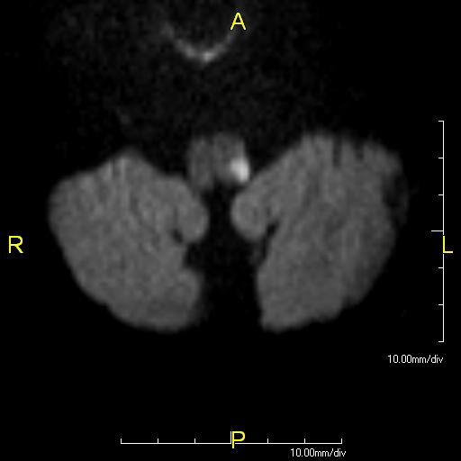
Lateral medullary (Wallenberg) syndrome is infarction of the dorsolateral medulla, usually from vertebral artery or PICA occlusion. Damage to the spinal trigeminal nucleus, spinothalamic tract, nucleus ambiguus (IX, X), vestibular nuclei, inferior cerebellar peduncle and descending sympathetic fibres gives ipsilateral facial pain and temperature loss, contralateral body pain and temperature loss, vertigo, ataxia, dysphagia, hoarseness and an ipsilateral Horner syndrome.
| Syndrome | Usual site | Notes |
|---|---|---|
| Pure motor hemiparesis | Posterior limb of internal capsule, corona radiata, ventral pons | Commonest (about 45%); face, arm and leg weakness, no sensory loss |
| Sensorimotor | Thalamus, internal capsule | Second commonest (about 20%) |
| Ataxic hemiparesis | Internal capsule, pons, corona radiata | Weakness with limb ataxia on the same side (10–18%) |
| Pure sensory | Thalamus | About 7%; may later cause thalamic pain |
| Dysarthria–clumsy hand | Pons or internal capsule | Dysarthria with a clumsy contralateral hand, strength preserved |
Lacunes are caused mainly by lipohyalinosis and microatheroma of small penetrating arteries; hypertension and diabetes are the key risk factors. Lacunar strokes make up about a quarter of ischaemic strokes.
How does management differ in haemorrhagic stroke?
| Point | Intracerebral haemorrhage | Subarachnoid haemorrhage |
|---|---|---|
| BP | Presenting SBP 150–220: lower toward 140, keep 130–150; below 130 may harm | Lower marked hypertension with short-acting titratable drugs until the aneurysm is secured; no single target |
| Key drug | Stop and reverse anticoagulants at once | Nimodipine 60 mg every 4 h for 21 days (prevents delayed ischaemia) |
| Procedure | Cerebellar ICH with deterioration, hydrocephalus or volume ≥ 15 mL → surgical evacuation | Coil or clip the aneurysm, ideally within 24 h |
| Steroids | Not used | Not used |
What are the common exam traps in stroke management?
- The clock starts at last known well, not when the patient was found. A patient who woke with symptoms has an unknown onset.
- Tenecteplase is now first-line alongside alteplase (0.25 mg/kg, max 25 mg) — older answer keys list alteplase only.
- Thrombolysis then thrombectomy — giving one does not cancel the other.
- BP thresholds: 185/110 to start thrombolysis, 180/105 for 24 h after it, 220/120 when no reperfusion therapy is given.
- No antithrombotics for 24 h after thrombolysis.
- DAPT is for minor stroke or high-risk TIA only, for 21 days — not long term.
- CEA is for symptomatic 50–99% stenosis, ideally within 2 weeks — the benefit falls sharply after that.
- Face and arm > leg = MCA; leg > arm = ACA; macular-sparing hemianopia = PCA.