Written & reviewed by: Kinase Medical Team · Last reviewed: 30 September 2026 · Editorial policy
Key features
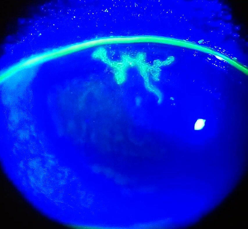
- Branching (dendritic) epithelial lesion with terminal bulbs and swollen borders on slit-lamp examination.
- Fluorescein shows the dendritic ulceration; rose bengal or lissamine green also stain the lesion.
- Usually HSV-1; the virus stays latent in the trigeminal ganglion and reactivates, so disease is often recurrent.
- Repeated attacks can reduce corneal sensation and cause scarring; HSV keratitis is a leading infectious cause of corneal blindness.
- Treatment: topical antivirals, cycloplegics and oral acyclovir.
Differential diagnosis
| Condition | How to tell it apart |
|---|---|
| Varicella-zoster keratitis | Can look similar clinically; PCR of corneal scrapings identified VZV in some lesions labelled HSV. |
| Acanthamoeba or fungal keratitis | Amoebic and fungal keratitis have been misdiagnosed as HSV keratitis; consider them when the lesion is atypical or does not respond to antivirals. |
How it appears in NEET PG
Presented as a fluorescein-stained cornea under cobalt blue light; questions ask for the organism, the characteristic shape or the drug to avoid.
Frequently asked questions
Why does a dendritic ulcer stain with fluorescein?
Fluorescein pools wherever the corneal epithelium is broken, so the branching epithelial defect lights up green under cobalt blue light.
Where does herpes simplex virus stay latent in ocular herpes?
In the trigeminal ganglion. Reactivation sends virus back along the sensory nerve to the cornea.
Can steroids be used for a dendritic ulcer?
Antivirals are the treatment. Topical steroids can prolong the infection and risk corneal melting and perforation, so they are used only with extreme caution.
Sources
- Azher TN et al. Herpes simplex keratitis: challenges in diagnosis and clinical management. Clin Ophthalmol 2017 (PMC review)
- Harrison's Principles of Internal Medicine (textbook)
Keep practising
- NEET PG Ophthalmology previous-year questions
- Kayser-Fleischer Ring
- Cherry-Red Spot (Central Retinal Artery Occlusion)
- Papilloedema
- Horner Syndrome
- All clinical images
For exam preparation and medical education only — not a substitute for clinical judgement, diagnosis or treatment of any patient.