Written & reviewed by: Kinase Medical Team · Last reviewed: 30 September 2026 · Editorial policy
Key features
- Optic disc swelling due to raised intracranial pressure; usually bilateral, though it can be asymmetric.
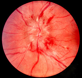
- Fundus: blurred disc margins, hyperaemia, elevated disc, vessels obscured at the disc edge, peripapillary haemorrhages, cotton-wool spots, Paton lines.
- Spontaneous venous pulsations are lost (not specific — some normal people lack them).
- Visual acuity is preserved early; fields show an enlarged blind spot. Transient visual obscurations are a classic symptom.
- Causes: idiopathic intracranial hypertension, hydrocephalus, intracranial mass, venous sinus thrombosis, diffuse cerebral oedema.
Differential diagnosis
| Condition | How to tell it apart |
|---|---|
| Optic neuritis (papillitis) | Usually one eye, early loss of acuity and a muted pupillary light reaction. |
| Pseudopapilloedema (optic disc drusen) | Elevated-looking disc without raised ICP; drusen can mimic papilloedema on imaging too. |
| Foster Kennedy syndrome | Papilloedema in one eye with optic atrophy in the other, classically from a frontal tumour or olfactory groove meningioma. |
How it appears in NEET PG
Shown as a fundus photograph with a raised-intracranial-pressure vignette (headache, vomiting) or idiopathic intracranial hypertension; follow-ups ask for the field defect or the likely cause.
Frequently asked questions
Is vision affected in papilloedema?
Visual acuity is usually normal early on. The typical field defect is an enlarged blind spot; acuity falls only when papilloedema is severe, long-standing or involves the macula.
Is papilloedema unilateral or bilateral?
Usually bilateral because the raised intracranial pressure affects both optic nerve sheaths, although the swelling can be asymmetric.
What is the difference between papilloedema and optic disc oedema?
Papilloedema is reserved for disc swelling caused by raised intracranial pressure; swelling from other causes such as optic neuritis or ischaemic optic neuropathy is called optic disc oedema.
Sources
- Radiopaedia — Papilloedema
- Harrison's Principles of Internal Medicine (textbook)
- Adams and Victor's Principles of Neurology (textbook)
Keep practising
- NEET PG Ophthalmology previous-year questions
- Kayser-Fleischer Ring
- Cherry-Red Spot (Central Retinal Artery Occlusion)
- Dendritic Ulcer (Herpes Simplex Keratitis)
- Horner Syndrome
- All clinical images
For exam preparation and medical education only — not a substitute for clinical judgement, diagnosis or treatment of any patient.