Written & reviewed by: Kinase Medical Team · Last reviewed: 30 September 2026 · Editorial policy
Key features
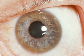
- Golden-brown to dark red-brown ring at the periphery of the cornea, from copper deposited in Descemet membrane (the deepest corneal layer).
- Early rings may be incomplete and visible only on slit-lamp examination, especially in brown eyes.
- Frequency depends on presentation: present in >95–99% of patients with neurological/psychiatric Wilson disease, but in only about 30–50% of those diagnosed in the hepatic or presymptomatic stage.
- Not pathognomonic: also reported in primary biliary cholangitis, chronic cholestasis and cryptogenic cirrhosis.
- Ring density tracks disease activity; rings shrink and may vanish on chelation therapy (e.g. penicillamine).
Differential diagnosis
| Condition | How to tell it apart |
|---|---|
| Arcus senilis | Yellow-white lipid ring, common in the elderly; at a young age it can signal hyperlipidaemia. |
How it appears in NEET PG
Typically shown as an eye photograph with a young patient who has liver disease or a movement disorder; the question may ask for the corneal layer involved or the underlying metabolic disorder.
Frequently asked questions
In which layer of the cornea is copper deposited in a KF ring?
Descemet membrane — the deepest layer of the cornea — at the corneal periphery.
Is a Kayser-Fleischer ring diagnostic of Wilson disease?
It is strongly suggestive but not pathognomonic; it has also been reported in chronic cholestatic liver diseases such as primary biliary cholangitis.
Do KF rings disappear with treatment?
Yes. With copper-chelating therapy the rings usually shrink and may disappear completely.
Sources
- Radiopaedia — Kayser-Fleischer rings
- Harrison's Principles of Internal Medicine (textbook)
- Adams and Victor's Principles of Neurology (textbook)
Keep practising
- NEET PG Ophthalmology previous-year questions
- Cherry-Red Spot (Central Retinal Artery Occlusion)
- Papilloedema
- Dendritic Ulcer (Herpes Simplex Keratitis)
- Horner Syndrome
- All clinical images
For exam preparation and medical education only — not a substitute for clinical judgement, diagnosis or treatment of any patient.