How is the brainstem organised, and why do its lesions cause crossed signs?
The brainstem joins the cerebrum to the spinal cord and cerebellum. From above down it has three parts — midbrain, pons and medulla oblongata — and it carries the nuclei of 10 of the 12 cranial nerves (all except I and II). Every level contains three kinds of structure: cranial nerve nuclei and fascicles, long tracts passing through (corticospinal, medial lemniscus, spinothalamic), and local nuclei (red nucleus, substantia nigra, olive, vestibular nuclei).
The corticospinal tract decussates at the lower end of the medulla (pyramidal decussation), and the dorsal column pathway crosses as the medial lemniscus in the lower medulla. A brainstem lesion therefore sits above these crossings. It damages a cranial nerve on its own side but the limb pathway destined for the opposite side. That gives the hallmark of a brainstem lesion: ipsilateral cranial nerve palsy + contralateral hemiplegia — a crossed or alternating hemiplegia.
What does a cross-section of the midbrain show at each level?
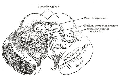
The midbrain surrounds the cerebral aqueduct, which joins the third and fourth ventricles and is ringed by periaqueductal grey. Behind the aqueduct lies the tectum with the paired superior and inferior colliculi (corpora quadrigemina); in front lies the tegmentum; the most anterior part is the crus cerebri (cerebral peduncle base), carrying corticospinal, corticonuclear and corticopontine fibres, with the substantia nigra between crus and tegmentum.
| Feature | Superior colliculus level | Inferior colliculus level |
|---|---|---|
| Tectum | Superior colliculi — visual reflexes, saccades | Inferior colliculi — auditory pathway |
| Cranial nerve nucleus | Oculomotor (III) nucleus with Edinger-Westphal nucleus | Trochlear (IV) nucleus |
| Tegmentum | Red nucleus, MLF, medial lemniscus | Decussation of superior cerebellar peduncles, MLF, lemnisci |
| Nerve exit | III leaves through the interpeduncular fossa on the medial crus | IV leaves dorsally, just below the inferior colliculi |
| Base | Substantia nigra + crus cerebri | Substantia nigra + crus cerebri |
What are Weber, Benedikt, Claude and Parinaud syndromes?
All the ventral and paramedian midbrain syndromes share an ipsilateral third nerve palsy — ptosis, an eye that rests down and out (unopposed lateral rectus and superior oblique) and, if the upper fascicles are involved, a dilated pupil. What is added depends on how far back the lesion reaches: the crus (hemiplegia), the red nucleus (tremor, ataxia) or the dorsal midbrain (vertical gaze).
| Syndrome | Site | Structures | Features | Usual artery |
|---|---|---|---|---|
| Weber (ventral midbrain; superior alternating hemiplegia) | Base of midbrain | III fascicles + cerebral peduncle (± substantia nigra) | Ipsilateral III palsy; contralateral hemiplegia incl. lower face and tongue | Paramedian branches of the posterior cerebral artery |
| Benedikt (paramedian midbrain) | Tegmentum | III fascicles + red nucleus (± peduncle) | Ipsilateral III palsy; contralateral tremor (Holmes), choreoathetosis, ataxia ± hemiparesis | PCA and paramedian basilar branches |
| Claude | Dorsal tegmentum | III + rubrodentate fibres | Ipsilateral III palsy; contralateral ataxia | PCA |
| Nothnagel | Tectum / superior cerebellar peduncle | III + superior cerebellar peduncle | Ipsilateral III palsy; ipsilateral limb ataxia; often bilateral (quadrigeminal tumours) | — |
| Parinaud (dorsal midbrain) | Pretectum at superior colliculus | Vertical gaze centres (riMLF, interstitial nucleus of Cajal), posterior commissure | Upgaze palsy, convergence-retraction nystagmus, light-near dissociation, lid retraction (Collier sign) | PCA / paramedian basilar; often compression |
Parinaud syndrome is classically caused by a pineal gland tumour pressing on the dorsal midbrain, though midbrain infarction, haemorrhage, multiple sclerosis and obstructive hydrocephalus also cause it. Neoplastic causes dominate in children and young adults; vascular causes in older patients. Downgaze is classically preserved and the vestibulo-ocular reflex is spared.
What lies at each level of the pons?
The pons has a bulging basilar (ventral) part — corticospinal fibres broken into bundles by transverse pontocerebellar fibres, with the basilar artery in the basilar groove on its surface — and a dorsal tegmentum forming the upper floor of the fourth ventricle. It joins the cerebellum through the middle cerebellar peduncles, the largest of the three.
- Upper pons: trigeminal (V) motor and principal sensory nuclei; CN V, the largest cranial nerve, exits the anterolateral upper pons.
- Lower pons: abducens (VI) and facial (VII) nuclei in the tegmentum near the pontomedullary junction. This region underlies the facial colliculus in the floor of the fourth ventricle.
- Pontomedullary groove: VI, VII and VIII emerge from medial to lateral.
- Throughout: medial lemniscus behind the corticospinal fibres, spinothalamic tract more laterally, MLF next to the midline.

What are Millard-Gubler, Foville, Raymond and lateral pontine syndromes?
Millard-Gubler syndrome (ventral pontine syndrome, 1858) is the textbook crossed palsy of the lower pons: a lesion of the basis pontis involving the facial (VII) fascicles and the corticospinal tract gives an ipsilateral lower motor neuron facial palsy with contralateral hemiplegia. The abducens (VI) is often involved too (ipsilateral lateral rectus palsy and diplopia), though it was not part of the original description. Sensory tracts are usually spared. In older patients the cause is mostly vascular — occlusion of basilar artery branches to the caudal pons; in the young, tumours, infections such as tuberculosis and neurocysticercosis, and demyelination.
| Syndrome | Structures | Features | Artery |
|---|---|---|---|
| Millard-Gubler (ventral, caudal pons) | VII ± VI fascicles + corticospinal tract | Ipsilateral LMN facial palsy (± lateral rectus palsy); contralateral hemiplegia | Paramedian/short circumferential branches of the basilar artery |
| Raymond | VI fibres + corticospinal tract | Ipsilateral lateral rectus palsy; contralateral hemiparesis | Basilar branches |
| Foville (inferior medial / lower dorsal pontine) | VI and VII nuclei, corticospinal tract, medial lemniscus, middle cerebellar peduncle | Ipsilateral facial palsy and lateral gaze palsy; contralateral hemiparesis and loss of proprioception; ipsilateral ataxia | Basilar branches |
| Marie-Foix (lateral pontine) | VII and VIII nuclei, corticospinal, spinothalamic and cerebellar tracts | Ipsilateral facial palsy, hearing loss, vertigo, nystagmus, limb ataxia; contralateral hemiparesis | AICA and basilar perforators |
| Locked-in (bilateral ventral pons) | Both corticospinal and corticobulbar tracts, VI nuclei | Quadriplegia, bilateral facial palsy, horizontal gaze palsy; consciousness, vertical eye movement and blinking preserved | Proximal–middle basilar artery |
What is locked-in syndrome and how is it different from coma?
Locked-in syndrome is quadriplegia and bulbar palsy (and often loss of body sensation) from brainstem damage — most commonly the anterior (ventral) pons — with preserved consciousness, vertical eye movements, blinking and hearing. The ventral pons is supplied by the basilar artery, so basilar thrombosis is the classic cause; trauma with vertebrobasilar injury is another.
| Type | What is preserved |
|---|---|
| Classical | Total immobility except vertical eye movements and blinking; normal consciousness |
| Incomplete | As classical, plus small remnants of other voluntary movement |
| Total | Complete paralysis including eye movements, with consciousness preserved |
The sparing makes anatomical sense: the lesion is ventral, so the reticular activating system in the dorsal tegmentum (consciousness) and the midbrain vertical gaze centres above the lesion survive, while horizontal gaze — organised in the pons around the abducens nucleus — is lost. Patients communicate with vertical eye movements or blinks. The diagnosis is easily missed because it looks like coma or akinetic mutism; a 'motor' score on the Glasgow Coma Scale will badly underestimate awareness.
What structures are seen in a cross-section of the medulla?
The anterior medulla carries the pyramids on either side of the anterior median fissure; at its lower end most corticospinal fibres cross in the pyramidal decussation to form the lateral corticospinal tract. Lateral to the pyramids are the olives. The hypoglossal nerve (XII) emerges from the sulcus between the pyramid and the olive; IX, X and XI emerge from the postolivary groove in that order from above down. The medulla meets the spinal cord at the foramen magnum.
| Medial medulla | Lateral (dorsolateral) medulla |
|---|---|
| Pyramid (corticospinal) | Spinothalamic tract |
| Medial lemniscus | Spinal trigeminal nucleus and tract |
| Hypoglossal nucleus and fibres | Nucleus ambiguus (IX, X) |
| MLF | Vestibular nuclei, inferior cerebellar peduncle |
| Supplied by anterior spinal artery / vertebral paramedian branches | Descending sympathetic fibres; supplied by vertebral artery / PICA |
How do lateral medullary (Wallenberg) and medial medullary (Dejerine) syndromes differ?
Lateral medullary syndrome (Wallenberg) is ischaemia of the dorsolateral medulla, most often from occlusion of the vertebral artery or the posterior inferior cerebellar artery (PICA). It is the commonest brainstem stroke syndrome you will be asked about, and its signs map neatly onto lateral structures.
| Structure damaged | Sign (side) |
|---|---|
| Spinal trigeminal nucleus and tract | Loss of pain and temperature on the face — ipsilateral |
| Lateral spinothalamic tract | Loss of pain and temperature on the body and limbs — contralateral |
| Descending sympathetic fibres | Horner syndrome — ipsilateral |
| Nucleus ambiguus | Dysphagia, dysarthria, hoarseness, impaired cough; persistent hiccups — ipsilateral |
| Vestibular nuclei | Vertigo, nausea, vomiting, nystagmus |
| Inferior cerebellar peduncle, spinocerebellar fibres | Limb ataxia — ipsilateral |
| Nucleus tractus solitarius | Impaired taste — ipsilateral |
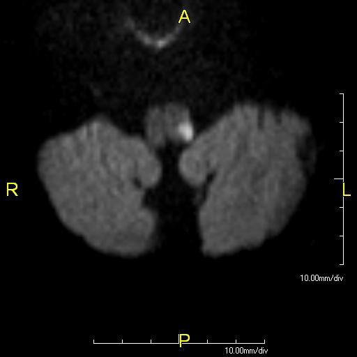
Medial medullary syndrome (Dejerine) is infarction of the medial medulla, most often atherothrombotic occlusion of paramedian branches of the anterior spinal artery or the vertebral artery. Its triad is ipsilateral hypoglossal palsy (the tongue deviates towards the lesion, a lower motor neuron sign) + contralateral hemiparesis (pyramid) + contralateral loss of vibration and proprioception (medial lemniscus). It is rare — about 1% of cerebral infarcts — and the full triad is seen in only a minority.
Acute management follows the usual posterior-circulation stroke pathway — see stroke management. Bulbar weakness makes swallow assessment and aspiration prevention an early priority in lateral medullary stroke.
What is the quickest way to revise all brainstem lesions?
| Level | Syndrome | Ipsilateral | Contralateral |
|---|---|---|---|
| Midbrain, ventral | Weber | III palsy | Hemiplegia (incl. lower face, tongue) |
| Midbrain, tegmentum | Benedikt | III palsy | Tremor/chorea, ataxia ± hemiparesis |
| Midbrain, dorsal | Parinaud | — (bilateral vertical gaze signs) | — |
| Lower pons, ventral | Millard-Gubler | VII (± VI) palsy | Hemiplegia |
| Lower pons, dorsal | Foville | VII palsy, horizontal gaze palsy, ataxia | Hemiparesis, loss of proprioception |
| Pons, lateral | Marie-Foix | VII, VIII, limb ataxia | Hemiparesis |
| Pons, bilateral ventral | Locked-in | Bilateral: quadriplegia, bulbar palsy | Vertical gaze and blinking preserved |
| Medulla, lateral | Wallenberg | Facial pain/temperature loss, Horner, IX–X palsy, ataxia | Body pain/temperature loss |
| Medulla, medial | Dejerine | XII palsy (tongue to lesion side) | Hemiparesis, loss of vibration/proprioception |